Clear cell renal cell carcinoma (ccRCC) remains a challenging malignancy to treat, with immune checkpoint inhibitors (ICIs) revolutionizing patient management. This pilot study, evaluated the efficacy and safety of combination therapy comprising camrelizumab, an anti-PD-1 antibody, and autologous cytokine-induced killer (CIK) cell therapy in patients with refractory ccRCC. Twenty-one patients with refractory ccRCC were randomly assigned to receive either camrelizumab monotherapy (control group, n = 12) or camrelizumab combined with CIK cell re-transfusion (trial group, n = 9). Due to early termination (21 of 60 planned patients), all endpoints were exploratory. The objective response rate (ORR) was numerically higher in the combination group (55.6% vs. 41.7%; odds ratio 1.75, 95% confidence interval [CI]: 0.32-9.51), but not statistically significant. Median progression-free survival (PFS) was 28.5 vs. 8.67 months (hazard ratio [HR] 0.40, 95% CI: 0.12-1.34), and median overall survival (OS) was not reached vs. 57.47 months (HR 0.48, 95% CI: 0.09-2.53). One patient in the trial group achieved a complete metabolic response (CMR). The combination was well-tolerated without new safety signals. Exploratory analysis suggested that higher baseline PD-1 expression on CD8+ T cells might be associated with a better response, and the frequency of PD-1 positive cells tended to decrease after camrelizumab administration. The addition of CIK cell therapy to anti-PD-1 antibody showed signals of potential benefit in refractory ccRCC with a tolerable safety profile. This pilot study suggests the combination approach appears feasible and warrants investigation in larger trials in pretreated ccRCC patients.Registry: ClinicalTrials.gov, TRN: NCT03987698, Registration date: 17 June 2019.
Anti-programmed cell death 1 (PD-1)/programmed death-ligand 1 (PD-L1) monoclonal antibody therapy has been approved as second-line treatment for advanced non-small-cell lung cancer (NSCLC). Unfortunately, the objective response rate (ORR) is not satisfied. T lymphocytes obtained by co-culture with personalized tumor-specific antigenic peptide-pulsed dendritic cells (DC), also known as multiple target cytotoxic T lymphocytes (MCTL) can restore the antitumor immunity and potentially improve clinical outcomes. We conducted a clinical study evaluating MCTL immunotherapy combined with toripalimab in patients with advanced NSCLC. This single-center, open-label, phase Ib trial (NCT04193098) evaluated the combination of MCTL and toripalimab as second-line therapy in 21 patients with advanced NSCLC. Peripheral blood samples were collected for antigenic peptide analysis, and patient immune status was assessed. The primary and secondary endpoints were to evaluate safety and clinical outcomes, respectively. Among the 21 patients, the ORR and disease control rate (DCR) were 33.3 and 76.2
E74-like ETS transcription factor 3 (ELF3) has been implicated in various tumorigenesis and inflammatory diseases. However, its expression profile and role in lung adenocarcinoma (LUAD) remain poorly defined. In the present study, through comprehensive clinical and experimental analyses, we aim to clarify the association between ELF3 overexpression in LUAD tissues and poor prognosis. Functional assays reveal that ELF3 knockdown inhibits the proliferation, migration, and invasion of LUAD cells, while ELF3 overexpression enhances these functions. Pathway enrichment analysis indicates that ELF3 influences the metabolic processes of LUAD. Mechanistically, ELF3 exerts oncogenic effects by regulating the transcription of hexokinase 2 (HK2) and glucose transporter type 1 (GLUT1). High-throughput screening reveals that dacinostat, by targeting the active site of the ELF3 protein, attenuates the glycolytic, proliferative, and metastatic abilities of LUAD cells. Additionally, ubiquitin-specific peptidase 18 (USP18) strengthens the stability of the ELF3 protein and influences the malignant biological behavior of LUAD through ELF3. In conclusion, the USP18/ELF3/HK2 and USP18/ELF3/GLUT1 axes play critical roles in glucose metabolism, proliferation, and metastasis of LUAD cells. Dacinostat inhibits the malignant progression of LUAD by targeting ELF3, providing strong evidence for developing novel therapeutic strategies targeting ELF3.
Cancer stem cells (CSC) and the immunosuppressive microenvironments are key drivers of breast cancer (BC) progression and drug resistance. However, the molecular mechanisms by which One Cut Homeobox 2 (ONECUT2) governs CSC and the tumor immune microenvironment (TIME) remain largely unknown. Given the critical knowledge gap, we sought to investigate ONECUT2's regulatory impact on CSC properties and TIME profiles using BC cell lines, animal models, and clinical specimens. Here, we demonstrated that inhibition of ONECUT2, a core transcription factor, potently drives CSC characteristics and reprograms the TIME to favor macrophage polarization to the M2-type, a tumor-promoting state. Mechanistically, ONECUT2 inhibition transcriptionally activated POU6F2, which subsequently triggered beta-catenin, thereby enhancing CSC properties and chemoresistance in BC. With respect to modulating the immune microenvironment, suppression of ONECUT2 governs macrophage polarization toward the immunosuppressive M2 phenotype. CCL28 is identified as a transcriptional target of ONECUT2 required for M2-type macrophage polarization, and CCR10 as a key receptor involved in this immune modulation. These findings highlight the critical involvement of ONECUT2 in modulating BC stemness via targeting the POU6F2-beta-catenin axis and managing macrophage polarization to M2 phenotype through the CCL28-CCR10 pathway. Our study suggests that ONECUT2 modulates cancer stemness and the TIME, and that targeting its downstream POU6F2-beta-catenin axis and CCL28-CCR10 pathway may provide an effective approach for BC treatment.
SUMOylation is characterized as a ubiquitin-like post-translational modification that exhibits mechanistic similarities with ubiquitination. Importantly, it modulates the ubiquitination levels of some target proteins, thereby influencing protein stability. Dysregulation of this modification pathway plays a pivotal role in the pathogenesis and progression of various tumors. While recent studies have demonstrated that protein SUMOylation regulates tumor progression, the specific function and mechanism of Ran-binding protein 2 (RanBP2), an E3 SUMO ligase, remain to be elucidated in lung adenocarcinoma (LUAD). This study demonstrates that RanBP2 exhibits elevated expression in LUAD and correlates with poor prognosis. Functionally, RanBP2 inhibits ferroptosis and enhances the growth of LUAD cells. Mechanistically, RanBP2 augments the SUMOylation of E3 ubiquitin ligase leucine-rich repeat and sterile alpha motif-containing protein 1 (LRSAM1), facilitating its subsequent degradation in the ubiquitin-proteasome pathway, thereby reducing SLC7A11 degradation. The stabilized SLC7A11 protein suppresses ferroptosis and promotes LUAD cell growth. The identified RanBP2 inhibitor, amodiaquine (AQ), in combination with ferroptosis inducer sulfasalazine (SAS), effectively triggered ferroptosis and suppressed LUAD cell growth in vitro and in vivo. This combination enhanced T-cell infiltration and improved anti-PD-1 immunotherapy efficacy. The research identifies a novel RanBP2-LRSAM1-SLC7A11 axis that promotes ferroptosis resistance and LUAD progression. The combination of AQ and SAS represents a promising therapeutic strategy for LUAD.
Introduction:Peripheral T-cell lymphoma (PTCL) is a heterogeneous and highly aggressive subtype of non-Hodgkin lymphoma. Approximately 30% of patients develop relapsed or refractory PTCL (R/R PTCL) due to disease recurrence or failure to achieve complete remission after first-line therapy. Despite therapeutic advances, the molecular and cellular mechanisms underlying treatment resistance in R/R PTCL remain unclear. Methods:Single-cell RNA sequencing and single-cell T-cell receptor sequencing were performed on seven tumor samples from six patients with R/R PTCL. These approaches were used to systematically characterize the transcriptional profiles of malignant T-cell clones and reactive T lymphocytes, define the transcriptomic landscape of R/R PTCL, and identify potential epigenetic biomarkers associated with drug response. Results:We observed significant upregulation of genes associated with cell proliferation, oncogenic signaling, and immune modulation in R/R PTCL. Within the tumor microenvironment, specific protumorigenic ligand-receptor interactions were identified, including CXCL13-CXCR5, CCL5-CCR5, and CD74-MIF interactions, which may facilitate immune evasion by malignant T cells. Longitudinal analysis of a patient who progressed following dual epigenetic therapy revealed marked downregulation of immune response-related genes, including HLA-DRA/DPA1/DRB5, CD74, C1QC, and LYZ, as well as functional reprogramming of tumor-associated macrophages. Enhanced LGALS9-HAVCR2 and CSF1-CSF1R interactions were also observed following combination treatment with chidamide and azacitidine. Discussion:This study delineates the transcriptional heterogeneity of malignant T-cell clones in R/R PTCL and suggests that this heterogeneity may contribute to resistance to epigenetic therapies. These findings provide novel insights into the molecular mechanisms underlying treatment resistance and highlight potential avenues for therapeutic intervention in R/R PTCL.
Clear cell renal cell carcinoma (ccRCC) remains a challenging malignancy to treat, with immune checkpoint inhibitors (ICIs) revolutionizing patient management. This clinical study, involving a small cohort, evaluated the efficacy and safety of combination therapy comprising camrelizumab, an anti-PD-1 antibody, and autologous cytokine-induced killer (CIK) cell therapy in patients with refractory ccRCC. Twenty-one patients with refractory ccRCC were randomly assigned to receive either camrelizumab monotherapy (control group, n = 12) or camrelizumab combined with CIK cell re-transfusion (trial group, n = 9). The objective response rate (ORR), progression-free survival (PFS), overall survival (OS), and safety were evaluated. Biomarker exploration was performed using peripheral blood samples from patients. A trend towards a higher ORR was observed in the trial group (55.6%) compared to the control group (41.7%). Favorable trends were also noted for median PFS (28.50 vs. 8.67 months) and median OS (not reached vs. 57.47 months) in the trial and control groups, respectively. One patient in the trial group achieved a complete metabolic response (CMR). No new safety signals were observed, and CIK cell re-transfusion did not appear to increase the frequency of adverse effects. Exploratory analysis suggested that higher baseline PD-1 expression on CD8 + T cells might be associated with a better response, and the frequency of PD-1 positive cells tended to decrease after camrelizumab administration. The addition of CIK cell therapy to anti-PD-1 antibody showed encouraging signals of antitumor activity in refractory ccRCC with a tolerable safety profile. These preliminary findings warrant further investigation, which could focus on the combination of ICIs, antiangiogenic agents, and CIK cell re-transfusion as a potential first-line therapy for metastatic or advanced ccRCC.Registry: ClinicalTrials.gov, TRN: NCT03987698, Registration date: 17 June 2019.
Clinical trials have shown that neoadjuvant anlotinib combined with PD-1 blockade therapy can prolong the survival of patients with driver gene negative non-small cell lung cancer (NSCLC), but some patients fail to benefit from the combination therapy. To explore the potential drug resistance mechanism and predict the efficacy of neoadjuvant therapy in NSCLC patients, we used scRNA-seq to observe and analyze the dynamic changes of immune cells, stromal cells and cancer cells in NSCLC patients who received neoadjuvant combination therapy. We analyzed transcriptome data of 47,000 single cells from 9 NSCLC patients, including 3 treatment naïve patients, 3 post-treatment patients with major pathological response (MPR), and 3 Non-MPR patients. Subsequently, the infiltration of immune cells was detected by immunohistochemistry and multiplex immunofluorescence in NSCLC. In MPR patients, we found that neoadjuvant therapy reduced the expression of the T cell exhausted signature, reduced the transition of T_THEMIS cells to Tregs, and enhanced the positive feedback between CD4+ T cells and PAX5+ memory B cells. In Non-MPR patients, tumor-associated macrophages (TAMs) dampen therapeutic efficiency by being the hub of cell communication. TAMs and fibroblasts stimulate endothelial cells via VEGF, endothelial ZEB1 may up-regulate FLT1 (VEGFR) expression in response to anlotinib, and VEGFR+ endothelial cell signature can predict survival of NSCLC cohort in TCGA. In addition, PLA2G4A, the key enzyme in the VEGF pathway, was highly expressed in the tumor cells of Non-MPR patients after anlotinib treatment. In 135 NSCLC patients, we confirmed by immunohistochemistry that PLA2G4A was positively correlated with poor prognosis and Tregs infiltration. In conclusion, VEGF signaling dependent dynamic changes in endothelial and epithelial cells are deeply involved in the formation of anlotinib resistance and immunosuppression phenotypes in NSCLC patients.
Background:This study aimed to investigate the risk factors associated with grade ≥3 diarrhea and neutropenia, which are the most common adverse events (AEs) leading to discontinuation and dose reduction in patients with hormone receptor-positive (HR+)/human epidermal growth factor 2-negative (HER2-) breast cancer treated with cyclin-dependent kinase 4/6 (CDK4/6) inhibitor abemaciclib combined with endocrine therapy (ET). Subsequently, two prediction nomograms were developed to serve as a foundation for enhancing the management of patients' side effects and improving treatment quality. Methods:A retrospective cohort analysis was conducted to explore the clinical characteristics and treatment variables of breast cancer patients treated with abemaciclib combined with ET in Yunnan Cancer Hospital from December 2021 to December 2022. Logistic regression was used to determine the risk factors for the occurrence of grade ≥3 diarrhea and neutropenia, and two kinds of nomograms were established. An external validation group of patients from three additional centers was used to validate the constructed nomograms. The area under the receiver operating characteristic (ROC) curve (AUC), calibration curve, and decision curve analysis (DCA) were used to assess the predictive performance and clinical applicability of the two nomograms. Results:A total of 497 patients were included, including 403 in the modeling group and 94 in the external validation group. The results of the multifactorial analysis revealed that age ≥70 years, Eastern Cooperative Oncology Group (ECOG) score ≥1, and underlying gastrointestinal diseases were independent risk factors for grade ≥3 diarrhea. ECOG score ≥1, radiotherapy in the same period/within 1 month, and neutrophils ≤2.0×10⁹/L before treatment were independent risk factors for grade ≥3 neutropenia. Two nomogram models were used to predict risk based on the above independent factors. The AUCs for the developmental and external validation groups were 0.747(95%CI:0.687-0.806) and 0.803(95%CI:0.702-0.918) for the diarrhea prediction nomogram and 0.765(95%CI:0.711-0.818) and 0.783(95%CI:0.691-0.892) for the neutropenia prediction nomogram, respectively. Calibration curves and DCA of both models also showed good predictive performance and clinical applicability. Conclusion:We identified risk factors for grade ≥3 diarrhea and neutropenia in patients treated with abemaciclib combined with ET, and established a risk prediction nomogram, providing a scientific basis for safety assessment.
Colorectal tumorigenesis generally progresses from adenoma to adenocarcinoma, accompanied by dynamic changes in the tumor microenvironment (TME). A randomized controlled trial has confirmed the efficacy and safety of Shen-Bai-Jie-Du decoction (SBJDD) in preventing colorectal tumorigenesis. However, the mechanism remains unclear. In this study, we employed single-cell RNA sequencing (scRNA-seq) to investigate the dynamic evolution of the TME and validated cell infiltration with multiplex immunohistochemistry and flow cytometry. Bulk RNA sequencing was utilized to assess the underlying mechanisms. Our results constructed the mutually verifiable single-cell transcriptomic atlases in ApcMin/+ mice and clinical patients. There was a marked accumulation of CCL22+ dendritic cells (DCs) and an enhanced immunosuppressive action, which SBJDD and berberine reversed. Combined treatment with cholesterol and lipopolysaccharide induced characteristic gene expression of CCL22+ DCs, which may represent “exhausted DCs”. Intraperitoneal injection of these DCs after SBJDD treatment eliminated its therapeutic effects. TMEM131 derived CCL22+ DCs generation by TNF signaling pathway and may be a potential target of berberine in retarding colorectal tumorigenesis. These findings emphasize the role of exhausted DCs and the regulatory mechanisms of SBJDD and berberine in colorectal cancer (CRC), suggesting that the multi-component properties of SBJDD may help restore TME homeostasis and offer novel cancer therapy.
Background: Clear cell renal cell carcinoma (ccRCC) remains a challenging malignancy to treat, with immune checkpoint inhibitors (ICIs) revolutionizing patient management. This clinical study, involving a small cohort, evaluated the efficacy and safety of combination therapy comprising camrelizumab, an anti-PD-1 antibody, and autologous cytokine-induced killer (CIK) cell therapy in patients with refractory ccRCC. Methods: Twenty-one patients with refractory ccRCC were randomly assigned to receive either camrelizumab monotherapy (control group, n=12) or camrelizumab combined with CIK cell re-transfusion (trial group, n=9). The objective response rate (ORR), progression-free survival (PFS), overall survival (OS), and safety were evaluated. Biomarker exploration was performed using peripheral blood samples from patients. Results: The ORR was higher in the trial group (55.6%) compared to the control group (41.7%). The median PFS and OS in the trial group were 28.50 months and not reached, respectively, whereas in the control group, these values were 8.67 months and 57.47 months, respectively. One patient in the trial group achieved a complete metabolic response (CMR). No new safety signals were observed, and CIK cell re-transfusion did not increase the frequency of adverse effects. Higher baseline PD-1 expression on CD8+ T cells in the peripheral blood was associated with a better response, and PD-1 positive frequency decreased after camrelizumab administration. Conclusions: CIK cell therapy enhances the antitumor efficacy of anti-PD-1 antibody therapy in refractory ccRCC with tolerable adverse effects. Further investigation should focus on the combination of ICIs, antiangiogenic agents, and CIK cell re-transfusion as first-line therapy for metastatic or advanced ccRCC. Registry: ClinicalTrials.gov, TRN: NCT03987698, Registration date: 17 June 2019.
This study aimed to examine the spatial distribution of immune cells by application of Gcross function in 170 patients with stage I to IIIA lung adenocarcinoma (LUAD) and explore its prognostic value. A total of 170 stage I to IIIA LUAD patients who underwent radical surgery were enrolled. Paraffinized tumor sections were collected for 2 panels of multicolor immunofluorescence staining (panel 1: CD4, CD8, FOXP3, CD69, CD39, CD73, and DAPI; panel 2: CD68, CD163, CD20, CD11c, PDL1, IDO, and DAPI). The immune cells were categorized as CD8(+), CD4(+) T helper cell (CD4Th), regulatory T cell, macrophage type 1 (M1), M2, dendritic cell (DC), and B cell. The immune cell numbers were enumerated, and the immune cell proximity score was calculated employing the Gcross function. The correlation between immune cell variables and disease-free survival (DFS) was explored through univariate Cox regression analyses. Factors with P < 0.05 were subjected to multivariate analyses. According to univariate Cox regression analyses, total PDL1(+) and PDL1(+) DC counts were negative factors (P = 0.003 and 0.031, respectively). CD4Th and IDO-DC counts were positive factors (P = 0.022 and 0.024, respectively). The proximity score (M1 to M2) was a positive factor for DFS (P = 0.032), and the proximity score (PDL1 (+) DC to M1) was a negative factor (P = 0.009) according to univariate Cox analyses. In multivariate analyses, stage (IIIA vs I + II) (hazard ratio [HR]: 1.77 [95% confidence interval (CI): 1.18-2.64], P = 0.006) and proximity score (PDL1 (+) DC to M1) (HR: 1.60 [95% CI: 1.07-2.37], P = 0.021) were independent negative factors and CD4Th counts (HR: 0.60 [95% CI: 0.40-0.90], P = 0.013) was an independent positive factor. Our study indicated that a higher level of tumor-infiltrating CD4Th cells predicted longer DFS, and a closer proximity of PDL1(+) DCs to M1 cells was associated with dismal DFS in stage I to IIIA LUAD patients.
8554 Background: The safety and efficacy of combination of gefitinib with chemotherapy and bevacizumab in treatment patients with epidermal growth factor receptor (EGFR) mutations are currently unknown. This phase Ib clinical trial was designed to evaluate the safety and preliminary efficacy of a combination therapy consisting of gefitinib, bevacizumab, pemetrexed, and carboplatin in patients with advanced non-squamous non–small cell lung cancer (NSCLC) harboring EGFR mutations. Methods: Eligible patients with EGFR-mutated advanced non-squamous NSCLC were recruited and received gefitinib combination with bevacizumab plus pemetrexed and carboplatin treatment. The primary endpoints were safety and progression-free survival (PFS). Secondary endpoints included objective response rate (ORR), disease control rate (DCR), duration of response (DOR), and overall survival (OS). Results: From June 2019 to June 2021, 20 patients were enrolled in this trial. The median follow-up was 33.8 months (95% CI, 31.0-36.6). Grade ≥3 adverse events was 65%, including neutropenia (30%), thrombocytopenia (20%), nausea (20%), skin rash (20%), bleeding (10%), and increased ALT (10%). There was no death related to toxicity occurred. The median PFS was 28 months (95% CI, 20.4-35.6). the ORR was 95% (95% CI, 75.1%-99.9%), the DCR was 100% (95% CI, 83.2%-100%), and the median DOR was 26.4 months (95% CI, 18.9-33.9). The median OS has not been reached. Conclusions: The results of this study demonstrate that the four-drug combination regimen, led by gefitinib, is well-tolerated and effective for patients with EGFR-mutated advanced non-squamous NSCLC.
AbstractObjectiveImmunotherapy, specifically immune checkpoint inhibitors (ICIs), has revolutionized cancer treatment. However, it can also cause immune‐related adverse events (irAEs). This study aimed to develop a clinically practical animal model of irAEs using BALB/c mice.MethodsSubcutaneous tumors of mouse breast cancer 4T1 cells were generated in inbred BALB/c mice. The mice were treated with programmed death‐1 (PD‐1) and cytotoxic t‐lymphocyte antigen 4 (CTLA‐4) inhibitors once every 3 days for five consecutive administration cycles. Changes in tumor volume and body weight were recorded. Lung computed tomography (CT) scans were conducted. The liver, lungs, heart, and colon tissues of the mice were stained with hematoxylin–eosin (H&E) staining to observe inflammatory infiltration and were scored. Serum samples were collected, and enzyme‐linked immunosorbent assay (ELISA) was used to detect the levels of ferritin, glutamic‐pyruvic transaminase (ALT), tumor necrosis factor‐α (TNF‐α), interferon‐gamma (IFN‐γ), and interleukin‐6 (IL‐6). Mouse liver and lung cell suspensions were prepared, and changes in macrophages, T cells, myeloid‐derived suppressor cells (MDSCs), and regulatory (Treg) cells were detected by flow cytometry.ResultsMice treated with PD‐1 and CTLA‐4 inhibitors showed significant reductions in tumor volume and body weight. The tissue inflammatory scores in the experimental group were significantly higher than those in the control group. Lung CT scans of mice in the experimental group showed obvious inflammatory spots. Serum levels of ferritin, IL‐6, TNF‐α, IFN‐γ, and ALT were significantly elevated in the experimental group. Flow cytometry analysis revealed a substantial increase in CD3+T cells, Treg cells, and macrophages in the liver and lung tissues of mice in the experimental group compared with the control group, and the change trend of MDSCs was opposite.ConclusionsThe irAE‐related animal model was successfully established in BALB/c mice using a combination of PD‐1 and CTLA‐4 inhibitors through multiple administrations with clinical translational value and practical. This model offers valuable insights into irAE mechanisms for further investigation.
Objective: The combination of epithelial growth factor receptor tyrosine kinase inhibitors (EGFR-TKIs) and immune checkpoint inhibitors (ICIs) leads to an increased incidence of severe immune-related adverse events (irAEs). However, the mechanisms underlying macrophages in irAEs have not been elucidated. Methods: An osimertinib and ICI-induced irAE mouse model was constructed. Lung micro-CT scans were used to assess the degree of inflammatory infiltration. Hematoxylin-eosin staining was used to analyze the histopathologic inflammatory infiltration in mouse liver and lung tissues. Flow cytometry was used to detect the percentages of T cells, NK cells, and macrophages and the expression of EGFR. Enzyme-linked immunosorbent assay (ELISA) was used to detect the serum interleukin (IL)-6, alanine transaminase (ALT), ferritin, and tumor necrosis factor (TNF)-α levels. Total RNA extracted from mouse liver macrophages was analyzed by RNA-seq. Simple Western blot analysis was used to detect the IL-6/JAK/STAT3 pathway activation state. Results: Osimertinib combined with ICIs upregulated EGFR expression on macrophages with increased serum IL-6, ALT, and ferritin levels. RNA-seq and simple Western blot analysis of mouse liver macrophages confirmed that that the IL-6/JAK/STAT3 pathway was activated in the combination treatment group. Ruxolitinib blocked the IL-6/JAK/STAT3 pathway and significantly decreased the serum IL-6, ALT, and ferritin levels in the combination treatment group. Conclusions: An osimertinib and ICI-induced irAE mouse model was constructed that showed osimertinib combined with ICIs inhibited EGFR phosphorylation and activated the IL-6/JAK/STAT3 signaling pathway in mouse liver macrophages, which led to the release of relevant cytokines.
Mature T-and natural killer(NK)-cell lymphomas are heterogeneous groups of malignant lymphoid neoplasms arising from T and NK cells. The incidence of mature T-and NK-cell lymphomas is 2.1 per 100,000 people, according to a US report~1.
The combination of epithelial growth factor receptor tyrosine kinase inhibitors (EGFR-TKIs) and immune checkpoint inhibitors (ICIs) leads to an increased incidence of severe immune-related adverse events (irAEs). Macrophages express epidermal growth factor receptor (EGFR), and play an important role in irAEs. However, the underlying mechanisms of EGFR-positive macrophages in irAEs have not been elucidated. In this study, we constructed an irAE mouse model induced by osimertinib combined with ICIs, and explore the role of EGFR-positive macrophages in irAEs. Our results found there was no significant difference in the degree of histopathological inflammatory infiltration and the percentage of macrophages in the ICIs group or the osimertinib + ICIs group, but osimertinib + ICIs increased the expression of EGFR on macrophages, accompanying with increased serum levels of interleukin (IL)-6, alanine transaminase (ALT) and ferritin. RNA-seq and Simple Western blot confirmed that IL-6/JAK/STAT3 pathway was activated in macrophages of osimertinib + ICIs group. Ruxolitinib could significantly decrease serum levels of IL-6, ALT and ferritin in oxitinib + ICIs group. Taken together, we successfully constructed an irAE mouse model induced by osimertinib combined with ICIs, and found that osimertinib increased immune checkpoint inhibitors-related severe adverse events via activating the IL-6/JAK/STAT3 pathway of EGFR-positive macrophages.
Murine cytokine-induced killer (CIK) cells are heterologous cells that kill various allogeneic and isogenic tumors and have functional and phenotypic characteristics of natural killer cells and T lymphocytes. However, the effect of CIK cells alone on solid tumor therapy is only limited. To enhance the therapeutic effect, it is vital to discover a mix of several therapy approaches. Immune cell function is inhibited by abnormal tumor vessels and the tumor microenvironment, which block lymphocyte entry into tumor tissue. To increase the effectiveness of CIK cells' antitumor activity, antivascular therapy and CIK cell therapy can be combined. Furthermore, anlotinib is a tiny drug with multitarget tyrosine kinase inhibitors that can block cell migration, delay angiogenesis, and decrease blood vessel density. Compared with other antiangiogenesis drugs, anlotinib stands out due to the wider target of action and lower effective dose. In this work, anlotinib and murine CIK cells were coupled to boost CD3(+) T cell infiltration, CD3(+)CD4(+) T cell infiltration, and expression of granzyme B and interferon gamma from CD3(+)CD8(+) T cells, which increased the antitumor activity. Through the generation of cytotoxic cytokines by T lymphocytes, the therapeutic group using anti-PD-1 monoclonal antibodies in conjunction with anlotinib and CIK cells was more successful than the group receiving dual therapy. The preclinical study contributes to exploring the therapeutic alternatives for patients with lung adenocarcinoma, thus prolonging their lives.
Background: Antihistamines alleviate the side effects of antitumor drugs and exert antitumor effects. This study aimed to investigate the potential impact of short-term concomitant use of antihistamines with immune checkpoint inhibitor (ICI) therapy on the efficacy and immune-related adverse events (irAEs) of immunotherapy for patients with advanced lung cancer. Methods: We retrospectively analyzed the medical records of 211 patients diagnosed with advanced primary lung cancer and treated with immunotherapy at Tianjin Medical University Cancer Institute and Hospital between January 1, 2018, and January 1, 2022. Among these patients, 109 who received H1 antihistamine during the infusion of anti-programmed cell death-1 (PD-1) and anti-programmed cell death ligand 1 (PD-L1) antibodies were assigned to the experimental group; meanwhile, the remaining 102 patients who did not receive H1 antihistamines were assigned to the control group. Balancing was achieved through inverse probability of treatment weight (IPTW) estimation. The data were analyzed using Kaplan- Meier curves and Cox regression analyses. Results: The median progression-free survival (mPFS) was 12.7 months in the experimental group and 4.3 months in the control group, while the median overall survival (mOS) was 32.8 months in the experimental group and 18.1 months in the control group. In the experimental group, patients treated with only H1 antihistamines had longer mPFS and mOS compared with those who received H1 plus H2 antihistamines. Similarly, in the control group, patients who did not receive antihistamines had a longer mPFS and mOS than those who only received H2 antihistamines. After conducting multivariate analyses, we found that H1 and H2 antihistamines were respectively identified as good and poor independent prognostic factors for both progression-free survival (PFS) and overall survival (OS). The rates of irAEs in the experimental and control groups were 52.4% and 69.2%, respectively, and grade >= 3 irAEs occurred in 4.5% and 25.9% of patients, respectively. Conclusions: Concomitant use of H1 antihistamines can improve immunotherapy efficacy and reduce irAEs. Meanwhile, concomitant use of H2 antihistamines is associated with reduced PFS and OS time.
 Xiubao Ren (任秀宝)合作论文数Tianjin Medical University Cancer Institude & Hospital38
 Wei Yan (闫卫)合作论文数Department of Cell Biology, College of Life Sciences, Wuhan University15
 Emily Wang合作论文数Department of Immunology and Biotherapy, Tianjin Medical University Cancer Institute and Hospital/Department of Pathology, University of California, San Diego14
 Liang Liu (刘亮)合作论文数Tumor Hospital, Tianjin Medical University;Key Laboratory of Cancer Prevention and Therapy of Tianjin,Tianjin ,China;Tumor Hospital, Tianjin Medical University11
 Lin Yang (杨林)合作论文数Cancer Hospital Chinese Academy of Medical Sciences11
 Donald Pizzo合作论文数10Minghui Cao合作论文数Dept Pathol, Univ Calif San Diego8Yuan Meng (孟元)合作论文数8
 Xiwei Wu合作论文数8Qian Sun (孙倩)合作论文数天津医科大学肿瘤医院8RUAN Xianhui (阮先辉)合作论文数7Wenwen Yu (于文文)合作论文数7Yanjuan Xiong合作论文数6
 Chuan Dong (董川)合作论文数School of Chemistry and Chemical Engineering, Shanxi University;School of Environmental & Resource Sciences, Shanxi University6
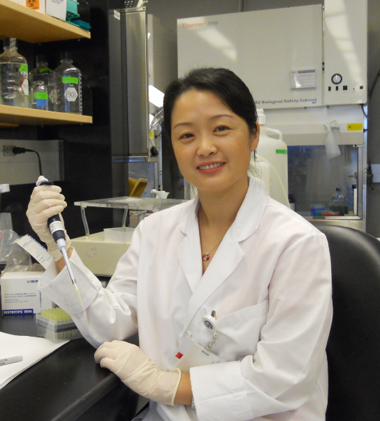 Lili Yang (杨莉莉)合作论文数Department of Biochemistry,Institute of Frontier Medical Sciences,Jilin University;Deparment of Immunology,Tianjin Cancer Hospital and Institute;Tianjin Medical University6Runmei Li (李润美)合作论文数6Weijiao Du合作论文数5Jiali Zhang合作论文数5Yang Wang合作论文数5jing zhai合作论文数5Xinwei Zhang合作论文数5
 George Somlo合作论文数5
 Jian Wang (王健)合作论文数Tianjin Medical University Cancer Institute & Hospital5Chin Andrew R合作论文数5Donald P. Pizzo合作论文数5
 Peiguo Chu合作论文数Department of Anatomic Pathology, City of Hope National Medical Center5Zhenzhen Hui合作论文数4
 Feng Wei合作论文数4Shiyong Zhou (周世勇)合作论文数4Lihua Qiu (邱立华)合作论文数4Zhengzi Qian (钱正子)合作论文数4Shui Cao (曹水)合作论文数天津市肿瘤医院4Ying Han (韩颖)合作论文数4Lan-Fang Li合作论文数4Xianhuo Wang合作论文数4
 Huilai Zhang (张会来)合作论文数Tianjin Medical University Cancer Institute and Hospital4Yingge Lv合作论文数3Li Zhou合作论文数3Wenchen Gong合作论文数3Baihui Li合作论文数3Fan Yang合作论文数3
 Bin Meng (孟斌)合作论文数Department of Pathology, Tianjin Medical University Cancer Institute and Hospital3Baozhu Ren合作论文数3Jingwei Yu合作论文数2Tianyuan Ren合作论文数2Yuheng Hong合作论文数2Yue Fei合作论文数2




