Prenatal diagnosis (PND) is an effective measure to prevent births with transfusion-dependent β-thalassemia (TDT) and other severe hemoglobinopathies. However, multicentric consolidated data from high-volume Indian referral laboratories is limited. The authors collated a multi-center experience of invasive PND for β-thalassemia and the major hemoglobinopathies, including center-wise volumes, affected-fetus rates, trends in primigravida referrals, and diagnostic challenges from seven Indian tertiary-care referral centers with a minimum of 10 y PND services. Retrospective data were contributed by SGRH-New Delhi, ICMR-NIIH-Mumbai, PGIMER-Chandigarh, AIIMS-New Delhi, NIMS-CDFD-Hyderabad, CMC-Vellore and SGPGI-Lucknow. Carrier evaluation was based on blood counts and Hb-HPLC. Fetal diagnosis utilized chorionic villus sampling or amniocentesis with molecular testing by ARMS-PCR, reverse dot-blot and/or Sanger sequencing as per local practices. Descriptive analyses summarized volumes, proportions of fetuses with biallelic HBB variants, and primigravida referral trends. Across centers, 6,780 PND procedures were performed over 2015–2024, with ICMR-NIIH and SGRH contributing 58.2
BackgroundKaryotyping is the standard confirmatory test for identifying chromosomal abnormalities, such as Trisomy 21, which requires amniotic fluid from pregnant women. The present study investigated the potential of methylated cell-free DNA (mcf-DNA) and methylated extracellular vesicle-derived DNA (mev-DNA) as an early, non-invasive epigenetic approach, specifically focusing on fetal-specific methylated regions (FSMRs) of RASSF1A, ERG, and UMODL1 (U1 and U2) to assess Trisomy 21 risk in maternal plasma.MethodsBlood samples were collected from pregnant women (n = 120) between 10th and 24th weeks of gestation who were at higher risk for Trisomy 21. Of the 120 women, 3 were found to be positive for Trisomy 21 through karyotyping method. Moreover, mcf-DNA and mev-DNA were isolated from Trisomy-positive and age-matched healthy pregnant women (n = 8) and non-pregnant women (n = 8). FSMRs were analyzed using qPCR to compare the cycle threshold (Ct) values. Gene copy number analysis was performed using the plasmid standards method to assess Trisomy 21 detection sensitivity.ResultsTrisomy pregnancies had significantly lower mean Ct values for RASSF1A and UMODL1 (U1 and U2) in both mcf-DNA and mev-DNA than healthy pregnancies, which was further confirmed by higher copy numbers in trisomy pregnancies than in healthy pregnancies. Moreover, the gene copy number in mcf-DNA was significantly higher than that in mev-DNA for the RASSF1A, ERG, and UMODL1 (U1 and U2) genes in trisomy pregnancies.ConclusionThis pilot study demonstrates the feasibility of using mcf-DNA and mev-DNA for detecting Trisomy 21-associated fetal methylation signatures in maternal plasma. While consistent with earlier findings, these results validate the applicability of methylated DNA immunoprecipitation (MeDIP)-based qPCR methylation assays in a North Indian cohort and support their potential integration into population-specific non-invasive prenatal screening strategies.
Van den Ende-Gupta syndrome (VDEGS) is an autosomal recessive disorder involving self-resolving joint contractures that are present since birth. Because of characteristic facial features, clinical diagnosis may be suspected by experienced clinicians or identified using facial recognition-based software. Confirmation by identification of disease-causing biallelic variants in SCARF2 can be helpful to definitively provide a clinico-molecular diagnosis to differentiate VDEGS from other disorders with overlapping phenotypes with blepharophimosis, arachnodactyly, and camptodactyly. Most affected individuals do not have major malformations or intellectual disability. However, occasional patients with ophthalmological, genitourinary, or laryngeal abnormalities are reported, indicating the need for a detailed evaluation. The causative gene, SCARF2, is located within the region of the 22q11.2 microdeletion syndrome. Two cases with microdeletion of the region on one chromosome and a SCARF2 variant on the other chromosome have been reported. Two cases resembling the phenotype but with no causative variants in SCARF2 have been reported, supporting the possibility of another causative gene with overlapping phenotype. Though prognosis can often be positive, there is a need for better long-term outcome data.
OBJECTIVES:To evaluate the short- and long-term clinical and laboratory outcomes of Enzyme Replacement Therapy (ERT) in Indian patients with Gaucher disease (GD) and to identify factors influencing the therapeutic response. METHODS:This retrospective, multicentre cohort study included 204 patients with confirmed GD across 13 Indian centres. Eligible participants had received at least one year of ERT and provided both baseline and follow-up data. Patients were stratified by splenectomy status. Longitudinal outcomes assessed over 1-5, 10, and 15 y included hemoglobin levels, platelet counts, liver and spleen volumes, chitotriosidase activity, bone pain, bone mineral density (BMD), and height/weight Z-scores. Subgroup analyses evaluated the impact of age at ERT initiation, GD subtype, genotype, and baseline disease severity. RESULTS:Of the 204 patients, 173 were non-splenectomised and 31 were post-splenectomy; notably, 136 (66.7%) presented with the GD3 phenotype. The median age at symptom onset, diagnosis, and ERT initiation was 1.5 y, 2.6 y, and 4.3 y respectively. The p.L483P allele was the predominant variant in the cohort. Within the first 1-5 y of ERT, non-splenectomised patients demonstrated marked improvements in hematological parameters, organomegaly, biomarker activity, growth metrics, and bone pain. These improvements were sustained through 10-15 y, characterized by stabilized visceral parameters, persistently low biomarker levels, steady growth, and a low incidence of new skeletal complications. Early initiation of ERT was associated with a greater magnitude of hematological response as early as 1 y. CONCLUSIONS:ERT facilitates rapid clinical and laboratory improvements within 1-5 y, with benefits sustained over 10-15 y in Indian GD patients. Despite significant diagnostic delays and the advanced stage of disease at presentation common in this population, long-term outcomes remain highly favourable. Early initiation of ERT is critical for optimizing treatment response and preventing irreversible sequelae.
Van den Ende-Gupta syndrome (VDEGS) is an autosomal recessive disorder involving self-resolving joint contractures that are present since birth. Because of characteristic facial features, clinical diagnosis may be suspected by experienced clinicians or identified using facial recognition-based software. Confirmation by identification of disease-causing biallelic variants in SCARF2 can be helpful to definitively provide a clinico-molecular diagnosis to differentiate VDEGS from other disorders with overlapping phenotypes with blepharophimosis, arachnodactyly, and camptodactyly. Most affected individuals do not have major malformations or intellectual disability. However, occasional patients with ophthalmological, genitourinary, or laryngeal abnormalities are reported, indicating the need for a detailed evaluation. The causative gene, SCARF2, is located within the region of the 22q11.2 microdeletion syndrome. Two cases with microdeletion of the region on one chromosome and a SCARF2 variant on the other chromosome have been reported. Two cases resembling the phenotype but with no causative variants in SCARF2 have been reported, supporting the possibility of another causative gene with overlapping phenotype. Though prognosis can often be positive, there is a need for better long-term outcome data.
Next Generation Sequencing (NGS) is being widely used for preconception carrier screening of couples, newborn screening, and personalized and preventive treatments, although its knowledge is still limited in laypersons. In this study, we have assessed the understanding, attitudes, and concerns related to NGS in 103 educated laypersons in India. The study participants were contacted by a combination of methods, including offline and online platforms. An information sheet was provided to them detailing the basics of DNA and the use of NGS in identifying genetic disorders with the possible types of results. Those participants who answered all the questions were included in the study. The participants in this study demonstrated a good general understanding of the information sheet (80.3% average correct response). We also observed that the majority wished to know the results of secondary findings related to actionable and unactionable conditions. Most of them wanted to sequence all their genes in their body, given a chance, and they had a general willingness to share the results with their relatives. The majority also felt that they would be better prepared mentally if they had a genetic diagnosis and would take necessary health measures as directed by their physician. About half of them felt that knowing the result may cause them distress. About half of them also wished to undergo prenatal testing based on an uncertain result, which stresses the need for better education about the limitations of NGS.
Childhood-onset dystonia, a clinically and genetically diverse group of disorders, can be challenging to diagnose. Information on the genotype and phenotype spectrum in the Indian population is limited. This study reports the clinical and molecular findings of monogenic childhood-onset dystonia in 22 individuals from 19 Indian families. Complex dystonia was the most frequent type, followed by combined and isolated forms. A total of 23 variants across 17 genes were identified, including nine novel ones. These disorders include four autosomal dominant, one X-linked recessive, one mitochondrial, and the remaining 11 autosomal recessive conditions. Five potentially treatable disorders were identified, and treatment was initiated in three families, showing satisfactory responses, particularly in dopa-responsive dystonias. Our study contributes four additional genes-CYP27A1, NDUFAF3, FUCA1, and FIG4-to the list of genes associated with complex dystonia. Exome sequencing proved crucial in diagnosing the etiology of dystonia, identifying treatable forms, and aiding genetic counseling. This study emphasizes the significance of using NGS for early genetic diagnosis to enable timely targeted therapies, offer precise genetic counseling to families, and prevent recurrence in the family.
This issue of "Genetic Clinics" is a milestone in the journey of our endeavor to spread awareness about medical genetics among clinicians. "Genetic Clinics" is 16 years/67 issues old. The publication of this quarterly journal was started in July 2008, and since its inception, each of the four issues per year has been published on the 1st day of each quarter. The objective was empowering clinicians at any stage of their career and from any medical background with information about the latest developments in medical genetics. My colleagues in medical genetics in India and I have strived to write about issues at the forefront of genetics in an easy-to-understand format so that medical practitioners, scientists, and researchers alike can benefit from it, and the knowledge spreads far and wide. Complex information is presented in a simplified format to help remove the fear of genetics from their minds. This platform has brought like-minded people from basic science backgrounds and physicians together. With the expansion of the community of medical geneticists, the Society for Indian Academy of Medical Genetics (SIAMG) came into existence in January 2012, and "Genetic Clinics" became the official publication of SIAMG. The size of the publication remained small, but the quality of the content always remained high and was always appreciated by many including Dr. John Carey, past editor of the American Journal of Medical Genetics. The scope of the journal includes review articles on various genetic disorders, clinical presentations, techniques, diagnostics and therapies of rare disorders, and interesting case reports documenting rare phenotypes and genotypes in the Indian population. This has been in keeping with our objectives of creating awareness and spreading knowledge about medical genetics. Now, the number of centers of medical genetics in India is increasing and this is reflected in the research work in the area. In this issue, which is the first issue published by Wolters Kluwer-Medknow, there are original research articles representing research work in India. Basic and clinical scientists are requested to submit their research work to "Genetic Clinics" and use this open-access but free platform to publish and increase the reach of their work. The new website for the journal is also more user-friendly. The previous issues continue to remain available. The new form of "Genetic Clinics" will continue to remain freely accessible. The cover page now has a new look but is also reminiscent of the previous version. We hope that the interesting and useful articles under "GenExpress," "PhotoQuiz," and "HearToHearTalk" will continue to increase the popularity of "Genetic Clinics" in their different ways. GenExpress is a window to the advanced research going on in the field of medical genetics and futuristic medicine, across the world. The GenExpress in this issue highlights the role of organoids in studying the functional effects of genetic variants as well as in drug development. My colleagues and I will continue to endeavor to enhance medical genetics-related patient care and research by disseminating knowledge through the medium of this journal. This journey of one and half decades was possible and could reach this milestone because of the full support of Dr. Girisha KM, Dr. Ashwin Dalal, and Dr. Prajnya Ranganath. During the early phase of the journal, proofreading was tedious and needed many rounds, partly because of our limited experience and expertise. Dr. Girisha and Dr. Ashwin had been doing all those time-consuming jobs without complaints. Dr. Prajnya with her meticulous nature has been a big pillar of support for "Genetic Clinics" and me. All of them are very modest but strive for the best. Dr. Prajnya and Dr. Ashwin will continue in their capacities as editors. We are happy to invite two new editors into our team: Dr. Sunita Bijarnia-Mahay and Dr. Anju Shukla, who have joined us to help with the expected expansion of work. We thank our editorial advisory board members: Dr. Madhulika Kabra, Dr. Ratna Dua Puri, Dr. John Carey, and Dr. Madhuri Hegde, for their advice and encouragement to improve the level and quality of the journal. The expanded editorial board includes Dr. Neerja Gupta, Dr. Sumita Danda, Dr. Sankar V H, Dr. S J Patil, and Dr. Shagun Aggarwal, and we will continue to get their support. Hope the readers continue to enjoy "Genetic Clinics" and also contribute prolifically!
Genomic sequencing identifies both primary findings related to a patient's phenotype and secondary findings (SFs) which are actionable with early intervention. In the Indian population, there is limited data about types and frequencies of SFs as well as on the understanding of patients' perspectives on receiving these findings. We identified P/LP variants in SF genes (ACMG v3.2) from 500 families who underwent research-based exome sequencing at our center. Families were recontacted, and phenotypic diagnostic tests were done for at-risk individuals. In a separate group of families undergoing diagnostic exome sequencing, a questionnaire-based survey assessed parents' perspectives of these findings for themselves and their extended family members. Frequency of SF is 2.2% with 12 P/LP variants identified in 11 families predominantly in genes of the cardiovascular group (6) and cancer group (5). One family had a positive family history compatible with the associated disease. In response to the questionnaire survey of families undergoing diagnostic exome sequencing, the majority of parents supported SF inclusion in proband tests (97.2%) and in themselves (84.7%). While thorough knowledge of all SFs was favored, financial constraints reduced willingness to pay for additional tests. A comprehensive study from Medical Genetics Center in India reporting 2.2% frequency of SF in 81 SF genes. Clinicians should ensure thorough pretest counseling and clear communication on long-term follow-up for phenotype-negative individuals, variable penetrance, expressivity, and differential severity of SFs and stringent variant interpretation. With the surge in genomic diagnostic tests, it is imperative to assess preparedness of the healthcare systems to ensure equitable and sustainable care for all patients.
Osteogenesis imperfecta (OI) is a heritable disorder characterized by bone fragility and marked genetic and phenotypic heterogeneity. This study explores the molecular and clinical spectrum of OI, with a focus on autosomal recessive (AR) forms, therapeutic outcomes, and bone mineral density (BMD) in carriers of AR OI-associated gene variants from the Indian population. A total of 78 clinically suspected OI patients were analyzed, yielding a high diagnostic rate of 92.3%. Exome sequencing was performed in all cases, with whole-genome sequencing in selected exome-negative cases. Autosomal dominant (AD) and AR OI accounted for 66% and 34% of cases, respectively. P3H1 ( n = 11) was the most frequently implicated AR gene causing OI, followed by SERPINF1 ( n = 5) and WNT1 ( n = 4), with 79% of AR variants being novel. Phenotypic evaluation ( n = 67) revealed fractures, short stature (87%), and bony deformities (84%) as predominant features. A rare homozygous COL1A1 variant was identified in one patient, while another patient harbored additional variants in AD OI genes, suggesting a potential digenic or modifier effect. Phenotypic severity followed the order from most to least severe: AR genes > COL1A2 (substitution and non-substitution) > COL1A1 (substitution > non-substitution). A self-designed, preliminary clinical severity scoring system ranked CRTAP followed by P3H1, as the AR genes associated with the most severe phenotypes. Therapeutic assessment showed a significant reduction in fracture incidence following zoledronate therapy only in the COL1A1 group, with no notable improvements in the COL1A2 or AR groups. Additionally, BMD evaluation in carrier parents of AR gene causing OI indicated a higher predisposition to low BMD among WNT1 gene carriers. However, these findings are preliminary and limited by small sample size. This study provides an extensive genotypic and phenotypic characterization of OI in the Indian population, with a focus on AR OI. It documents differential therapeutic responses among genetic subgroups and provides preliminary observations on BMD in carrier parents of AR OI—an aspect that has been less explored previously and suggest the need for tailored management strategies. The findings in this study also raise the possibility of genetic modifiers contributing to phenotypic variability, warranting further investigation.
Abstract Background: Genetic diagnosis of DSD (Disorders of Sex Development) is a challenge as it is a heterogeneous group of conditions. The availability of next-generation sequencing (NGS) has made the genetic diagnosis of these disorders easy. Objectives: This study describes the clinical, biochemical, and molecular characteristics of five cases of 46, XY DSD. Materials and Methods: Data of clinical, biochemical and molecular characteristics of five cases of 46, XY, DSD were collected retrospectively. Results: Out of the 5 cases, three had variants in the AR gene and one each had variants in the NR5A1 and DHX37 gene respectively. All five variants were pathogenic/likely pathogenic missense variants and two were novel variants. Conclusion: This study shows the genotypic and phenotypic heterogeneity of DSDs and the value of NGS-based testing in the diagnosis and management of DSD. NGS-based testing should be incorporated in the first-tier testing of DSDs. This study also highlights the challenges and complexities in the management of DSD including gender disclosure and sex of rearing.
Reference genomes serve as a baseline criterion for comparison of personal genomes to deduce clinical variants. The widely used reference genome, GRCh38, contains stretches of gaps and unresolved bases particularly in complex regions which could obscure variant discovery. In contrast, the gapless telomere-to-telomere CHM13 (T2T-CHM13) reference genome can be used to assess difficult regions of the genome. Optical genome mapping (OGM), an imaging technique for structural variation identification has improved resolution compared to traditional cytogenetic methods. Our study showcases the utility of the T2T-CHM13 reference genome for enhanced structural variant (SV) detection in complex regions. We illustrate this through two clinical cases, where improved alignment with T2T-CHM13 led to significantly higher confidence scores for critical SVs. We demonstrate improved clinical diagnostic outcomes with the updated T2T-CHM13 reference and advocate its adoption.
The COQ7 gene is one of the causative genes for primary COQ10 deficiency-related disorders. OMIM-related phenotypes include severe encephalo-myo-nephrocardiopathy and distal hereditary motor neuronopathy. In the present study, we performed the exome sequencing analysis on the proband of a single family with two siblings affected by hereditary spastic paraparesis (HSP). Segregation analysis was conducted on the affected siblings and parents using the Sanger sequencing. In silico secondary and tertiary pre-mRNA structure analysis and protein modelling were carried out. Exome sequencing identified a homozygous splice site variant in the COQ7 gene (NM_016138.5: c.367+5G>A) in the proband. Sanger sequencing confirmed the homozygous status in the affected sibling and heterozygous status in both parents, consistent with autosomal recessive inheritance. In silico secondary and tertiary premRNA structure analysis and protein modelling predicted the deleterious nature of the variant. This case highlights a distinct intermediate phenotype of COQ7 related disorders comprising early-onset spastic paraparesis due to a novel splice site variant in the COQ7 gene. This expands the spectrum of clinical manifestations associated with COQ7 deficiency and underscores the importance of considering COQ7 gene mutations in the differential diagnosis of HSP.
Background: Primary immune regulatory disorders (PIRDs) are inborn errors of immunity resulting in severe and recurrent infections as well as traits like autoinflammation, autoimmunity, lymphoproliferation, and susceptibility to malignancies. Due to diverse clinical presentations, they present to multiple specialities, often leading to delayed diagnoses and morbidity. Early detection and initiating targeted therapies are crucial. The spectrum of these disorders is expanding and data from North India is sparse. This study is aimed to describe the clinical spectrum and outcomes of immune regulatory defects from North India. Objectives: To describe the clinical spectrum, genetic etiology and outcomes of patients with primary immune regulatory defects. Methods: A retrospective analysis of patients suspected of having immune regulatory defects at our hospital between 2013 and 2023 was conducted. Patient data, including clinical profiles and genetic test results were compiled from case records and electronic databases. Results: Of the 25 suspected cases, 12 had a confirmed genetic diagnosis. 19 patients had the characteristic phenotype (Male: Female= 2.1:1) presenting at a median age of 12 months (3-54). The median age of onset was 36 months (50-120), with a median delay in diagnosis of 6 (1.75- 24) months. A history of consanguinity and sibling deaths due to similar illnesses was reported in 6 cases each (31.5%). Among the 14 individuals who underwent whole exome sequencing, the diagnosis was confirmed in 12 cases. One patient was admitted for evaluation with suspected congenital hemophagocytic lymphohistiocytosis (HLH) due to death of 3 siblings with history of fever, rash and cytopenia, whose genetic analysis report is awaited. Another patient with Very Early Onset Inflammatory Bowel Disease (VEOIBD) exhibited a NOD2 heterozygous mutation categorized as a Variant of Uncertain Significance (VUS) (Table 1). Primary manifestations included fever (12,63.1%), hepatosplenomegaly (9,47.3%), lymphadenopathy (5,26.3%), and inflammatory Bowel Disease (IBD) (6,31.5%). Notably, 8 patients (42.1%) each experienced infections and had cytopenia, 12 (63.1%) exhibited failure to thrive, while 5 (26.3%) had HLH and 2 (10.5%) had interstitial lung disease. 2 patients (10.5%) each had recurrent pneumonia, septicaemia and infective diarrhoea while subcutaneous abscess and urinary tract infection were seen in 1 patient (5.2%) each. Diagnoses varied from Autoimmune Lymphoproliferative Syndrome (ALPS), Immune Dysregulation, Polyendocrinopathy, Enteropathy, X-linked (IPEX) syndrome, SOCS1 haploinsufficiency, to familial Macrophage Activation Syndrome (MAS) (Refer to Table 1). Regarding treatment response, 10 patients (52.6%) received immunosuppression, 3 (15.7%) received chemotherapy according to HLH protocol and 1 patient (5.2%) underwent HSCT. 7 patients (36.8%) positively responded, while 7 (36.8%) succumbed to the illness, and 5 (26.3%) were lost to follow-up. (Table 1). Survival at 1 year and 5 years was 71.2% (52.5%-96.6%) and 56.9% (36.9%-87.9%) respectively. Patients with any episode of HLH had worse survival, however no significant association of increased mortality was seen with infections. Conclusion: The spectrum of immune regulatory defects is rapidly expanding. Maintaining a heightened clinical suspicion and a low threshold for genetic testing is crucial for early identification. This early recognition is pivotal for initiating targeted treatment strategies and facilitating genetic counselling. REFERENCES: NIL. Acknowledgements: NIL. Disclosure of Interests: None declared.
Objectives To study clinical response to treatment with enzyme replacement therapy (ERT) and substrate reduction therapy (SRT) in a cohort of Gaucher disease. Methods Retrospective data of 8 patients of Gaucher disease was compiled. The treatment included all three currently available enzyme replacement therapies as well as substrate reduction therapy with Eliglustat. The relevant blood investigations were done in follow-up visits. The assessment of the effects of long-term treatment over varying periods up to 13 y was done with various issues related to the course of therapy documented. Results Improvement in hematological parameters was seen in all patients. Reduction of spleen size occurred in 7 of 8 patients (87.5%). One patient had 2 successful pregnancies while on therapy. A distinct patient with type 3 Gaucher disease developed complication in the form of Gaucheroma within the spleen. Conclusions Awareness about the disease and the efficacy of the therapies amongst pediatricians will help in early diagnosis and better outcomes. The available therapies have changed the outcome of the patients and improved the quality of life in patients with Gaucher disease. The data of Indian patients is important at this juncture when under Rare Disease Policy, government funding has become available for ERT for Gaucher disease patients in India.
To evaluate the molecular aberrations at 11p15.5 locus in thirty-two patients with isolated lateralized overgrowth (ILO). Among selected 32 cases of ILO, methylation-sensitive multiplex ligation-dependent probe amplification (MS-MLPA) was performed initially followed by short tandem repeats (STR) marker analysis to confirm uniparental disomy (UPD). In those patients with normal MLPA reports, cyclin dependent kinase inhibitor 1C (CDKN1C) gene and whole exome sequencing was performed. Molecular analysis by MS-MLPA showed methylation aberrations in 28
Skeletal dysplasias are a clinically and genetically heterogeneous group of rare disorders. Studies from large cohorts are essential to provide insights into the disease epidemiology, phenotypic spectrum, and mutational profiles. Here we enumerate additional 248 Indians from 197 families with a skeletal dysplasia, following a similar study earlier. We achieved a clinical-molecular diagnosis in 145 families by targeted analysis in 37 and next generation sequencing (exomes and genomes) in 108 families that resulted in a diagnostic yield of 73.6% (145 of 197 families). We identified 149 causal variants, of which 85 were novel, across 73 genes. Eighty-one distinct monogenic forms of skeletal dysplasia were observed with a high proportion of autosomal recessive skeletal dysplasias (60%, 84 families). We observed consanguinity in 35% of the families. Lysosomal storage diseases with skeletal involvement, FGFR3-related skeletal dysplasia and disorders of bone mineralisation were most frequent in this cohort. We expand the phenotypic and genotypic spectrum of rarely reported conditions (RAB33B, TRIP11, NEPRO, RPL13, COL27A1, PTHR1, EXOC6B, PRKACA, FUZ and RSPRY1) and noted novel gene-disease relationships for PISD, BNIP1, TONSL, CCN2 and SCUBE3 related skeletal dysplasia. We successfully implemented genomic testing for skeletal dysplasia in clinical and research settings. Our study provides valuable information on the spectrum of skeletal dysplasia and disease-causing variants for Asian Indians.
 Kausik Mandal合作论文数Department of Medical Genetic, Sanjay Gandhi Post Graduate Institute of Medical Science53
 Dalal Ashwin B合作论文数Diagnostics Division, Centre for DNA Fingerprinting and Diagnostics, Hyderabad, India. ashwindalal@gmail.com44
 Amita Moirangthem合作论文数37Neerja Gupta合作论文数37
 Srivastava Priyanka合作论文数Department of Urology and Renal Transplantation, Sanjay Gandhi Post Graduate Institute of Medical Sciences35
 Katta Girisha合作论文数33
 Deepti Saxena合作论文数Department of Medical Genetics, Sanjay Gandhi Post Graduate Institute of Medical Sciences31
 Madhulika Kabra合作论文数31
 Prajnya Ranganath合作论文数30
 Ratna Dua Puri合作论文数Sir Ganga Ram Hospital27
 Anju Shukla合作论文数25Sheela Nampoothiri合作论文数23
 Shagun Aggarwal合作论文数Department of Genetics, Institute of Liver and Biliary Sciences, Vasant Kunj, New Delhi, India. shagun.genetics@gmail.com20Agarwal Meenal合作论文数20Patil Siddaramappa J合作论文数18K M Girisha合作论文数17Sumita Danda合作论文数Clinical Genetics Unit, Christian Medical College and Hospital16Narayanan Dhanya Lakshmi合作论文数15
 Ishwar Chander Verma合作论文数Sir Ganga Ram Hospital14
 Sarita Agarwal合作论文数Department of Medical Genetics;Post Graduate;Institute for Medical Sciences;Department of Medical Genetics, Institute for Medical Sciences14
 Haseena Sait合作论文数13
 Archana Rai合作论文数13Balraj Mittal合作论文数Department of Genetics, Sanjay Gandhi Post Graduate Institute of Medical Sciences13V H Sankar合作论文数12
 Suraksha Agrawal合作论文数12Arya Shambhavi合作论文数11Suzena Masih合作论文数11Shah Hitesh H合作论文数Hofstra North Shore-LlJ School of Medicine, North Shore University Hospital11Gandham SriLakshmi Bhavani合作论文数Genetics Department, Sultan Qaboos University Hospital11Gowrishankar Kalpana合作论文数11
 Inusha Panigrahi合作论文数Postgraduate Institute of Medical Education and Research (PGIMER) Genetic-Metabolic Unit, Department of Pediatrics Chandigarh India11Preeti Dabadghao合作论文数10Anita Sharma合作论文数9
 Parag M Tamhankar合作论文数Department of Medical Genetics, Sanjay Gandhi Postgraduate Institute of Medical Sciences9
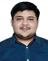 Divya Agarwal合作论文数LOreal Res Innovat9
 Seema Kapoor合作论文数9
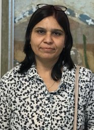 Amita Pandey合作论文数Jawaharlal Nehru University School of Life Sciences9Joshi Stephen合作论文数Department of Medical Genetics, Sanjay Gandhi Postgraduate Institute of Medical Sciences8
 Vijay Raju Boggula合作论文数Genetic Resources and Biotechnology Division, Central Institute of Medicinal and Aromatic Plant8S. S. Agarwal合作论文数8
 Rajendra Phadke合作论文数8Sheetal Sharda合作论文数7Poonam Singh Gambhir合作论文数7Meenakshi Lallar合作论文数Dept Med Genet, Sanjay Gandhi Post Grad Inst Med Sci7
 Neha Agrawal合作论文数7
 Sunita Bijarnia-Mahay合作论文数7
 Chaitanya Datar合作论文数Rare Genetic Disorder Clinic, Sahyadri Hospital7
 Mandakini Pradhan合作论文数Department of Medical Genetics, Sanjay Gandhi Postgraduate Institute of Medical Sciences7
 Mamta N Muranjan合作论文数7




