MORHEOLOGY. Conversely, a communication on theoretical hemorheo-
This issue contains, in addition to the Papers accepted by our Editors, a number of communications and abstracts concerning three conferences pertaining to clinical hemorheology.They were held during the past two years in Stuttgart (F.R.
Das Wachstumsverhalten zweier in das Gehirn von Goldhamstern implantierter menschlicher Tumoren (H. Ad. Nr. 1 und H. S. Nr. 1) wurde untersucht. Bei allen Tieren konnte ein Tumorwachstum histologisch nachgewiesen werden. Cortisonvorbehandlung der Wirtstiere begünstigte bei H. Ad. Nr. 1 die Tumorausbreitung und führte frühzeitiger zum Tode. Das intracerebrale Tumorwachstum führte jedoch auch unabhängig von der Wirtsvorbehandlung bei allen Tieren zum Tode innerhalb von 4 Wochen nach der Transplantation. Beide Geschwulst-typen wuchsen am besten in den liquorhaltigen Räumen, weniger in der Hirnsubstanz. Die Bevorzugung der Meningen durch beide Tumortypen entspricht der allgemeinen Erfahrung über die cerebrale Metastasierung entdifferenzierte Sarkome und Adenocarcinome.
Ladies and Gentlemen, dear Colleagues and Friends, It is indeed a great honour to receive the Fahraeus Award, and I would like to thank our European Society very much for this. I would now like to give you some insight into my experiences in the field of hemorheology and intravital microscopy over the years. In principal, hemorheology and microcirculation are two different scientific fields of study: with the microscope we examine small objects, especially cells, as well as the construction of tissues, that means more or less stationary structures are examined microscopically. In hemorheology, on the other hand, the dynamic flowing phenomena are investigated. For this reason both fields of science have developed independently for a long time from one another. In my clinical career, however, I encountered a continuous connection between these two areas. My clinical tutor, Norbert Henning, was a scholar of Paul Morawitz (Leipzig). Both were hematologists and gastroenterologists. Morawitz developed the classical scheme of blood coagulation and described the “agastric anaemia”, the relations between blood formation and the stomach. Henning developed the endoscopy and the cytodiagnostics of gastroenterological diseases, whereas I focused on the newer optical methods like phase contrast and fluorescence microscopy. During my internship I introduced blood coagulation diagnostics in our hospital, so I had to develop my own thromboplastin for the Quick Test. During those days this was only possible when one had good personal connections with a pathologist, as well as a “kitchen nurse”. The pathologist was able to supply us with grey matter of the brain, while the “kitchen nurse” had at her disposal the only deep freezer of the hospital, which was necessary for storage. The deep freeze was necessary for maintaining the stability of the thromboplastin preparations. During this time I started to develop an interest for blood coagulation. I realized later, that even before these early clinical contact with blood coagulation, I already had a certain connection with our teacher and founder Alfred Lewin Copley (Fig. 1). This was at the physiological institute of the University of Wurzburg, where I studied medicine. This institute was headed by Edgar Wohlisch, who in his day was an authority on coagulation physiology. A.L. Copley was a student of his, performing the coagulation tests. For these tests he required large amounts of ox
BACKROUND: Research findings in clinical hemorheology must be established in the optimum way to keep medical research and practice currently informed.OBJECTIVE: Critical presentation of current possibilities for publication.RESULTS: Hemorheology supplies results which are of interest to many clinical fields. These results include: diagnostic questions connected in particular with clinical laboratories and blood transfusion centres; pathophysiological problems; the fields of epidemiology and prognosis; the most important fields of prophylaxis and therapy in all branches of medicine and surgery; pharmacology and drug research. Therefore, a wide spectrum must be covered by publication. It has to reach the medical practitioner, the laboratory doctor, the pharmacologist, and the industrial partner in diagnosis and therapy. The protection of a high, scientific standard is the main duty of the inpendent journal. the editorial policy of the international journal ''Clinical Hemorheology'' is presented in its actual form and future development is discussed.CONCLUSION: Hemorheological publications are in essential means to present and spread this science. Quality control is based on the independence and international competence of a publisher and on the peer review policy of a multidisciplinary editorial board.
Microcirculation research preferentially uses fluorescent microscopic techniques. This applies in particular to experimental investigations. In the example of the everted rat mesentery, our own results obtained with the following vital microscopic methods are presented: fluorescent labelling of proteins, in particular of the components of blood plasma, detection with the image amplifier TV technique, quantitative detection by microfluorometry with rapid scanning facilities, image analysis techniques with computer control. The results on the relationships between plasma proteins and the vessel wall, accumulation of individual proteins on the endothelium (fibrinogen, fibronectin, polypeptide from factor VIII, annexin PP4), their permeation through the vessel wall and its experimental modification by blood clotting and by the vessel wall collagen are described. Finally, results on fluorochroming of cells (endothelium, leukocytes) and microspheres with which phenomena of adherence on the endothelium can be demonstrated are reported.
A survey is given which describes the actual situation of the rather new science of Perihemorheology, a term introduced by ALFRED L. COPLEY. Perihemorheology includes the exchange of rheological processes between the vessel-blood organ and its surrounding tissues as well as in reverse. The article summarizes the anatomical and physiological basis and includes own methodical approaches to the experimental and clinical pathophysiology of the Perihemorheology: the permeation through the blood vessel wall, the transport in the interstitium, the lymph production.
Diagnostic cytology has recently enjoyed increased attention and significance in modern research. Essential information on latest developments in methods and applications in cytology is provided by this book. Chapters review methodological advances, such as in cancer detection, and explore potential relationships to molecular biology. Also discussed are: viral infection, fundamentals of quantitative methods, and the revolutionary role of immunocytochemistry in diagnostic cell typing. The new insights offered by transmission and scanning electron microscopy into cellular structure and function are discussed, and the connections between cytology and histology are highlighted. Epidemiology in connection with cytology is incorporated in special reports. The current developments described here will become routine methods of the cytology of tomorrow.
Our own development of fluorometric scanning techniques in intravital microscopy of the microcirculation is described. Very tiny amount of fluorometric substances are detected with a high temporal and locational resolution. The everted small intestinal mesentery of the rat serves as a model. We have given a detailed description of the microscopes used, the optical systems, the conditions of measurement of the microfluorometry, the scanning techniques and the evaluation of the measurement data. The present state of technical development detects 10−12 g of a fluorochromed plasma protein in 8 ms in a measurement field of 2 µm2. The four-digit measurement data of a scanning line of 200 µm length in 0.25 µm locational resolution are registered in about 2 s.
The dependence on hematocrit of whole blood viscoelasticity must be considered in order to compare pathological blood samples to normal ones. If one wants to calculate the measured values to a standard hematocrit value, the hematocrit dependence for the pathological sample must be available. As the latter however is unknown, the same dependence is assumed for both normal and pathological blood samples. To prove the validity of this assumption, hematocrit dependence of random blood samples from different diseases (cerebral and coronary vascular and myocardial disorders) were investigated. A statistical analysis showed the assumption as invalid. Therefore, it will be recommended to evaluate pathological blood samples at the measured hematocrit.
 Paul Dirnberger合作论文数9Dietrich Bressel合作论文数6
 David M. Goldenberg合作论文数Center for Molecular Medicine and Immunology;eMedEvents5Alfred L. Copley合作论文数5Suse Zenzes-Geprägs合作论文数4Horst Chmiel合作论文数4I. Anadere合作论文数4Erich Müller合作论文数3K. Th. Schricker合作论文数3E. Walitza合作论文数3H. Dehlinger合作论文数2Käthe Wilmes合作论文数2Suse Geprägs合作论文数2E.R. Movitt合作论文数1H.K.v. Rechenberg合作论文数1G.H. Lubeck合作论文数1T Gahm合作论文数1W J Janssens合作论文数1Ef Luscher合作论文数1Lars H. Breimer合作论文数1Gregorio Caimi合作论文数1Jj Friedman合作论文数1
 Francesco Gentile合作论文数1F. Scheiffarth合作论文数1H. G. Lasch合作论文数1A SERRA合作论文数1M Rasia合作论文数1B Walzl合作论文数1Alejandra Luquita合作论文数1D'Asaro S合作论文数1Reinhardt E R合作论文数1
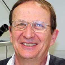 Gerard Nash合作论文数Institute of Cardiovascular Sciences, College of Medical and Dental Sciences, University of Birmingham1Anna Catania合作论文数1T Fonk合作论文数1Dirk Heinrich合作论文数1
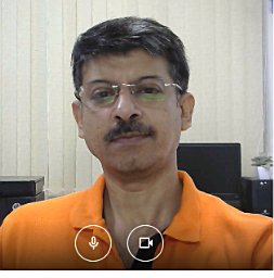 Arindam Mukherjee合作论文数University of North Carolina at Charlotte1
 Shu Chien (钱煦)合作论文数Jacobs School of Engineering, University of California, San Diego;Institute of Engineering in Medicine, University of California, San Diego1Gualtiero Palareti合作论文数University of Bologna, Bologna, Italy1
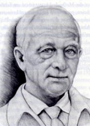 Klaus Goerttler合作论文数Institute of Experimental Pathology;German Cancer Research Centre;Institute of Experimental Pathology, German Cancer Research Centre1L. Zicha合作论文数1P. Lechner合作论文数1G. Kleyensteiber合作论文数1Brunhilde Schmidt合作论文数1Flemming Kissmeyer-Nielsen合作论文数1Antonio Sarno合作论文数1Georg Feichter合作论文数1K. T. Schricker合作论文数1




