Human mesenchymal stem cells (MSCs) have been studied in over 1500 clinical trials to treat over 30 diseases. However, the understanding towards MSC stemness remains under studied. So far, little is known about how MSCs maintain undifferentiated state or commit to specific lineages under different microenvironmental cues. The lack of comprehensive understanding regarding MSC stemness greatly hampers the translation of research findings into successful clinical application due to unclear mechanism of action. Emerging evidence shows that a variety of genetic factors delicately regulate MSC self-renewal and differentiation. In this review, we summarize the role of transcriptional factors, cell cycle regulators, genomic stability genes, cellular quality control, epigenetic regulators, non-coding RNAs, mitochondrial function, growth factors and m6A modification in regulating the stemness of MSCs. Strategies to maintain MSC stemness during ex-vivo expansion are also discussed. This review will deepen understanding of MSC stemness for advancing clinical applications and provide insights into future directions for research aimed at improving MSC-based therapies.
Regenerative dental medicine continuously expands to improve treatments for prevalent clinical problems in dental and oral medicine. Stem cell based translational opportunities include regenerative therapies for tooth restoration, root canal therapy, and inflammatory processes (e.g., periodontitis). The potential of regenerative approaches relies on the biological properties of dental stem cells. These and other multipotent somatic mesenchymal stem cell (MSC) types can in principle be applied as either autologous or allogeneic sources in dental procedures. Dental stem cells have distinct developmental origins and biological markers that determine their translational utility. Dental regenerative medicine is supported by mechanistic knowledge of the molecular pathways that regulate dental stem cell growth and differentiation. Cell fate determination and lineage progression of dental stem cells is regulated by multiple cell signaling pathways (e.g., WNTs, BMPs) and epigenetic mechanisms, including DNA modifications, histone modifications, and non-coding RNAs (e.g., miRNAs and lncRNAs). This review also considers a broad range of novel approaches in which stem cells are applied in combination with biopolymers, ceramics, and composite materials, as well as small molecules (agonistic or anti-agonistic ligands) and natural compounds. Materials that mimic the microenvironment of the stem cell niche are also presented. Promising concepts in bone and dental tissue engineering continue to drive innovation in dental and non-dental restorative procedures.
Background & AimCurrent efforts to manufacture and bank clinical-grade mesenchymal stem cells (MSCs) necessitate large-scale and prolonged ex vivo expansion due to the small number of adherent MSCs in a typical bone marrow aspirate (0.01%–0.0001%). Such expansion places MSCs at risk of replicative senescence-related changes that ultimately impact the quality and limit the number of functionally competent cells. Here, we investigate the influence of glycosaminoglycan (GAG) capable of enhancing the stability and bioactivity of endogenously produced fibroblast growth factor-2 (FGF-2) as media additives to replenish lost molecular signals crucial to MSC growth and aging during long-term culture.Methods, Results & ConclusionHuman bone marrow-derived MSCs were grown up to passage 20 with or without an affinity-isolated heparan sulfate (HS) GAG variant (termed HS8). The influence of this GAG as a stem cell culture adjuvant on the proliferation, colony formation, differentiation, telomere length, and senescence of MSCs was assessed. Array-based gene expression profiling and Western blot analyses were utilized to identify the underlying molecular mechanism regulating the action of HS8 on MSCs. In this study, we demonstrate that media supplementation with HS8 delays in vitro senescence of MSCs during serial passaging while concurrently enhancing their proliferation (Figures A and B). The data showed increased expression of the epigenetic marker EZH2, known to suppress p16 gene expression and inhibit cell senescence, in both early-passage MSCs and late-passage MSCs supplemented with HS8. Expression levels of the proliferation marker PCNA decreased in late-passage senescent MSCs and remained elevated in cells expanded in the HS8-supplemented media (Figure C). Notably, the HS8-expanded MSCs showed enhanced differentiation and colony-forming efficiency compared to non-supplemented cells (Figure D). Serial passaging of MSCs in media supplemented with an HS variant that has increased affinity for FGF-2 can delay senescence, supporting the manufacturing of larger numbers of cells over a reduced time period. Importantly, HS8 supplementation did not adversely affect MSC differentiation potential. These findings warrant further exploration of GAG components within the MSC niche that can regulate cell fate and, thus, therapeutic efficacy.
Heparan sulfate (HS) is a glycosaminoglycan (GAG) found throughout nature and is involved in a wide range of functions including modulation of cell signalling via sequestration of growth factors. Current consensus is that the specificity of HS motifs for protein binding are individual for each protein. Given the structural complexity of HS the synthesis of libraries of these compounds to probe this is not trivial. Herein we present the synthesis of an HS decamer, the design of which was undertaken rationally from previously published data for HS binding to the growth factor BMP-2. The biological activity of this HS decamer was assessed in vitro, showing that it had the ability to both bind BMP-2 and increase its thermal stability as well as enhancing the bioactivity of BMP-2 in vitro in C2C12 cells. At the same time no undesired anticoagulant effect was observed. This decamer was then analysed in vivo in a rabbit model where higher bone formation, bone mineral density (BMD) and trabecular thickness were observed over an empty defect or collagen implant alone. This indicated that the HS decamer was effective in promoting bone regeneration in vivo.
Bioactive polyethylene incorporating hydrophobic PE-bearing macromonomers and hydrophilic PEGylated-peptide macromonomers was synthesized via ROMP. 3D-printed sheets of it with UHMWPE showed enhanced osteogenic activity for potential orthopaedic applications.
Bone morphogenetic protein 2 (BMP-2) is an osteoinductive protein and a potent inducers of bone formation, playing an essential role during bone fracture repair. Heparan sulfate (HS), a highly charged and linear poly-saccharide, is known to interact with and enhance BMP-2 bioactivity. Despite showing potential as a potent adjuvant of the endogenous bone healing response, commercially available HS is derived from animal sources which are less desirable when considering translation into the clinic. In the present study, we screen twenty glycomimetics against BMP-2 to determine if fully synthetic analogues of HS can enhance the bioactivity of BMP-2 in vitro and bone healing in vivo. We found that a four-armed dendrimer harboring oversulfated maltose res-idues could bind BMP-2 with high affinity, enhance BMP-2 bioactivity in vitro and enhance bone regeneration in vivo. These data suggest fully synthetic glycomimetics are viable alternatives to naturally derived HS and offer an attractive alternative for clinical translation.
It is challenging to regenerate periodontal tissues fully. We have previously reported a heparan sulfate variant with enhanced affinity for bone morphogenetic protein-2, termed HS3, that enhanced periodontal tissue regeneration in a rodent model. Here we seek to transition this work closer to the clinic and investigate the efficacy of the combination HS3 collagen device in a non-human primate (NHP) periodontitis model. Wire-induced periodontitis was generated in ten Macaca fascicularis, and defects were treated with Emdogain or collagen (CollaPlug) loaded with (1) distilled water, (2) HS low (36 µg of HS3), or (3) HS high (180 µg of HS3) for 3 months. At the endpoint, microscopic assessment showed significantly less epithelial down-growth, greater alveolar bone filling, and enhanced cementum and periodontal ligament regeneration following treatment with the HS-collagen combination devices. When evaluated using a periodontal regeneration assessment score (PRAS) on a scale of 0–16, collagen scored 6.78 (± 2.64), Emdogain scored 10.50 (± 1.73) and HS low scored 10.40 (± 1.82). Notably, treatment with HS high scored 12.27 (± 2.20), while healthy control scored 14.80 (± 1.15). This study highlights the efficacy of an HS-collagen device for periodontal regeneration in a clinically relevant NHP periodontitis model and warrants its application in clinical trials.
The growing interest in regenerative medicine has opened new avenues for novel cell therapies using stem cells. Bone marrow aspirate (BMA) is an important source of stromal mesenchymal stem cells (MSCs). Conventional MSC harvesting from BMA relies on archaic centrifugation methods, often leading to poor yield due to osmotic stress, high centrifugation force, convoluted workflow, and long experimental time (∼2-3 hours). To address these issues, we have developed a scalable microfluidic technology based on deterministic lateral displacement (DLD) for MSC isolation. This passive, label-free cell sorting method capitalizes on the morphological differences between MSCs and blood cells (platelets and RBCs) for effective separation using an inverted L-shaped pillar array. To improve throughput, we developed a novel multi-chip DLD system that can process 2.5 mL of raw BMA in 20 ± 5 minutes, achieving a 2-fold increase in MSC recovery compared to centrifugation methods. Taken together, we envision that the developed DLD platform will enable fast and efficient isolation of MSCs from BMA for effective downstream cell therapy in clinical settings.
Commercial porcine intestinal mucosal heparan sulfate (HS) is a valuable material for research into its biological functions. As it is usually produced as a side-stream of pharmaceutical heparin manufacture, its chemical composition may vary from batch to batch. We analysed the composition and structure of nine batches of HS from the same manufacturer. Statistical analysis of the disaccharide compositions placed these batches in three categories: group A had high GlcNAc and GlcNS, and low GlcN typical of HS; group B had high GlcN and GlcNS, and low GlcNAc; group C had high di- and trisulfated, and low unsulfated and monosulfated disaccharide repeats. These batches could be placed in the same categories based on their 1H NMR spectra and molecular weights. Anticoagulant and growth factor binding activities of these HS batches did not fit within these same groups but were related to the proportions of more highly sulfated disaccharide repeats.
The multilineage differentiation potential of human mesenchymal stem cells (hMSCs) underpins their clinical utility for tissue regeneration. Control of such cell-fate decisions is tightly regulated by different growth factors/cytokines and their cognate receptors. Fibroblast growth factors (FGFs) are among such factors critical for osteogenesis. However, how FGF receptors (FGFRs) help to orchestrate osteogenic progression remains to be fully elucidated. Here, we studied the protein levels of FGFRs during osteogenesis in human adult bone marrow-derived MSCs and discovered a positive correlation between FGFR2 expression and alkaline phosphatase (ALP) activity, an early marker of osteogenesis. Through RNA interference studies, we confirmed the role of FGFR2 in promoting the osteogenic differentiation of hMSCs. Knockdown of FGFR2 resulted in downregulation of pro-osteogenic genes and upregulation of pro-adipogenic genes and adipogenic commitment. Moreover, under osteogenic induction, FGFR2 knockdown resulted in upregulation of Enhancer of Zeste Homolog 2 (EZH2), an epigenetic enzyme that regulates MSC lineage commitment and suppresses osteogenesis. Lastly, we show that serial-passaged hMSCs have reduced FGFR2 expression and impaired osteogenic potential. Our study suggests that FGFR2 is critical for mediating osteogenic fate by regulating the balance of osteo-adipogenic lineage commitment. Therefore, examining FGFR2 levels during serial-passaging of hMSCs may prove useful for monitoring their multipotency.
The ability to repair critical-sized long-bone injuries using growth factor and cell delivery was investigated using hydrogel biomaterials. Physiological doses of the recombinant human bone morphogenic protein-2 (rhBMP2) were delivered in a sustained manner from a biodegradable hydrogel containing peripheral human blood-derived endothelial progenitor cells (hEPCs). The biodegradable implants made from polyethylene glycol (PEG) and denatured fibrinogen (PEG-fibrinogen, PF) were loaded with 7.7 μg/ml of rhBMP2 and 2.5 × 10⁶ cells/ml hEPCs. The safety and efficacy of the implant were tested in a rodent model of a critical-size long-bone defect. The hydrogel implants were formed ex-situ and placed into defects in the tibia of athymic nude rats and analyzed for bone repair after 13 weeks following surgery. The hydrogels containing a combination of 7.7 μg/ml of rhBMP2 and 2.5 × 10⁶ cells/ml hEPCs were compared to control hydrogels containing 7.7 μg/ml of rhBMP2 only, 2.5 × 10⁶ cells/ml hEPCs only, or bare hydrogels. Assessments of bone repair include histological analysis, bone formation at the site of implantation using quantitative microCT, and assessment of implant degradation. New bone formation was detected in all treated animals, with the highest amounts found in the treatments that included animals that combined the PF implant with rhBMP2. Moreover, statistically significant increases in the tissue mineral density (TMD), trabecular number and trabecular thickness were observed in defects treated with rhBMP2 compared to non-rhBMP2 defects. New bone formation was significantly higher in the hEPC-treated defects compared to bare hydrogel defects, but there were no significant differences in new bone formation, trabecular number, trabecular thickness or TMD at 13 weeks when comparing the rhBMP2 + hEPCs-treated defects to rhBMP2-treated defects. The study concludes that the bone regeneration using hydrogel implants containing hEPCs are overshadowed by enhanced osteogenesis associated with sustained delivery of rhBMP2.
We report the modular synthesis of bioactive brush-type polycaprolactone-peptide and polylactide-peptide copolymers for applications in bone tissue engineering. The brush copolymers containing pendant side chains of polycaprolactone (PCL) or polylactide (PLA) and PEGylated peptides, including linear Arg-Gly-Asp and collagen-like peptide (Gly-Pro-Hyp)3, were synthesized by ring-opening metathesis polymerization with high conversions and low dispersities (<1.5). These PCL-peptide and PLA-peptide copolymers exhibited good thermal stability for material processing using melt-extrusion-based methods. The copolymers were blended with commercial PCL or PLA, extruded into filaments, and 3D printed using fused filament fabrication methods. These bioactive PCL and PLA materials promoted osteogenic differentiation in vitro and showed good biocompatibility in in vivo murine model study. The promising results presented herein will serve as a useful guide for the design and functionalization of PCL or PLA materials for use in bone tissue engineering.
Therapies using mesenchymal stromal cells (MSCs) to treat immune and inflammatory conditions are now at an exciting stage of development, with many MSC-based products progressing to phase II and III clinical trials. However, a major bottleneck in the clinical translation of allogeneic MSC therapies is the variable immunomodulatory properties of MSC products due to differences in their tissue source, donor heterogeneity and processes involved in manufacturing and banking. This variable functionality of MSC products likely contributes to the substantial inconsistency observed in the clinical outcomes of phase III trials of MSC therapies; several trials have failed to reach the primary efficacy endpoint. In this review, we discuss various strategies to consistently maintain or enhance the immunomodulatory potency of MSCs during ex vivo expansion, which will enable the manufacture of allogeneic MSC banks that have high potency and low variability. Biophysical and biochemical priming strategies, the use of culture additives such as heparan sulfates, and genetic modification can substantially enhance the immunomodulatory properties of MSCs during in vitro expansion. Furthermore, robust donor screening, the use of biomarkers to select for potent MSC subpopulations, and rigorous quality testing to improve the release criteria for MSC banks have the potential to reduce batch-to-batch heterogeneity and enhance the clinical efficacy of the final MSC product. Machine learning approaches to develop predictive models of individual patient response can enable personalized therapies and potentially establish correlations between in vitro potency measurements and clinical outcomes in human trials.
INTRODUCTION:Culture conditions and differentiation cocktails may facilitate cell maturation and extracellular matrix (ECM) secretion and support the production of engineered fibroblastic tissues with applications in ligament regeneration. The objective of this study is to investigate the potential of two connective tissue-related ligands (i.e., BMP6 and GDF5) to mediate collagenous ECM synthesis and tissue maturation in vitro under normoxic and hypoxic conditions based on the hypothesis that BMP6 and GDF5 are components of normal paracrine signalling events that support connective tissue homeostasis. METHODS:Human adipose-derived MSCs were seeded on 3D-printed medical-grade polycaprolactone (PCL) scaffolds using a bioreactor and incubated in media containing GDF5 and/or BMP6 for 21 days in either normoxic (5% oxygen) or hypoxic (2% oxygen) conditions. Constructs were harvested on Day 3 and 21 for cell viability analysis by live/dead staining, structural analysis by scanning electron microscopy, mRNA levels by RTqPCR analysis, and in situ deposition of proteins by immunofluorescence microscopy. RESULTS:Pro-fibroblastic gene expression is enhanced by hypoxic culture conditions compared to normoxic conditions. Hypoxia renders cells more responsive to treatment with BMP6 as reflected by increased expression of ECM mRNA levels on Day 3 with sustained expression until Day 21. GDF5 was not particularly effective either in the absence or presence of BMP6. CONCLUSIONS:Fibroblastic differentiation of MSCs is selectively enhanced by BMP6 and not GDF5. Environmental factors (i.e., hypoxia) also influenced the responsiveness of cells to this morphogen.
Introduction High concentrations of bone morphogenetic protein 2 (BMP2) in bone regeneration cause adverse events (e.g, heterotopic bone formation and acute inflammation). This study examines novel epigenetic strategies (i.e., EZH2 inhibition) for augmenting osteogenesis thereby aiming to reduce the required BMP2 dose in vivo for bone regeneration and minimize these adverse effects. Methods Human bone marrow-derived mesenchymal stem cells (BMSCs) were grown on 3D-printed medical-grade PCL scaffolds and incubated in osteogenic media containing 50ng/mL BMP2 and/or 5μM GSK126 (EZH2 inhibitor) for 6 days (n=3 per group and time point). Constructs were harvested for RTqPCR analysis at Day 10 and immunofluorescence (IF) microscopy at Day 28. After pre-treating for 6 days and maintaining in osteogenic media for 4 days, BMSC-seeded scaffolds were also implanted in an immunocompromised subcutaneous murine model (n=39; 3/group/donor and 3 control scaffolds) for histological analysis at 8 weeks. Results Pre-treatment of BMSCs with BMP2 and BMP2/GSK126 co-stimulated expression of osteoblast-related genes (e.g., IBSP, SP7, RUNX2 and DLX5), as well as protein accumulation (e.g., collagen Type 1/COL1A1 and osteocalcin/BGLAP) based on immunofluorescence staining. While in vivo implantation for 8 weeks did not result in bone formation, increased angiogenesis was observed in BMP2 and BMP2/GSK126 groups. Conclusions This study finds that BMP2 and GSK126 co-stimulate osteogenic differentiation of MSCs on 3D scaffolds in vitro and may contribute to enhanced vascularization when implanted in vivo to support bone formation. Thus, epigenetic priming with EZH2 inhibitors may have translational potential in bone healing by permitting a reduction of BMP2 dosing in vivo to mitigate its side effects.
Human mesenchymal stem/stromal cell (hMSC)-based cell therapies are promising for treating a variety of diseases. The unique immunomodulatory properties of hMSCs have extended their therapeutic potential beyond tissue regeneration. However, extensive pre-clinical culture expansion inevitably drives cells toward replicative ?aging?and a consequent decline in quality. These ?in vitro-aged?hMSCs resemble biologically aged cells, which have been reported to show senescence signatures, diminished immunosuppressive capacity, and weakened regenerative potential as well as pro-inflammatory features. In this review, we have surveyed the literature to explore the intimate relationship between the inflammatory status of hMSCs and their in vitro aging process. We posit that a shift from an anti-inflammatory to a pro-inflammatory phenotype of culture-expanded hMSCs contributes to a deterioration in their therapeutic efficacy. Potential molecular and cellular mechanisms underpinning this phenomenon have been discussed. We have also highlighted studies that leverage these mechanisms to make culture-expanded hMSCs more amenable for clinical use.
High doses bone morphogenetic protein 2 (BMP‐2) have resulted in a series of complications in spinal fusion. We previously established a polyelectrolyte complex (PEC) carrier system that reduces the therapeutic dose of BMP‐2 in both rodent and porcine spinal fusion models. This study aimed to evaluate the safety and efficacy of the combination of bone marrow mesenchymal stem cells (BMSCs) and low dose BMP‐2 delivered by PEC for bone regeneration in a porcine model of anterior lumbar interbody spinal fusion (ALIF) application. Six Yorkshire pigs underwent a tri‐segmental (L2/L3; L3/L4; L4/L5) ALIF in four groups, namely: (a) BMSCs + 25 μg BMP‐2/PEC (n = 9), (b) 25 μg BMP‐2/PEC (n = 3), (c) BMSCs (n = 3), and (d) 50 μg BMP‐2/absorbable collagen sponge (n = 3). Fusion outcomes were evaluated by radiography, biomechanical testing, and histological analysis after 12 weeks. Mean radiographic scores at 12 weeks were 2.7, 2.0, 1.0, and 1.0 for Groups 1 to 4, respectively. μ‐CT scanning, biomechanical evaluation, and histological analysis demonstrated solid fusion and successful bone regeneration in Group 1. In contrast, Group 2 showed inferior quality and slow rate of fusion, and Groups 3 and 4 failed to fuse any of the interbody spaces. There was no obvious evidence of seroma formation, implant rejection, or any other complications in all groups. The results suggest that the combination of BMSCs and low dose BMP‐2/PEC could further lower down the effective dose of the BMP‐2 and be used as a bone graft substitute in the large animal ALIF model.
Periodontitis is the most common inflammatory disease that leads to periodontal defects and tooth loss. Regeneration of alveolar bone and soft tissue in periodontal defects is highly desirable but remains challenging. A heparan sulphate variant (HS3) with enhanced affinity for bone morphogenetic protein-2 (BMP2) that, when combined with collagen or ceramic biomaterials, enhances bone tissue regeneration in the axial and cranial skeleton in several animal models was reported previously. In the current study, establishing the efficacy of a collagen/HS3 device for the regeneration of alveolar bone and the adjacent periodontal apparatus and related structures was sought. Collagen sponges loaded with phosphate-buffered saline, HS3, BMP2, or HS3 + BMP2 were implanted into surgically-created intra-bony periodontal defects in rat maxillae. At the 6 week end- point the maxillae were decalcified, and the extent of tissue regeneration determined by histomorphometrical analysis. The combination of collagen/HS3, collagen/BMP2 or collagen/HS3 + BMP2 resulted in a three to four-fold increase in bone regeneration and up to a 1.5 × improvement in functional ligament restoration compared to collagen alone. Moreover, the combination of collagen/HS3 + BMP2 improved the alveolar bone height and reduced the amount of epithelial growth in the apical direction. The implantation of a collagen/ HS3 combination device enhanced the regeneration of alveolar bone and associated periodontal tissues at amounts comparable to collagen in combination with the osteogenic factor BMP2. This study highlights the efficacy of a collagen/HS3 combination device for periodontal regeneration that warrants further development as a point-of-care treatment for periodontitis-related bone and soft tissue loss.
BACKGROUNDIncreasing kyphosis of the spine in a human is a well-recognized clinical phenomenon that has been associated with back pain, poor physical performance and disability. The pathophysiology of age-related kyphosis is complex and has been associated with physiological changes in vertebrae, intervertebral disc (IVD) and paraspinal musculature, which current cross-sectional studies are unable to demonstrate. Creating an in vivo, paraspinal myopathic animal model for longitudinal study of these changes under controlled conditions is thus warranted.PURPOSETo confirm the TSC1 gene knockout effect on paraspinal muscle musculature; to analyze the development of spinal kyphosis, IVD degeneration and vertebra structural changes in a longitudinal manner to gain insights into the relationship between these processes.STUDY DESIGNA prospective cohort study of 28 female mice, divided into 4 groups—9-month-old TSC1mKO (n=7), 9-month-old control (n=4), 12-month-old TSC1mKO (n=8), and 12-month-old controls (n=9).METHODSHigh resolution micro-computed tomography was used to measure sagittal spinal alignment (Cobb's angle), vertebral height, vertebral body wedging, disc height index (DHI), disc wedge index (DWI), histomorphometry of trabecular bone and erector spinae muscle cross-sectional area. Paraspinal muscle specimens were harvested to assess for myopathic features with H&E stain, muscle fiber size, density of triangular fiber and central nucleus with WGA/DAPI stain, and percentage of fibers with PGC-1α stain. Intervertebral discs were evaluated for disc score using FAST stain.RESULTSCompared to controls, paraspinal muscle sections revealed features of myopathy in TSC1mKO mice similar to human sarcopenic paraspinal muscle. While there was significantly greater presence of small triangular fiber and density of central nucleus in 9-and 12-month-old TSC1mKO mice, significantly larger muscle fibers and decreased erector spinae muscle cross-sectional area were only found in 12-month-old TSC1mKO mice compared to controls. TSC1mKO mice developed accelerated thoracolumbar kyphosis, with significantly larger Cobb angles found only at 12 months old. Structural changes to the trabecular bone in terms of higher bone volume fraction and quality, as well as vertebral body wedging were observed only in 12-month-old TSC1mKO mice when compared to controls. Disc degeneration was observed as early as 9 months in TSC1mKO mice and corresponded with disc wedging. However, significant disc height loss was only observed when comparing 12-month-old TSC1mKO mice with controls.CONCLUSIONSThis study successfully shows the TSC1 gene knockout effect on the development of paraspinal muscle myopathy in a mouse which is characteristic of sarcopenia. The TSC1mKO mice is by far the best model available to study the pathological consequence of sarcopenia on mice spine. With paraspinal muscle myopathy established as early as 9 months, TSC1mKO mice developed disc degeneration and disc wedging. This is followed by kyphosis of the spine at 12 months with concomitant disc height loss and vertebral body wedging due to bone remodeling. Age-related bone loss was not found in our study, suggesting osteoporosis and myopathy-induced vertebral body wedging are likely two independent processes.CLINICAL SIGNIFICANCEThis is the first study to provide key insights on the early and late consequences of paraspinal myopathy on intervertebral disc degeneration, spinal kyphosis, and vertebral body changes. With this new understanding, future studies evaluating therapies for spinal degeneration may be performed to develop time-sensitive interventions.
 Victor Nurcombe合作论文数SMC Biotechnology Inc.;Institute of Medical Biology, Agency for Science, Technology and Research146
 Andre Van Wijnen合作论文数The Stein/Lian Laboratory, Department of Biochemistry, Larner College of Medicine, University of Vermont48
 Sadasivam Murali合作论文数23
 Lisbeth Grondahl合作论文数School of Chemistry and Molecular Biosciences, The University of Queensland22
 C. Dombrowski合作论文数20Bina Rai合作论文数Department of Biomedical Engineering, National University of Singapore20
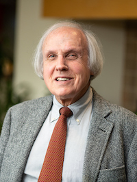 Gary S Stein合作论文数Department of Biochemistry, Larner College of Medicine, University of Vermont;Department of Surgery, Larner College of Medicine, University of Vermont;University of Vermont Cancer Center20
 Samsonraj Rebekah M合作论文数Glycotherapeutics Group.19
 Smith Raymond A A合作论文数Stem Cell Glycobiology Group, University of Manchester17
 James Hui合作论文数Department of Orthopaedic Surgery, National University Hospital;Department of Orthopaedic Surgery, Yong Loo Lin School of Medicine, National University of Singapore;Alexandra Hospital16
 Matt Trau合作论文数Australian Institute for Bioengineering and Nanotechnology, The University of Queensland16
 Dietmar W Hutmacher合作论文数School of Mechanical, Medical and Process Engineering, Faculty of Engineering, Queensland University of Technology16Larisa M. Haupt合作论文数Institute of Molecular and Cell Biology Stem Cells and Tissue Repair Group Proteos, 61 Biopolis Drive Singapore 138673 Singapore16L. Ling合作论文数14
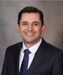 Dudakovic Amel合作论文数the Department of Orthopedic Surgery., From14
 Zophia Lim合作论文数13
 Kishore Bhakoo合作论文数Department of Orthopaedic Surgery, National University of Singapore;University of Madras;UHI Millennium Institute12Tuan Chun Tan合作论文数11
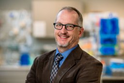 Allan B Dietz合作论文数Mayo Clinic10
 James Goh合作论文数Department of Biomedical Engineering, College of Design and Engineering, National University of Singapore10
 Gajadhar Bhakta合作论文数Department of Chemistry, University of Delhi9Imelda Keen合作论文数8Birch William R合作论文数8Abbah Sunny Akogwu合作论文数8
 Titmarsh Drew M合作论文数Australian Institute for Bioengineering and Nanotechnology, The University of Queensland8
 Michael Raghunath合作论文数Dept. of Dermatology and Ludwig Boltzmann Institute for Cell Biology and Immunobiology of the Skin, University of Münster8Emma Luong-Van合作论文数8A Kumarasuriyar合作论文数7Oh Steve Kah-Weng合作论文数7
 Emily Camilleri合作论文数7Hu Tao合作论文数7
 Lee Jaslyn Jie Lin合作论文数Nanyang Environment and Water Research Institute, Nanyang Technological University7David A. Rider合作论文数6Torben Helledie合作论文数6
 Ming Wang合作论文数Dept Orthopaed Surg, Natl Univ Singapore6Raggatt Liza J合作论文数6raymond wing moon lam合作论文数6
 Le Bach Q合作论文数MIRA Institute for Biomedical Technology and Technical Medicine, University of Twente6Sathiyanathan Padmapriya合作论文数6
 Jun Li合作论文数Department of Biomedical Engineering, College of Design and Engineering, National University of Singapore;Environmental Research Institute, National University of Singapore;Research Institute, National University of Singapore(Suzhou);Research Institute, National University of Singapore(Chongqing)6
 Simon F R Hinkley合作论文数Department of Chemistry, University of Otago6Rebecca A. Jackson合作论文数School of Medicine, University of California, San Francisco6H Chipperfield合作论文数5Kerry J Manton合作论文数5
 Christopher Robert Paradise合作论文数Department of Orthopedic Surgery, Mayo Clinic5
 Jennifer J. Westendorf合作论文数Department of Biochemistry and Molecular Biology, Mayo Clinic;Department of Orthopedic Surgery, Mayo Clinic5b kenny合作论文数5Toh Soo Yein合作论文数5Peter M. Fredericks合作论文数5




