You have accessJournal of UrologyCME1 Apr 2023MP76-14 ROBUST HEMOSTASIS OF A HEMOSTATIC URINARY CATHETER FOR TRANSURETHRAL SURGICAL APPLICATIONS James Jones, MacKayla Carolan, Shalini Gautam, Jason Hedges, Simon McCarthy, and Hua Xie James JonesJames Jones More articles by this author , MacKayla CarolanMacKayla Carolan More articles by this author , Shalini GautamShalini Gautam More articles by this author , Jason HedgesJason Hedges More articles by this author , Simon McCarthySimon McCarthy More articles by this author , and Hua XieHua Xie More articles by this author View All Author Informationhttps://doi.org/10.1097/JU.0000000000003350.14AboutPDF ToolsAdd to favoritesDownload CitationsTrack CitationsPermissionsReprints ShareFacebookLinked InTwitterEmail Abstract INTRODUCTION AND OBJECTIVE: Bleeding is the leading complication of outlet surgery for men with lower urinary tract symptoms, harming patients and increasing resource utilization. Our group has developed a urinary catheter which incorporates a specialized biodegradable chitosan endoluminal hemostatic dressing (CEHD) for transurethral deployment. Previous investigation focused on deployment feasibility with limited assessment of hemostatic efficacy. METHODS: Hemostatic efficacy of CEHD prototypes relative to standard of care Foley catheter was investigated using a heparinized swine hepatic injury model. 2.5 cm diameter concave injuries in hepatic lobes were created with a rotary cutting tool. CEHD prototype test treatments and Foley balloon catheter control treatments (N=40, each) were randomly paired to injuries in 4 swine. Tests consisted of a prototype placed on the wound bed followed by a 10 mL Foley balloon and 25 g gel block to apply 35 g of tamponade pressure. Controls did not include prototype dressing. Hemostasis was determined at 30 s. Failures at 30 s were re-applied for 90 s and hemostasis reassessed. Both 30 s and 90 s were followed by 60 s observation (up to 250 s total). Bleeding rate was determined at initiation and conclusion. Hemostasis was analyzed either as no bleeding or as an >85% reduction in bleed rate. RESULTS: Mean clotting time was 561 seconds and mean pre-treatment bleeding rate was 19.5 mL/min with no significant difference between test and control groups. The CEHD group demonstrated a significant difference in bleed rate reduction (97% vs 73%, p<0.001), and mean time to hemostasis (98 s vs 287 s, p<0.001). While the Foley Balloon alone did lead to a hemostatic reduction in bleed rate (≤85%) in 58% of cases, CEHD application provided rapid significantly improved hemostasis, with 38/40 CEHD prototypes achieving complete hemostasis by 3 minutes, versus only 3/40 controls (p<0.001). CONCLUSIONS: CEHD prototypes demonstrated superior hemostatic capability compared to conventional Foley balloon controls in the setting of high bleed rates. Previous investigation demonstrated in vivo feasibility of transurethral deployment, tissue adhesion and biodegradation of the device and dressing over a 7-day period. These results suggest robust hemostatic capability of the CEHD prototype in an anticoagulated in vivo model, supporting future clinical investigation. Source of Funding: NIH/NIDDK Award R42DK078400 © 2023 by American Urological Association Education and Research, Inc.FiguresReferencesRelatedDetails Volume 209Issue Supplement 4April 2023Page: e1096 Advertisement Copyright & Permissions© 2023 by American Urological Association Education and Research, Inc.MetricsAuthor Information James Jones More articles by this author MacKayla Carolan More articles by this author Shalini Gautam More articles by this author Jason Hedges More articles by this author Simon McCarthy More articles by this author Hua Xie More articles by this author Expand All Advertisement PDF downloadLoading ...
OBJECTIVE:To investigate a novel transurethral hemostatic catheter device with an integrated chitosan endoluminal hemostatic dressing (CEHD). Development and implementation of this technology may help address bleeding following surgery such as transurethral resection of prostate (TURP). Bleeding remains the most common complication following TURP, leading to increased morbidity and hospitalization.METHODS:Investigation of hemostasis, delivery, safety and efficacy of the CEHD device is conducted using Female Yorkshire swine (N = 23). Hemostatic efficacy of the CEHD (N = 12) is investigated against a control of gauze (N = 12) in a splenic injury model (3 swine). The delivery, safety, and efficacy of the CEHD device (N = 10) are investigated against Foley-catheter control (N = 10) for 7 days using a swine bladder-neck-injury model.RESULTS:In the splenic injury study, 9/12 CEHD dressings successfully achieved hemostasis within 150 seconds (mean 83 seconds) vs success of 6/12 (mean 150 seconds) for gauze (P = .04). In the 7-day study, the CEHD was successfully deployed in 10/10 animals and all dressings were tolerated without histologic or clinical adverse effect. Hemostasis of the CEHD device was found to be noninferior to control catheters. Noninferiority is attributed to low bleeding rates in the swine bladder neck injury model.CONCLUSION:This investigation successfully demonstrated the feasibility of transurethral deployment of the CEHD in vivo. Routine use of safe and slowly dissolvable CEHDs could reduce the rate of complications and hospitalizations associated with bleeding and blood loss in TURP procedures. Further investigation is warranted.
Synthetic polymer leaflets in prosthetic cardiac valves hold the potential to reduce calcification and thrombus, while improving blood flow, durability, and device economics. A recently developed siloxane poly(urethane‐urea) (LifePolymer, LP) exhibits properties essential for heart valve leaflets, including low dynamic modulus, high tensile strength, minimal creep, and excellent biostability. LP's properties result from carefully designed “linked co‐macrodiol” chemistry that maximizes silicone content and virtual crosslinks between soft and hard phases. Characterization of multiple commercial batches demonstrates a robust synthesis process with minimal variation. Extensive ISO 10993–based biocompatibility testing results in no observable toxicity or other adverse reactions. An ex vivo AV shunt thrombogenicity investigation reveals nearly undetectable levels of platelet attachment and thrombus formation on LP surfaces. Chronic ovine implantation of prototype heart valves with LP leaflets shows no differences in thrombogenicity or systemic tissue response when compared to a clinically standard tissue‐based valve. Toxicological risk assessment, based on extractables and leachables analysis of LP‐based heart valves, confirms minimal toxicological risk. Lastly, 24‐week, strain‐accelerated in vivo LP biostability testing confirms previous favorable in vitro biostability findings. These studies demonstrate that this newly developed elastomer exhibits ideal biomaterial properties for the flexible leaflets of a totally synthetic heart valve replacement.
Platelet factor 4 (PF4) is produced by platelets with roles in both inflammation and wound healing. PF4 is stored in platelet α-granules bound to the glycosaminoglycan (GAG) chains of serglycin. This study revealed that platelet serglycin is decorated with chondroitin/dermatan sulfate and that PF4 binds to these GAG chains. Additionally, PF4 had a higher affinity for endothelial-derived perlecan heparan sulfate chains than serglycin GAG chains. The binding of PF4 to perlecan was found to inhibit both FGF2 signaling and platelet activation. This study revealed additional insight into the ways in which PF4 interacts with components of the vasculature to modulate cellular events.
Implantation of a foreign material almost certainly results in the formation of a fibrous capsule around the implant however, mechanistic events leading to its formation are largely unexplored. Mast cells are an inflammatory cell type known to play a role in the response to material implants, through the release of pro-inflammatory proteases and cytokines from their α-granules following activation. This study examined the in vivo and in vitro response of mast cells to chitosan, through detection of markers known to be produced by mast cells or involved with the inflammatory response. Mast cells, identified as Leder stained positive cells, were shown to be present in response to material implants. Additionally, the mast cell receptor, c-kit, along with collagen, serglycin, perlecan and chondroitin sulphate were detected within the fibrous capsules, where distribution varied between material implants. In conjunction, rat mast cells (RBL-2H3) were shown to be activated following exposure to chitosan as indicated by the release of β-hexosaminidase. Proteoglycan and glycosaminoglycans produced by the cells showed similar expression and localisation when in contact with chitosan to when chemically activated. These data support the role that mast cells play in the inflammatory host response to chitosan implants, where mediators released from their α-granules impact on the formation of a fibrous capsule by supporting the production and organisation of collagen fibres.
Burns are a significant health challenge and healing can result in scar formation. Chitosan, a derivative of chitin, has been used to promote wound healing. In this study we used gene expression profiling in a mouse model of full thickness cutaneous burn to assess the benefits of treating with a chitosan lactate dressing. Three days after wounding mice treated with chitosan showed increased expression of genes associated with formation of granulation tissue. At a later time point, seven days after wounding, genes that initially showed increased expression were now down-regulated, and there was increased expression of genes involved in remodeling suggesting that the chitosan treatment results in accelerated healing. Quantitative RT-PCR showed modulated mRNA levels for TGFβ1 by the chitosan dressing. TGFβ1 initially promotes healing but extended activity can result in scarring. Importantly we found that expression was elevated at day three, but decreased at day seven suggesting that chitosan treatment will not result in scar formation, and may even be beneficial in preventing scar formation. Additionally, the biphasic regulation of expression of TGFβ1 could be a powerful biomarker for future studies of the wound-healing potential of chitosan based and other treatments for burn wounds.
Mast cells are derived from hematopoietic progenitors that are known to migrate to and reside within connective and mucosal tissues, where they differentiate and respond to various stimuli by releasing pro-inflammatory mediators, including histamine, growth factors, and proteases. This study demonstrated that primary human mast cells as well as the rat and human mast cell lines, RBL-2H3 and HMC-1, produce the heparan sulfate proteoglycan, perlecan, with a molecular mass of 640 kDa as well as smaller molecular mass species of 300 and 130 kDa. Utilizing domain-specific antibodies coupled with N-terminal sequencing, it was confirmed that both forms contained the C-terminal module of the protein core known as endorepellin, which were generated by mast cell-derived proteases. Domain-specific RT-PCR experiments demonstrated that transcripts corresponding to domains I and V, including endorepellin, were present; however, mRNA transcripts corresponding to regions of domain III were not present, suggesting that these cells were capable of producing spliced forms of the protein core. Fractions from mast cell cultures that were enriched for these fragments were shown to bind endothelial cells via the alpha(2)beta(1) integrin and stimulate the migration of cells in "scratch assays," both activities of which were inhibited by incubation with either anti-endorepellin or anti-perlecan antibodies. This study shows for the first time that mast cells secrete and process the extracellular proteoglycan perlecan into fragments containing the endorepellin C-terminal region that regulate angiogenesis and matrix turnover, which are both key events in wound healing.
Chitosan has been shown to promote initial wound closure events to prevent blood loss. Platelet adhesion and activation are crucial early events in these processes after traumatic bleeding leading to thrombus formation. Platelet adhesion to chitosan was found to be enhanced in the presence of adsorbed plasma and extracellular matrix proteins and was found to be primarily mediated by αIIbβ3 integrins, while α2β1 integrins were found to be involved in platelet adhesion to collagen and perlecan. Platelets were found to be activated by chitosan, as shown by an increase in the expression of αIIbβ3 integrins and P-selectin, while the extent of activation was modulated by the presence of proteins including perlecan and fibrinogen. Collagen-coated chitosan was found to activate platelets to the same extent as either chitosan or collagen alone. These data support the role of plasma and extracellular matrix proteins in promoting chitosan mediated platelet adhesion and activation supporting the hypothesis that chitosan promotes wound healing via these interactions.
 Gordon F. Meijs合作论文数28PA Gunatillake合作论文数17
 Raju Adhikari合作论文数Molecular and Health Technologies13Pathiraja A. Gunatillake合作论文数13
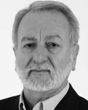 Klaus Schindhelm合作论文数Centre for Biomedical Engineering, University of New South Wales11
 Kenton W. Gregory合作论文数Department of Biomedical Engineering, School of Medicine, Oregon Health Sciences University;Center for Regenerative Medicine, Oregon Health Sciences University9
 Ezio Rizzardo合作论文数CSIRO7Ronald C. Chatelier合作论文数Cooperative Research Centre for Eye Research and Technology, University of New South Wales7E. Winata合作论文数6
 Arthur Brandwood合作论文数6
 John Whitelock合作论文数Graduate School of Biomedical Engineering, College of Engineering, University of New South Wales6
 Megan S. Lord合作论文数Graduate School of Biomedical Engineering, The University of New South Wales, Sydney, NSW 2052, Australia6L. Buckley合作论文数5K. E. Culbertson合作论文数4A. Egan合作论文数4
 Darren Martin合作论文数School of Chemical Engineering, Faculty of Engineering, Architecture and Information Technology, The University of Queensland4Ivan R. Wilson合作论文数4A. C. Wold合作论文数3MCGRATH BARBARA合作论文数3Baw Coller合作论文数3darren martin合作论文数3
 James Jones合作论文数Oregon Hlth & Sci Univ, Ctr Regenerat Med, 3181 SW Sam Jackson Pk Rd, Portland, OR 97239 USA3john morgan合作论文数3
 Bill Cheng合作论文数3Wiesmann William P合作论文数3Enn Elbing合作论文数3
 MoonSun Jung合作论文数Graduate School of Biomedical Engineering, The University of New South Wales3
 Jason C Hedges合作论文数Chicago3Lisa A. Buckley合作论文数3Clinton B. Pepper合作论文数3ajay ahuja合作论文数2KR NOBLE合作论文数2Hua Xie合作论文数Oregon Hlth & Sci Univ, Portland, OR 97239 USA2
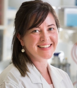 Brooke Farrugia合作论文数Department of Biomedical Engineering, Faculty of Engineering and Information Technology, University of Melbourne2Gordon M. Renwick合作论文数2David F. Sangster合作论文数2
 Renato V. Iozzo合作论文数College of Life Sciences, Thomas Jefferson University2
 Laura A. Poole Warren合作论文数Graduate School of Biomedical Engineering, Faculty of Engineering, The University of New South Wales2Nicole Sherriff合作论文数2kathryn r noble合作论文数1Radim Dvorak合作论文数1CLEON W. GOODWIN合作论文数1V. Rupp合作论文数1Ae Pusateri合作论文数1michael s radovan合作论文数1yichi chen合作论文数1S Kuhn合作论文数Department of Biomedical Engineering, Vanderbilt University1Jack Lazar合作论文数Portland, OR1




