All life at the cellular level has the capacity to induce an antiviral response. Here, we examine the result of imposing the antiviral response of one branch of life onto another and find evidence for conflict.
The effect of SARS-CoV-2 infection on placental function is not well understood. Analysis of placentas from women who tested positive at delivery showed SARS-CoV-2 genomic and subgenomic RNA in 22 out of 52 placentas. Placentas from two mothers with symptomatic COVID-19 whose pregnancies resulted in adverse outcomes for the fetuses contained high levels of viral Alpha variant RNA. The RNA was localized to the trophoblasts that cover the fetal chorionic villi in direct contact with maternal blood. The intervillous spaces and villi were infiltrated with maternal macrophages and T cells. Transcriptome analysis showed an increased expression of chemokines and pathways associated with viral infection and inflammation. Infection of placental cultures with live SARS-CoV-2 and spike protein-pseudo-typed lentivirus showed infection of syncytiotrophoblast and, in rare cases, endothelial cells mediated by ACE2 and Neuropilin-1. Viruses with Alpha, Beta, and Delta variant spikes infected the placental cultures at significantly greater levels.
SARS-CoV-2 infects less than 1% of cells in the human body, yet it can cause severe damage in a variety of organs. Thus, deciphering the non-cell-autonomous effects of SARS-CoV-2 infection is imperative for understanding the cellular and molecular disruption it elicits. Neurological and cognitive defects are among the least understood symptoms of COVID-19 patients, with olfactory dysfunction being their most common sensory deficit. Here, we show that both in humans and hamsters, SARS-CoV-2 infection causes widespread downregulation of olfactory receptors (ORs) and of their signaling components. This non-cell-autonomous effect is preceded by a dramatic reorganization of the neuronal nuclear architecture, which results in dissipation of genomic compartments harboring OR genes. Our data provide a potential mechanism by which SARS-CoV-2 infection alters the cellular morphology and the transcriptome of cells it cannot infect, offering insight to its systemic effects in olfaction and beyond.
Negative-sense RNA viruses (NSVs) rely on prepackaged viral RNA -dependent RNA polymerases (RdRp) to replicate and transcribe their viral genomes. Their replication machinery consists of an RdRp bound to viral RNA which is wound around a nucleoprotein (NP) scaffold, forming a viral ribonucleoprotein complex. NSV NP is known to regulate transcription and replication of genomic RNA; however, its role in maintaining and protecting the viral genetic material is unknown. Here, we exploited host microRNA expression to target NP of influenza A virus and Sendai virus to ascertain how this would impact genomic levels and the host response to infection. We find that in addition to inducing a drastic decrease in genome replication, the antiviral host response in the absence of NP is dramatically enhanced. Additionally, our data show that insufficient levels of NP prevent the replication machinery of these NSVs to process full-length genomes, resulting in aberrant replication products which form pathogen-associated molecular patterns in the process. These dynamics facilitate immune recognition by cellular pattern recognition receptors leading to a strong host antiviral response. Moreover, we observe that the consequences of limiting NP levels are universal among NSVs, including Ebola virus, Lassa virus, and measles virus. Overall, these results provide new insights into viral genome replication of negative-sense RNA viruses and highlight novel avenues for developing effective antiviral strategies, adjuvants, and/or live-attenuated vaccines. IMPORTANCE Negative-sense RNA viruses comprise some of the most important known human pathogens, including influenza A virus, measles virus, and Ebola virus. These viruses possess RNA genomes that are unreadable to the host, as they require specific viral RNA-dependent RNA polymerases in conjunction with other viral proteins, such as nucleoprotein, to be replicated and transcribed. As this process generates a significant amount of pathogen-associated molecular patterns, this phylum of viruses can result in a robust induction of the intrinsic host cellular response. To circumvent these defenses, these viruses form tightly regulated ribonucleoprotein replication complexes in order to protect their genomes from detection and to prevent excessive aberrant replication. Here, we demonstrate the balance that negative-sense RNA viruses must achieve both to replicate efficiently and to avoid induction of the host defenses.
COVID-19 has proven to be a metabolic disease resulting in adverse outcomes in individuals with diabetes or obesity. Patients infected with SARS-CoV-2 and hyperglycemia suffer from longer hospital stays, higher risk of developing acute respiratory distress syndrome (ARDS), and increased mortality compared to those who do not develop hyperglycemia. Nevertheless, the pathophysiological mechanism(s) of hyperglycemia in COVID-19 remains poorly characterized. Here we show that insulin resistance rather than pancreatic beta cell failure is the prevalent cause of hyperglycemia in COVID-19 patients with ARDS, independent of glucocorticoid treatment. A screen of protein hormones that regulate glucose homeostasis reveals that the insulin sensitizing adipokine adiponectin is reduced in hyperglycemic COVID-19 patients. Hamsters infected with SARS-CoV-2 also have diminished expression of adiponectin. Together these data suggest that adipose tissue dysfunction may be a driver of insulin resistance and adverse outcomes in acute COVID-19.
SARS-CoV-2 infects less than 1% of cells in the human body, yet it can cause severe damage in a variety of organs. Thus, deciphering the non-cell autonomous effects of SARS-CoV-2 infection is imperative for understanding the cellular and molecular disruption it elicits. Neurological and cognitive defects are among the least understood symptoms of COVID-19 patients, with olfactory dysfunction being their most common sensory deficit. Here, we show that both in humans and hamsters SARS-CoV-2 infection causes widespread downregulation of olfactory receptors (OR) and of their signaling components. This non-cell autonomous effect coincides with a dramatic reorganization of the neuronal nuclear architecture, which results in dissipation of genomic OR compartments and elimination of genomic contact domains genomewide. Our data provide a novel mechanism by which SARS-CoV-2 infection alters the cellular morphology and the transcriptome of cells it cannot infect, providing insight to its systemic effects in the nervous system and beyond.Funding Information: NIDCD 3R01DC018744-01S1 (SL, JO) National Institutes of Health grant, 4D Nucleome Consortium U01DA052783 (SL) Howard Hughes Medical Institute Faculty Scholar Award (SL), Zegar Family Foundation (SL).Ethics Approval Statement: The study was approved by the ethics and Institutional Review Board of Columbia University Medical Center (IRB AAAT0689, AAAS7370). LVG Golden Syrian hamsters (Mesocricetus auratus) were treated in compliance with the rules and regulations of IACUC under protocol number PROTO202000113-20-0743.
The rapid repurposing of antivirals is particularly pressing during pandemics. However, rapid assays for assessing candidate drugs typically involve in vitro screens and cell lines that do not recapitulate human physiology at the tissue and organ levels. Here we show that a microfluidic bronchial-airway-on-a-chip lined by highly differentiated human bronchial-airway epithelium and pulmonary endothelium can model viral infection, strain-dependent virulence, cytokine production and the recruitment of circulating immune cells. In airway chips infected with influenza A, the co-administration of nafamostat with oseltamivir doubled the treatment-time window for oseltamivir. In chips infected with pseudotyped severe acute respiratory syndrome coronavirus 2 (SARS-CoV-2), clinically relevant doses of the antimalarial drug amodiaquine inhibited infection but clinical doses of hydroxychloroquine and other antiviral drugs that inhibit the entry of pseudotyped SARS-CoV-2 in cell lines under static conditions did not. We also show that amodiaquine showed substantial prophylactic and therapeutic activities in hamsters challenged with native SARS-CoV-2. The human airway-on-a-chip may accelerate the identification of therapeutics and prophylactics with repurposing potential.
Rationale: While respiratory failure is a frequent and clinically significant outcome of coronavirus disease 2019 (COVID-19), cardiac complications are a common feature in hospitalized COVID-19 patients and are associated with worse patient outcomes. The cause of cardiac injury in COVID-19 patients is not yet known. Case reports of COVID-19 autopsy heart samples have demonstrated abnormal inflammatory infiltration of macrophages in heart tissues. Objective: Generate an immunocardiac coculture platform to model macrophage-mediated hyperinflammation in COVID-19 hearts and screen for drugs that can block the macrophage-mediated inflammation. Methods and Results: We systematically compared autopsy samples from non-COVID-19 donors and COVID-19 patients using RNA sequencing and immunohistochemistry. We observed strikingly increased expression levels of CCL2 (C-C motif chemokine ligand 2) and macrophage infiltration in heart tissues of COVID-19 patients. We generated an immunocardiac coculture platform containing human pluripotent stem cell-derived cardiomyocytes and macrophages. We found that macrophages induce increased reactive oxygen species and apoptosis in cardiomyocytes by secreting IL (interleukin)-6 and TNF-alpha (tumor necrosis factor alpha) after Severe acute respiratory syndrome coronavirus 2 (SARS-CoV-2) exposure. Using this immunocardiac coculture platform, we performed a high content screen and identified ranolazine and tofacitinib as compounds that protect cardiomyocytes from macrophage-induced cardiotoxicity. Conclusions: We established an immuno-host coculture system to study macrophage-induced host cell damage following SARS-CoV-2 infection and identified Food and Drug Administration-approved drug candidates that alleviate the macrophage-mediated hyperinflammation and cellular injury.
Recent clinical data have suggested a correlation between coronavirus disease 2019 (COVID-19) and diabetes. Here, we describe the detection of SARS-CoV-2 viral antigen in pancreatic beta cells in autopsy samples from individuals with COVID-19. Single-cell RNA sequencing and immunostaining from ex vivo infections confirmed that multiple types of pancreatic islet cells were susceptible to SARS-CoV-2, eliciting a cellular stress response and the induction of chemokines. Upon SARS-CoV-2 infection, beta cells showed a lower expression of insulin and a higher expression of alpha and acinar cell markers, including glucagon and trypsin1, respectively, suggesting cellular transdifferentiation. Trajectory analysis indicated that SARSCoV-2 induced eIF2-pathway-mediated beta cell transdifferentiation, a phenotype that could be reversed with trans-integrated stress response inhibitor (trans-ISRIB). Altogether, this study demonstrates an example of SARS-CoV-2 infection causing cell fate change, which provides further insight into the pathomechanisms of COVID-19.
The COVID-19 pandemic has caused significant mortality and morbidity around the world. Although effective vaccines have been developed, large parts of the world remain unvaccinated while new SARS-CoV-2 strains keep emerging.
SARS-CoV-2 infection during pregnancy leads to an increased risk of adverse pregnancy outcomes. Although the placenta itself can be a target of virus infection, most neonates are virus free and are born healthy or recover quickly. Here, we investigated the impact of SARS-CoV-2 infection on the placenta from a cohort of women who were infected late during pregnancy and had tested nasal swab positive for SARS-CoV-2 by qRT-PCR at delivery. SARS-CoV-2 genomic and subgenomic RNA was detected in 23 out of 54 placentas. Two placentas with high virus content were obtained from mothers who presented with severe COVID-19 and whose pregnancies resulted in adverse outcomes for the fetuses, including intrauterine fetal demise and a preterm delivered baby still in newborn intensive care. Examination of the placental samples with high virus content showed efficient SARS-CoV-2 infection, using RNA in situ hybridization to detect genomic and replicating viral RNA, and immunohistochemistry to detect SARS-CoV-2 nucleocapsid protein. Infection was restricted to syncytiotrophoblast cells that envelope the fetal chorionic villi and are in direct contact with maternal blood. The infected placentas displayed massive infiltration of maternal immune cells including macrophages into intervillous spaces, potentially contributing to inflammation of the tissue. Ex vivo infection of placental cultures with SARS-CoV-2 or with SARS-CoV-2 spike (S) protein pseudotyped lentivirus targeted mostly syncytiotrophoblast and in rare events endothelial cells. Infection was reduced by using blocking antibodies against ACE2 and against Neuropilin 1, suggesting that SARS-CoV-2 may utilize alternative receptors for entry into placental cells.
The host response to SARS-CoV-2, the etiologic agent of the COVID-19 pandemic, demonstrates significant inter-individual variability. In addition to showing more disease in males, the elderly, and individuals with underlying co-morbidities, SARS-CoV-2 can seemingly render healthy individuals with profound clinical complications. We hypothesize that, in addition to viral load and host antibody repertoire, host genetic variants also impact vulnerability to infection. Here we apply human induced pluripotent stem cell (hiPSC)-based models and CRISPR-engineering to explore the host genetics of SARS-CoV-2. We demonstrate that a single nucleotide polymorphism (rs4702), common in the population at large, and located in the 3’UTR of the protease FURIN, impacts alveolar and neuron infection by SARS-CoV-2 in vitro. Thus, we provide a proof-of-principle finding that common genetic variation can impact viral infection, and thus contribute to clinical heterogeneity in SARS-CoV-2. Ongoing genetic studies will help to better identify high-risk individuals, predict clinical complications, and facilitate the discovery of drugs that might treat disease.
Individuals infected with SARS-CoV-2 who also display hyperglycemia suffer from longer hospital stays, higher risk of developing acute respiratory distress syndrome (ARDS), and increased mortality. Nevertheless, the pathophysiological mechanism of hyperglycemia in COVID-19 remains poorly characterized. Here, we show that hyperglycemia is similarly prevalent among patients with ARDS independent of COVID-19 status. Yet among patients with ARDS and COVID-19, insulin resistance is the prevalent cause of hyperglycemia, independent of glucocorticoid treatment, which is unlike patients with ARDS but without COVID-19, where pancreatic beta cell failure predominates. A screen of glucoregulatory hormones revealed lower levels of adiponectin in patients with COVID-19. Hamsters infected with SARS-CoV-2 demonstrated a strong antiviral gene expression program in the adipose tissue and diminished expression of adiponectin. Moreover, we show that SARS-CoV-2 can infect adipocytes. Together these data suggest that SARS-CoV-2 may trigger adipose tissue dysfunction to drive insulin resistance and adverse outcomes in acute COVID-19.
Adipose-derived stem cells (ASCs) appear to be a useful stem cell population, which has been shown to possess multipotentiality. The aim of this study was to evaluate the utility of ASCs in tissue-engineered cartilage using a hybrid scaffold from fibrin-modified PLGA scaffold. ASCs were isolated from rabbit adipose tissue. The PLGA scaffold was prepared by low-temperature deposition technology and the hybrid scaffold was fabricated by a freeze-drying method. When ASCs were seeded onto fibrin-modified PLGA scaffold in vitro, enhanced cellular viability was observed compared to unmodified PLGA scaffold. The analysis of proteoglycan and collagen II revealed that fibrin-modified scaffold succeeded in inducing ASCs to differentiate into chondrocytes in vitro. A preliminary study on cartilage regeneration was also performed in vivo. Observation of histology and immunoblotting demonstrated that ASCs containing the hybrid scaffold promoted cartilage regeneration in the defects of articular cartilage much better than other groups. These results indicated that ASCs containing the hybrid scaffold are a more effective way to potentially enhance articular cartilage regeneration.
Autoinflammatory disease can result from monogenic errors of immunity. We describe a patient with early-onset multi-organ immune dysregulation resulting from a mosaic, gain-of-function mutation (S703I) in JAK1, encoding a kinase essential for signaling downstream of >25 cytokines. By custom single-cell RNA sequencing, we examine mosaicism with single-cell resolution. We find that JAK1 transcription was predominantly restricted to a single allele across different cells, introducing the concept of a mutational “transcriptotype” that differs from the genotype. Functionally, the mutation increases JAK1 activity and transactivates partnering JAKs, independent of its catalytic domain. S703I JAK1 is not only hypermorphic for cytokine signaling but also neomorphic, as it enables signaling cascades not canonically mediated by JAK1. Given these results, the patient was treated with tofacitinib, a JAK inhibitor, leading to the rapid resolution of clinical disease. These findings offer a platform for personalized medicine with the concurrent discovery of fundamental biological principles.
The SARS-CoV-2 virus has caused already over 3.5 million COVID-19 cases and 250,000 deaths globally. There is an urgent need to create novel models to study SARS-CoV-2 using human disease-relevant cells to understand key features of virus biology and facilitate drug screening. As primary SARS-CoV-2 infection is respiratory-based, we developed a lung organoid model using human pluripotent stem cells (hPSCs) that could be adapted for drug screens. The lung organoids, particularly aveolar type II cells, express ACE2 and are permissive to SARS-CoV-2 infection. Transcriptomic analysis following SARS-CoV-2 infection revealed a robust induction of chemokines and cytokines with little type I/III interferon signaling, similar to that observed amongst human COVID-19 pulmonary infections. We performed a high throughput screen using hPSC-derived lung organoids and identified FDA-approved drug candidates, including imatinib and mycophenolic acid, as inhibitors of SARS-CoV-2 entry. Pre- or post-treatment with these drugs at physiologically relevant levels decreased SARS-CoV-2 infection of hPSC-derived lung organoids. Together, these data demonstrate that hPSC-derived lung cells infected by SARS-CoV-2 can model human COVID-19 disease and provide a valuable resource to screen for FDA-approved drugs that might be repurposed and should be considered for COVID-19 clinical trials. ### Competing Interest Statement R.E.S. is on the scientific advisory board of Miromatrix Inc. The authors have no conflict of interest. L.C.C. is a founder and member of the board of directors of Agios Pharmaceuticals and is a founder and receives research support from Petra Pharmaceuticals. L.C.C. is an inventor on patents (pending) for Combination Therapy for PI3K-associated Disease or Disorder, and The Identification of Therapeutic Interventions to Improve Response to PI3K Inhibitors for Cancer Treatment. L.C.C. is a co-founder and shareholder in Faeth Therapeutics. T.M.Y. is a stockholder and on the board of directors of DESTROKE, Inc., an early-stage start-up developing mobile technology for automated clinical stroke detection.
Viruses are efficient metabolic engineers that actively rewire host metabolic pathways to support their lifecycle. Charting SARS-CoV-2 induced metabolic changes in lung cells could offer insight into COVID-19 pathogenesis while presenting new therapeutic targets. Here we show that the transcriptional response SARS-CoV-2 in primary lung epithelial cells and biopsies of COVID-19 patients is predominantly metabolic. This transcriptional signature was dominated by changes to lipid metabolism and the induction of IRE1 and PKR pathways of endoplasmic stress in a process regulated by several viral proteins. Transcriptional regulatory analysis of these changes reveals small clusters of transcription factors modulating key enzymes in each pathway. The upregulation of glycolysis and the dysregulation of the citric acid cycle was mediated by NFκB and RELA. While the upregulation of fatty acid and cholesterol synthesis showed a more complex control conditionally modulated by ER-stress activated PPARγ, C/EBP, and PPARα. Viral protein ORF3a appeared to interact with all three pathways suggesting both direct and indirect modulation of host metabolism. Finally, we show that PPARα-agonist fenofibrate reversed the metabolic changes induced by SARS-CoV-2 blocking viral replication. Taken together, our data suggest that elevated lipid metabolism may underlie aspects of COVID-19 pathogenesis, offering new therapeutic avenues in targeting this critical pathway on which the virus relies.Funding: Funding was provided by European Research Council Consolidator Grants OCLD (project no. 681870) and generous gifts from the Nikoh Foundation and the Sam and Rina Frankel Foundation (YN). This work was additionally funded by generous support from the Marc Haas Foundation, the National Institutes of Health, and DARPA's PREPARE Program (HR0011-20-2-0040). The views, opinions, and/or findings expressed are those of the author and should not be interpreted as representing the official views or policies of the Department of Defense or the U.S. government (BRT). Ethical Approval: Experiments using samples from human subjects were conducted in accordance with local regulations and with the approval of the institutional review board at the Icahn School of Medicine at Mount Sinai under protocol HS#12-00145.
 Benjamin TenOever合作论文数Department of Microbiology, Grossman School of Medicine, New York University;Bryan Cave Leighton Paisner LLP;Archean Biologics25
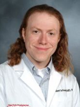 Robert Schwartz合作论文数Phyiology, Biophysics & Systems Biology, Weill Cornell Graduate School of Medical Sciences, Cornell University14
 Benjamin E Nilsson-Payant合作论文数TWINCORE Ctr Expt & Clin Infect Res13
 Yaron Bram合作论文数Weill Cornell Medicine12
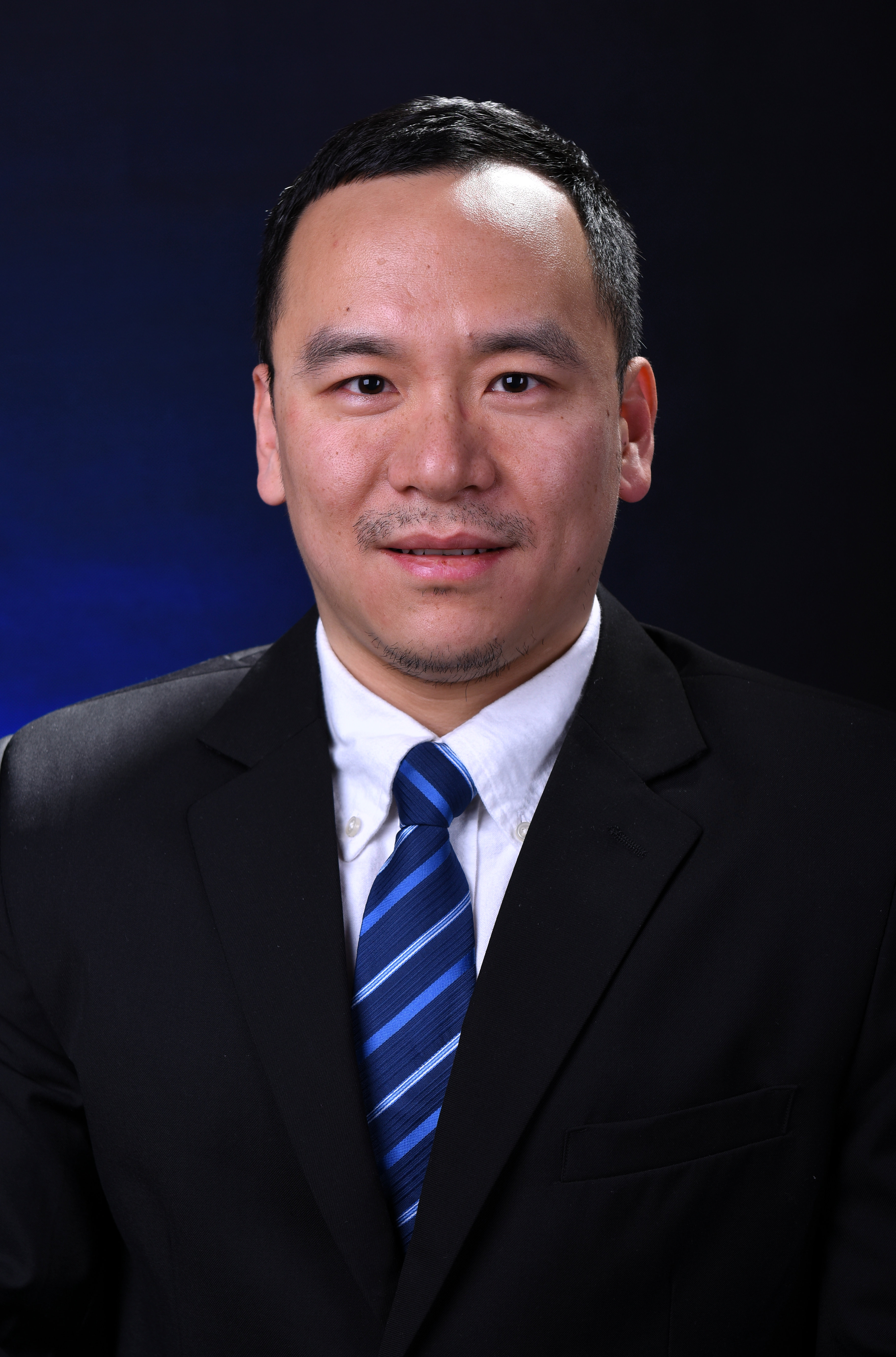 Tuo Zhang (张拓)合作论文数School of Automation, Northwestern Polytechnical University9
 Shuibing Chen合作论文数Department of Biochemistry & Biophysics, Weill Cornell Medical College;Graduate School of Medical Sciences, Weill Cornell Medical College9Vasuretha Chandar合作论文数8
 Daisy Hoagland合作论文数Department of Microbiology, Icahn School of Medicine at Mt. Sinai8
 Blanco-Melo Daniel合作论文数Icahn Sch Med Mt Sinai, Dept Microbiol, New York, NY 10029 USA8David Redmond合作论文数Joan and Sanford I. Weill Department of Medicine, Weill Cornell Medicine, Cornell University8
 Todd R. Evans合作论文数Cell and Developmental Biology Department, Weill Cornell Medicine, Cornell University7
 Jenny Xiang合作论文数Department of Plant and Microbial Biology, North Carolina State University7
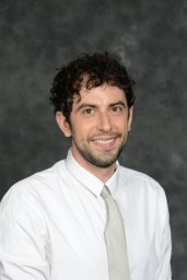 Sean Houghton合作论文数Weill Cornell Med, Ansary Stem Cell Inst6
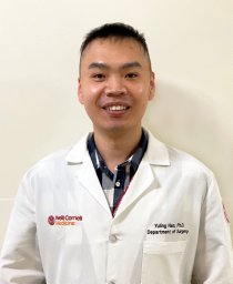 Yuling Han合作论文数Weill Cornell Med, Dept Surg, New York, NY 10065 USA6Rasmus Møller合作论文数Icahn Sch Med Mt Sinai, Dept Microbiol, New York, NY 10029 USA6Yang Liuliu合作论文数Chinese Academy of Sciences Key Laboratory of Infection and Immunity, Chinese Academy of Sciences6
 David D. Ho (何大一)合作论文数Aaron Diamond AIDS Research Center, Columbia University Irving Medical Center6
 Pengfei Wang (王鹏飞)合作论文数School of Life Sciences, Fudan University6
 Yaoxing Huang合作论文数Division of Infectious Diseases, Department of Medicine, Columbia University Irving Medical Center6Jean K. Lim合作论文数5Kohei Oishi合作论文数5Huanhuan Joyce Chen合作论文数Pritzker School of Molecular Engineering, The University of Chicago5
 Hui Wang (王慧)合作论文数School of Public Health, School of Medicine, Shanghai Jiao Tong University5
 Fong Cheng Pan合作论文数5Jordan Tristan X合作论文数5
 Dong Xu (许东)合作论文数Tongji Hospital, Tongji Medical College, Huazhong University of Science & Technology5Maryline Panis合作论文数5Chanel Richardson合作论文数4
 Fuyu Duan (段福宇)合作论文数Department of Engineering Biology, Faculty of Synthetic Biology, Shenzhen University of Advanced Technology4Shu Horiuchi合作论文数Icahn Sch Med Mt Sinai, Dept Microbiol, New York, NY 10029 USA4
 Xuming Tang合作论文数Center for Genomic Health, Weill Cornell Medicine4Zeping Zhao合作论文数4Lucía Carrau合作论文数CNRS, Inst Pasteur, Viral Populat & Pathogenesis Unit, UMR 3569, 28 Rue Dr Roux, F-75724 Paris 15, France4
 Xiaohua Duan (段晓华)合作论文数School of Pharmaceutical Sciences, East China Normal University4Justin Frere合作论文数Department of Microbiology, Icahn School of Medicine at Mount Sinai4
 Jared Lee Johnson合作论文数Lewis Cantley Lab, Meyer Cancer Center, Weill Cornell Medicine, Cornell University4
 Lewis Cantley合作论文数Department of Cell Biology, Harvard Medical School;Department of Cell Biology, Dana-Farber Cancer Institute4Jiajun Zhu (朱佳俊)合作论文数4
 Wen-Chun Liu合作论文数4
 Randy A Albrecht合作论文数Icahn School of Medicine at Mount Sinai4
 Alain Charles Borczuk合作论文数Department of Pathology, Northwell Health4Xing Wang合作论文数3Girija Goyal合作论文数3Melissa Rodas合作论文数3Danni Y. Zhu合作论文数3Kenneth E Carlson合作论文数3
 Atiq Nurani合作论文数Wyss Institute for Biologically Inspired Engineering, Harvard University3
 Luis Guillermo Gómez Escobar合作论文数Univ Los Andes3Robert E Haupt合作论文数3




