Abstract Chronic inflammation increases the susceptibility of the colon to neoplasia and cancer through establishment of an inflammatory microenvironment and persistent release of reactive oxygen species (ROS), inducing genome damage. Interleukin -1beta (IL-1beta), a pro-inflammatory cytokine, plays a pivotal role in the pathogenesis of acute and chronic intestinal inflammation, regulating both innate and adaptive immune responses. IL-1beta has been shown to promote accumulation of IL-17A through lymphoid and Th17 infiltration in the colon tumor microenvironment, supporting increased inflammation and correlating directly with poor patient prognosis. New insights into the signaling pathways supported by IL-1beta in the pathogenesis of colon cancer, including angiogenesis and metastasis, may provide downstream targets for treatments that minimally perturb intestinal immunity. Prior studies from our group have demonstrated significant up-regulation of IL-1, dual oxidase 2 (DUOX2) and its partner maturation factor, DUOXA2, in surgically resected colon cancer specimens compared with adjacent normal colonic epithelium. To elucidate a direct link between IL-1 family cytokines, DUOX2/DUOXA2 derived ROS, and colorectal cancer, we examined human colon cancer cells (HT29, Ls513, T84 and Colo205) stimulated with IL-1beta, in cooperation with IL-6. Co-stimulation resulted in a dramatic, synergistic up-regulation of a hydrogen peroxide producing (Amplex Red oxidizing) DUOX2 enzyme complex and was directly associated with enhanced histone H2AX phosphorylation (γH2AX), a marker of DNA double strand breaks. Interestingly, this concentration- and time-dependent induction of expression and oxidative response was not mediated by other IL-1 family members (IL-18, IL-33 or IL-37). Investigations with the interleukin-1 receptor antagonist anakinra established that signaling for IL-1beta/IL-6 co-treatments proceeded through the IL-1 receptor for all cell lines, though minimal receptor expression is present in T84 and Ls513 cells. Similarly, exposure to tocilizumab, an IL-6R antagonist, demonstrated that the IL-1beta/IL-6 synergistic increase in DUOX2/DUOXA2 complex expression is IL-6 receptor dependent. Perturbation of the IL-1 and IL-6 signaling pathway elements MYD88, IRAK1, STAT1, and STAT3 by siRNA knockdown demonstrated their significant contribution to DUOX2/DUOXA2 up-regulation, while dependence on RELA was absent. Knockdown facilitated by MAPK14 siRNA and SB203580 p38 alpha/beta inhibition also demonstrated a role for p38alpha in the regulation of DUOX2/DUOXA2 expression by the IL-1beta/IL-6 signaling axis. Current studies are focused on elucidating the DUOX2 promoter transcription factor binding site(s) responsible for DUOX2 up-regulation through chromatin immunoprecipitation. Citation Format: Jennifer L. Meitzler, Becky A. Diebold, David J. Mallick, Yongzhong Wu, Smitha Antony, Mariam M. Konaté, Krishnendu Roy, James H. Doroshow. IL-1 beta/ IL-6 co-stimulation synergistically mediates DUOX2/DUOXA2 complex up-regulation via JAK/STAT and p38 signaling events [abstract]. In: Proceedings of the American Association for Cancer Research Annual Meeting 2026; Part 1 (Regular Abstracts); 2026 Apr 17-22; San Diego, CA. Philadelphia (PA): AACR; Cancer Res 2026;86(7 Suppl):Abstract nr 3319.
Abstract Epigenetic reprogramming, particularly histone deacetylation (HDAC), and reactive oxygen species (ROS) play key roles in melanoma progression and the development of drug resistance. While various histone deacetylase inhibitors (HDACi) are already in clinical use for hematological cancers, recent studies indicate their potential effectiveness in melanoma models as well. Given the increasing interest in enhancing the therapeutic efficacy of HDACi, we investigated the molecular mechanisms underlying their association with ROS formation in melanomas. Uveal melanoma (UM), a rare and aggressive eye cancer, has been identified in the TCGA dataset as expressing the highest levels of the superoxide-producing enzyme NADPH oxidase 5 (NOX5) among all human cancers. Our study demonstrates that HDACi treatment increases NOX5 expression in UM cell lines. This upregulation is accompanied by elevated extracellular ROS, which is suppressed when NOX5 is silenced. The use of actinomycin D and cycloheximide significantly reduced HDACi-induced NOX5 expression, suggesting regulation at both transcriptional and translational levels. HDACi treatment also led to accumulation of acetylated histones H3 and H4. Since histone acetylation (particularly H4K5/8/12Ac) recruits BRD4 to facilitate transcription, we evaluated the effect of BRD4 knockdown or exposure to BRD4 inhibitors on NOX5 levels in UM cells. Both approaches reduced NOX5 expression induced by HDACi. Furthermore, dual silencing of the transcription factors Sp1 and Sp3 markedly attenuated both basal and HDACi-induced expression of NOX5. ChIP assays revealed that HDACi treatment increases chromatin-associated acetylated histone H3, phosphorylated RNA polymerase II (p-Ser2), BRD4, Sp1, and Sp3 at the NOX5 promoter region. Collectively, these findings suggest that HDACi-mediated upregulation of NOX5 in UM cells occurs, at least in part, via transcriptional activation of the NOX5 promoter. This process is driven by HDACi-induced histone hyperacetylation, which enhances the recruitment of BRD4, Sp1, and Sp3 to the promoter region, thereby facilitating RNA polymerase II-dependent transcription. Given that HDAC inhibition elevates both NOX5 expression and ROS production, co-targeting HDACs and NOX5 may offer a novel and effective therapeutic strategy in the treatment of UM. Citation Format: Smitha Antony, Ballachanda N. Devaiah, Mariam M. Konaté, Yongzhong Wu, Guojian Jiang, Jennifer L. Meitzler, Jiamo Lu, Becky A. Diebold, David J. Mallick, Krishnendu K. Roy, Dinah S. Singer, James H. Doroshow. HDAC inhibitors induce NOX5 expression in human uveal melanoma cells in a Brd4-Sp1/Sp3-dependent manner [abstract]. In: Proceedings of the American Association for Cancer Research Annual Meeting 2026; Part 1 (Regular Abstracts); 2026 Apr 17-22; San Diego, CA. Philadelphia (PA): AACR; Cancer Res 2026;86(7 Suppl):Abstract nr 4491.
Abstract Chronic pancreatitis and sustained pancreatic inflammation increase the risk of pancreatic ductal adenocarcinoma (PDAC), in part through the release of pro-inflammatory cytokines and excessive generation of reactive oxygen species (ROS). Interleukin-1 (IL-1), a major upstream cytokine secreted by malignant or microenvironmental cells, promotes pancreatic inflammation, tumorigenesis, invasiveness, and intratumoral heterogeneity. Previously, our laboratory has established that dual oxidase 2 (DUOX2), one of seven NADPH oxidase (NOX) family members, plays a critical role in H2O2-mediated host defense and chronic inflammation in the gastrointestinal tract. DUOX2 is upregulated and expressed along with its maturation factor, DUOXA2, in several human pancreatic cancer cell lines following exposure to various pro-inflammatory cytokines, including IFN-γ, IL-4, and IL-17A. In the present study, we show that IL-1α and IL-1β robustly upregulate DUOX2/DUOXA2 mRNA and protein expression in a panel of human PDAC cell lines and patient-derived PDAC organoids. IL-1 exposure triggers transient activation of STAT1 and STAT3, followed by sustained induction of DUOX2/DUOXA2, without increasing expression of other NOX family members. Activation of canonical IL-1 signaling in PDAC cell lines was supported by the observation of IL-1-related upregulation of IL-8 (CXCL8) mRNA, a chemokine essential for neutrophil recruitment and a potent promoter of angiogenesis, in IL-1-treated PDAC cells. Co-administration of Anakinra, an IL-1 receptor antagonist, markedly suppresses IL-1-induced DUOX2/DUOXA2 and CXCL8 expression in vitro. Knocking out DUOX2 in BxPC-3 cells established DUOX2 as the predominant source of IL-1-mediated ROS production. Targeted siRNA knockdown of proximal IL-1 pathway nodes, such as IL-1R1, MYD88 and IRAK1/2, significantly decreased expression of DUOX2/DUOXA2 and of CXCL8. We also found that STAT1 and STAT3, two transcription factors involved in downstream transcription in the IL-1 pathway, are crucial for the regulation of DUOX2/DUOXA2 in pancreatic cancer. Collectively, these findings define an IL-1-STAT1/STAT3 axis that drives DUOX2-dependent ROS production in PDAC, reinforcing a pro-oxidant, pro-inflammatory microenvironment. Citation Format: David J. Mallick, Yongzhong Wu, Mariam M. Konaté, Becky A. Diebold, Smitha Antony, Jennifer L. Meitzler, Goujian Jiang, Jiamo Lu, Krishnendu K. Roy, James H. Doroshow. Interleukin-1 upregulates dual oxidase 2 expression and ROS production in human pancreatic cancer cells in a STAT1/STAT3-dependent manner [abstract]. In: Proceedings of the American Association for Cancer Research Annual Meeting 2026; Part 1 (Regular Abstracts); 2026 Apr 17-22; San Diego, CA. Philadelphia (PA): AACR; Cancer Res 2026;86(7 Suppl):Abstract nr 2881.
Abstract Gastrointestinal inflammation is associated with an increased risk for colorectal cancer (CRC) and is associated with elevated expression of NADPH oxidase (NOX) isoforms, NOX1 and DUOX2, which catalyze the synthesis of superoxide anion radical (O2 ·-) and hydrogen peroxide (H2O2), respectively. To explore the mechanisms that regulate DUOX2, we investigated the effects of a combination of IL-6 and IL-17A on DUOX2 expression in CRC patient-derived cells (PDC) (T-280R, F725, F1126) vs. normal colon cell lines (CCD-112, CCD-841, CCD-18). DUOX2 mRNA levels were generally higher across CRC PDCs relative to the normal colon cell lines. Treatment of PDCs with IL-6 plus IL-17A for 4-6 days resulted in significant upregulation of DUOX2 mRNA and protein. This agrees with data demonstrating elevated DUOX2 levels in CRC tumors relative to corresponding non-malignant tissues. Treatment of HT-29 and Ls513 colon cancer cell lines with IL-6 plus IL-17A for 8-15 days yielded greater-than-additive increases in DUOX2 mRNA, protein, and DUOX2-dependent H2O2 production as measured by Amplex Red assays. RNA-Seq analysis of cytokine-treated HT-29 cells indicated that STAT3 and NFkB signaling pathways mediated the IL-6/IL-17A-induced DUOX2 expression. We also investigated the regulation of DUOX2 using a triple-cytokine cocktail of IL-6, IL-17A, and TNFα. The inclusion of TNFα increased the expression of DUOX2 50-fold more than treatment with IL-6 plus IL-17A in HT29 cells, and even more in LS513 cells. There were also substantial increases in DUOX2 activity. In addition, the triple-cytokine treatment shortened the time course of mRNA and protein expression from several days to 24-48 h. When IL-22 was substituted for IL-6 in the triple-cytokine treatment, the fold-increase was even greater. The STAT3 pathway was activated by IL-6 or IL-22, and the NFkB pathway was activated by IL-17A and TNFα in these cell lines. Silencing of STAT3 or RELA by siRNA nearly abolished DUOX2 expression. The triple-cytokine treatments could also induce cell death within 48-72 h, as well as DNA damage as evidenced by phosphorylation of γ-H2AX. In summary, DUOX2 expression and DUOX2-dependent H2O2 production in HT-29 and LS513 cells and PDCs could be finely tuned by synergism amongst several pro-inflammatory cytokines known to be overexpressed in CRC. Citation Format: Becky A. Diebold, Agnes Juhasz, Mariam M. Konaté, Jiamo Lu, Guojian Jiang, Jennifer L. Meitzler, Yongzhong Wu, Smitha Antony, David J. Mallick, Krishnendu K. Roy, James H. Doroshow. DUOX2 is finely tuned by synergism between cytokines IL-6, IL-22, IL-17A, and TNFα in colon cancer [abstract]. In: Proceedings of the American Association for Cancer Research Annual Meeting 2026; Part 1 (Regular Abstracts); 2026 Apr 17-22; San Diego, CA. Philadelphia (PA): AACR; Cancer Res 2026;86(7 Suppl):Abstract nr 6585.
Abstract Dual oxidase 2 (DUOX2), one of the seven NADPH oxidases family members, plays a critical role in both host defense and chronic inflammation-associated cancer in the gastrointestinal system. In vitro, pro-inflammatory cytokines, such as IFN- y, IFNα/β, IL-4 and IL-17A, enhance DUOX2/DUOXA2 expression through activation of STATs and NF-kB signaling pathway proteins; in vivo, DUOX protein and mRNA levels are substantially upregulated in chronic pancreatitis, pre-malignant pancreatic intraepithelial neoplasms and the early stages of pancreatic cancer patients compared to histologically normal pancreatic tissues. In pancreatic adenocarcinoma, increased DUOX2 expression is adversely correlated with overall patient survival. Heat-Shock Protein 90 (Hsp90), an important molecular chaperone involved in tumorigenesis, invasion and metastasis of cancer cells, is critical in folding, maturation and stability of many oncogenic client proteins, including kinases such as AKT and JAK1/2, and transcription factors, such as STAT3 and HIF-1α. Several STAT family members, along with Hsp90, are overexpressed in human pancreatic carcinomas. Using a panel of human pancreatic cancer cell lines (BxPC-3, AsPC-1 and CFPAC-1), we found that two different Hsp90 inhibitors, Tanespimycin (17-AAG) and Ganetespib (STA-9090), inhibit JAK1 and JAK2 kinases, blocking cytokine-induced, JAK-regulated STAT phosphorylation. Additionally, these Hsp90 inhibitors suppress cytokine-induced DUOX2, VEGF-A, MMP-7 and PD-L1 expression in human pancreatic cancer cell lines with varying sensitivity. Furthermore, the JAK1/2 inhibitor Ruxolitinib inhibits IL-4 induced and JAK-mediated STAT6 phosphorylation, and DUOX2 mRNA and protein expression in BxPC-3 cells. Similar results were observed with JAK1, STAT1, 2 and STAT6 specific siRNA knockdown. However, simultaneous knockdown of both isoforms of Hsp90, Hsp90α and Hsp90β, with specific siRNA did not inhibit JAK1 activity, cytokine-induced DUOX2 mRNA or protein expression. Either remaining Hsp90 protein or other isoforms of Hsp90 in cells may compensate decreased Hsp90 function after siRNA knockdown. Our data suggests that Hsp90 inhibitors, through blocking the cytokine-activated JAK-STATs oncogenic signaling pathway and their downstream genes such as DUOX2, VEGF-A, MMP-7 and PD-L1expression, may be a valuable therapeutic approach for inflammation-associated pancreatic cancer. Citation Format: Yongzhong Wu, David J. Mallick, Allan Di, Smitha Antony, Jennifer Meitzler, Mariam M. Konaté, Becky Diebold, Krishnendu Roy, James H. Doroshow. Heat shock protein inhibitors suppress cytokine-induced DUOX2 mRNA and protein expression in human pancreatic cancer cells in a JAK, STAT dependent manner [abstract]. In: Proceedings of the American Association for Cancer Research Annual Meeting 2026; Part 1 (Regular Abstracts); 2026 Apr 17-22; San Diego, CA. Philadelphia (PA): AACR; Cancer Res 2026;86(7 Suppl):Abstract nr 6578.
Abstract Chronic pancreatitis increases the risk of developing pancreatic ductal adenocarcinoma (PDAC) and is associated with enhanced expression of the NADPH oxidase (NOX) isoform dual oxidase 2 (DUOX2). Interleukin-1 (IL-1) is a major upstream pro-inflammatory cytokine secreted by malignant or microenvironmental cells that supports pancreatic inflammation, tumorigenesis, invasiveness, and tumor heterogeneity. Previously, our laboratory demonstrated that the expression of DUOX2, one of seven NADPH oxidase (NOX) family members, is significantly enhanced by a wide variety of pro-inflammatory cytokines, and that DUOX2-related ROS production may contribute to the development of an oxidative, pro-angiogenic microenvironment in PDAC. In the current studies, we have shown that DUOX2 is upregulated and expressed along with its maturation factor, DUOXA2, in several human pancreatic cancer cell lines following exposure to both IL-1α and IL-1ß, two major agonists of IL-1 signaling. Administration of the anti-inflammatory antibody anakinra (an IL-1R1 antagonist) either in conjunction with or after prolonged treatment with IL-1 in vitro ablated induction of DUOX2/DUOXA2 and of IL-8 (CXCL8), a chemokine essential for neutrophil recruitment and a potent promoter of angiogenesis. Increased expression of other NOX family members following IL-1 exposure was not observed. Using CRISPR/Cas9-mediated DUOX2 knockout models, we report that DUOX2 is the major contributor to IL-1-mediated ROS production in PDAC cells. In a panel of PDAC lines, siRNA knockdown of proximal IL-1 signaling nodes, such as MYD88, significantly decreased expression of DUOX2/DUOXA2 and of CXCL8. However, knockdown of IRAK1 or IRAK4, two critical signaling kinases downstream of MYD88, as well as known downstream mediators of IL-1-related transcription (such as NF-κB, STAT1, MEK/ERK), did not affect IL-1-induced DUOX2/DUOXA2 upregulation, suggesting that alternative pathways of IL-1 signaling might be involved in the enhancement of DUOX2 expression by the cytokine. Because IL-1 signaling is known to affect various acetylation/methylation events in epithelial cells, we evaluated the effect of administering the histone acetyltransferase (HAT) inhibitor A-485 with IL-1; we found that A-485 significantly diminished cytokine-induced DUOX2/DUOXA2 upregulation; however, CXCL8 induction was unchanged. These studies suggest that epigenetic modulation of PDAC cells by IL-1 could contribute to DUOX2-induced ROS production and ROS-related inflammatory stress in the PDAC microenvironment. Citation Format: David J. Mallick, Yongzhong Wu, Mariam M. Konaté, Becky Diebold, Smitha Antony, Jennifer L. Meitzler, Guojian Jiang, Jiamo Lu, Krishnendu Roy, James H. Doroshow. Interleukin-1 upregulates dual oxidase 2 expression and ROS production in human pancreatic cancer cells [abstract]. In: Proceedings of the American Association for Cancer Research Annual Meeting 2024; Part 1 (Regular Abstracts); 2024 Apr 5-10; San Diego, CA. Philadelphia (PA): AACR; Cancer Res 2024;84(6_Suppl):Abstract nr 5327.
Supplementary Legends 1-2 from Novel Indenoisoquinolines NSC 725776 and NSC 724998 Produce Persistent Topoisomerase I Cleavage Complexes and Overcome Multidrug Resistance
Supplementary Figure 2 from Novel Indenoisoquinolines NSC 725776 and NSC 724998 Produce Persistent Topoisomerase I Cleavage Complexes and Overcome Multidrug Resistance
Supplementary Figure 2 from Novel Indenoisoquinolines NSC 725776 and NSC 724998 Produce Persistent Topoisomerase I Cleavage Complexes and Overcome Multidrug Resistance
Supplementary Figure 1 from Novel Indenoisoquinolines NSC 725776 and NSC 724998 Produce Persistent Topoisomerase I Cleavage Complexes and Overcome Multidrug Resistance
Pro-inflammatory cytokines upregulate the expression of the H2O2-producing NADPH oxidase dual oxidase 2 (DUOX2)2 which, when elevated, adversely affects survival from pancreatic ductal adenocarcinoma (PDAC). Because the cGAS-STING pathway is known to initiate pro-inflammatory cytokine expression following uptake of exogenous DNA, we examined whether activation of cGAS-STING could play a role in the generation of reactive oxygen species by PDAC cells. Here, we found that a variety of exogenous DNA species markedly increased the production of cGAMP, the phosphorylation of TBK1 and IRF3, and the translocation of phosphorylated IRF3 into the nucleus, leading to a significant, IRF3-dependent enhancement of DUOX2 expression, and a significant flux of H2O2 in PDAC cells. However, unlike the canonical cGAS-STING pathway, DNA-related DUOX2 upregulation was not mediated by NF-κB. Although exogenous IFN-β significantly increased Stat1/2-associated DUOX2 expression, intracellular IFN-β signaling that followed cGAMP or DNA exposure did not itself increase DUOX2 levels. Finally, DUOX2 upregulation subsequent to cGAS-STING activation was accompanied by the enhanced, normoxic expression of HIF-1α and VEGF-A as well as DNA double strand cleavage, suggesting that cGAS-STING signaling may support the development of an oxidative, pro-angiogenic microenvironment that could contribute to the inflammation-related genetic instability of pancreatic cancer.
Supplementary Figure 1 from Novel Indenoisoquinolines NSC 725776 and NSC 724998 Produce Persistent Topoisomerase I Cleavage Complexes and Overcome Multidrug Resistance
Pancreatic inflammation contributes to increased risk of pancreatic adenocarcinoma (PDAC) development through the release of pro-inflammatory cytokines and generation of reactive oxygen species (ROS). Interleukin-1 (IL-1) is a major upstream pro-inflammatory cytokine secreted by malignant or microenvironmental cells that supports pancreatic tumorigenesis, invasiveness, and tumor heterogeneity. Previously, our laboratory has established that dual oxidase 2 (DUOX2), one of seven NADPH oxidase (NOX) family members, plays a critical role in H2O2-mediated host defense and chronic inflammation in the gastrointestinal tract. DUOX2 is upregulated and expressed along with its maturation factor, DUOXA2, in several human pancreatic cancer cell lines following exposure to various pro-inflammatory cytokines, including IFN-γ, IL-4, and IL-17A. Using a panel of PDAC cells lines, we report the induction of DUOX2/DUOXA2 mRNA and protein expression by both IL-1α and IL-1ß, two major agonists of IL-1. In BxPC-3, CFPAC-1 and Hs 766T (HTB-134) cells, prolonged treatment with IL-1α or IL-1ß results in transient activation of STAT1 and NF-kB signaling followed by prolonged induction of DUOX2/DUOXA2 mRNA and protein expression. Stimulation by IL-1α or IL-1ß results in a specific increase in the expression of DUOX2/DUOXA2; increased expression of other NOX family members was not observed. Activation of canonical IL-1 signaling in our PDAC cell lines was supported by the observation of IL-1-related upregulation of IL-8 (CXCL8) mRNA, a chemokine essential for neutrophil recruitment and a potent promoter of angiogenesis, in IL-1-treated PDAC cells. Moreover, when the anti-inflammatory compound anakinra (an IL-1R antagonist) was co-administered in vitro with IL-1, cytokine-induced DUOX2/DUOXA2 and CXCL8 induction was significantly suppressed. Finally, xenografts established with BxPC-3 cells demonstrated slower growth and significant downregulation of IL-1ß and DUOX2 when the immunosuppressive corticosteroid dexamethasone was administered. Given our previous findings that DUOX2 is highly expressed in the pancreas during episodes of chronic pancreatitis or pancreatic cancer, and contributes significantly to extracellular ROS formation, these studies suggest that IL-1 may play a role in promoting inflammation and tumorigenesis in pancreatic malignancies through upregulation of DUOX2. Citation Format: David J. Mallick, Yongzhong Wu, Mariam M. Konaté, Becky A. Diebold, Smitha Antony, Jennifer L. Meitzler, Guojian Jiang, Jiamo Lu, Krishnendu Roy, James H. Doroshow. Interleukin-1 promotes enhanced functional expression of dual oxidase 2 in human pancreatic cancer cells [abstract]. In: Proceedings of the American Association for Cancer Research Annual Meeting 2022; 2022 Apr 8-13. Philadelphia (PA): AACR; Cancer Res 2022;82(12_Suppl):Abstract nr 1318.
ABSTRACTPreviously, we demonstrated that pro-inflammatory cytokines enhance dual oxidase 2 (DUOX2)-dependent production of reactive oxygen species by human pancreatic ductal carcinoma (PDAC) cells, and that DUOX2 expression is significantly increased in patients with early stages of PDAC. In genetically-engineered mouse models of PDAC, dexamethasone (Dex) decreases formation of pancreatic intraepithelial neoplasia (PanIn) foci as well as PDAC invasiveness. Herein, we report that Dex, in a concentration- and time-dependent fashion, inhibited pro-inflammatory cytokine (IFN-γ/LPS/IL-17A/IL-4)-mediated enhancement of DUOX2 expression in BxPC-3, CFPAC-1, and AsPC-1 human PDAC cell lines, as well as DUOX2–induced DNA damage. The inhibitory effects of Dex were abolished by pre-treatment with the Dex antagonist RU-486. Examination of the human DUOX2 promoter in silico revealed a putative negative glucocorticoid receptor (GR) binding element (IRnGRE). Western analysis, using nuclear extracts from Dex-treated PDAC cells, demonstrated that Dex activated the glucocorticoid receptor in PDAC cell nuclei in the presence of certain co-repressors, such as NCoR-1/2 and histone deacetylases (HDAC1, 2, and 3). Dex produced no anti-proliferative effects on PDAC cellsin vitro. However, Dex significantly decreased the growth of BxPC-3 xenografts while decreasing inflammatory and immune cell infiltration of the microenvironment, as well as the mRNA expression of DUOX2 and VEGF-A, in BxPC-3 tumors. In contrast, Dex had no effect on the growth of xenografts developed from MIA-PaCa cells that are unresponsive to pro-inflammatory cytokines in culture. In summary, these studies suggest that suppression of inflammation-related DUOX2 expression by Dex could diminish the oxidative milieu supporting PDAC growth and development.
Abstract There is a growing appreciation that pancreatic cancers harbor higher numbers of bacteria & fungi compared to normal pancreatic tissue in both humans & mice. These microbes have been suggested to contribute to pancreatic carcinogenesis, & impact therapeutic sensitivity & resistance. Dual oxidase 2 (DUOX2), one of the seven NADPH oxidases, is expressed at the pancreatic cancer plasma membrane & generates extracellular hydrogen peroxide (H2O2). We previously demonstrated that DUOX protein was significantly upregulated in pancreatitis, pancreatic intra-epithelial neoplasia (PanIN), & some early stage pancreatic ductal adenocarcinomas (PDACs). Moreover, DUOX expression in these tissues was associated with H2O2-mediated DNA double strand breaks & upregulation of hypoxia markers VEGF & HIF-1α. Incidentally, we also discovered that mycoplasma-contaminated BxPC-3 human pancreatic cancer cells upregulated known oncogenes such as EGFR, MET, & VEGF-A, in concert with ~20-fold upregulation of DUOX2 mRNA. Recently, the cytosolic DNA-sensing cGAS-STING pathway has been implicated as a critical mediator of tumor-targeting immune responses. Using a panel of human colon & pancreatic cancer cell lines coupled with Western blot analysis & qPCR methods, we have demonstrated that the introduction of purified E. coli genomic DNA fragments, as well as several kinds of plasmid DNA, into the pancreatic cancer cell cytosol significantly enhanced DUOX2 RNA & protein expression, leading to an enhanced DNA damage response. For BxPC-3, CFPAC-1, & HTB134 human pancreatic cancer cells, enhanced DUOX2 expression following introduction of exogenous DNA was associated with the activation of the cGAS-STING pathway, as indicated by enhanced IRF-3 and TBK1 phosphorylation. Treatment of these cell lines with exogenous cGAMP for 24h also activated the cGAS-STING pathway and significantly increased DUOX2 expression. Correspondingly, in BxPC-3 cells treated with human cGAS-specific small interfering RNAs, we observed the significant suppression of cGAS RNA and protein expression & related downstream signaling, along with the significant attenuation of DNA-induced DUOX2 RNA & protein. In contrast, exogenous DNA did not upregulate DUOX2 in colon cancer cell lines. In summary, our data suggest that, in pancreatic cancers, a bacteria-containing microenvironment is likely to promote DUOX2-related H2O2 production as a consequence of the bacterial DNA-induced activation of the cGAS-STING innate immunity pathway, which, in turn, could enhance the oxidative milieu, augment leukocyte recruitment, & increase genetic instability, thus contributing to the molecular evolution of pancreatic tumors. Citation Format: Yongzhong Wu, Stephen L. Wang, Smitha Antony, Jennifer L. Meitzler, Jiamo Lu, Guojian Jiang, Becky Diebold, Mallick David, Mariam M. Konate, Roy Krishnendu, James H. Doroshow. E. coli genomic DNA fragments induce cGAS-STING-mediated DUOX2 expression in human pancreatic cancer cells that is associated with H2O2-related DNA damage [abstract]. In: Proceedings of the American Association for Cancer Research Annual Meeting 2021; 2021 Apr 10-15 and May 17-21. Philadelphia (PA): AACR; Cancer Res 2021;81(13_Suppl):Abstract nr 1755.
Abstract Inflammatory bowel diseases are states of long-term inflammation of the colon resulting from an interplay of environmental factors and dysregulated immune responses. As inflammatory exposure increases over the time frame of active disease, patients with these chronic conditions are at risk of developing colorectal cancers. Chronic inflammation is thought to contribute to cancer initiation through persistent release of reactive oxygen species (ROS), leading to genome damage. Accumulating evidence suggests inflammatory cells contribute to this process, as the secretion of cytokines and growth factors in response to gut inflammation also supports cancer cell progression. Specifically, IL-1 family members play a pivotal role in the pathogenesis of acute and chronic intestinal inflammation, regulating both innate and adaptive immune responses. To elucidate a direct link between IL-1 family cytokines, ROS generation and colorectal cancer, we observed that treatment of HT29 or T84 cells with IL-1α or IL-1β, in cooperation with IL-6, resulted in oxidant production through up-regulation of both NOX1 and DUOX2 enzymes. Up-regulation of the hydrogen peroxide generating DUOX2/DUOXA2 enzyme complex was directly associated with enhanced histone H2AX phosphorylation (γH2AX), a marker of DNA double strand breaks. Interestingly, this concentration and time-dependent induction of expression and oxidative response was absent in Caco2 cells and was not mediated by other IL-1 family members (IL-18, IL-33 or IL-37). Prior studies from our group have demonstrated significant up-regulation of DUOX2 and DUOXA2 in surgically resected colon cancer specimens compared with adjacent normal colonic epithelium. Future studies will focus on cytokine stimulation of colon organoids derived from patient tumor and adjacent non-malignant tissues to evaluate enzymatic expression and oxidant level changes in a novel patient-derived colon model system. Citation Format: Jennifer L. Meitzler, Yongzhong Wu, Agnes Juhasz, Annamaria Rapisarda, Petreena Campbell, Becky A. Diebold, Smitha Antony, Guojian Jiang, Jiamo Lu, David J. Mallick, Mariam M. Konaté, Krishnendu Roy, James H. Doroshow. Synergistic, cytokine mediated stimulation (IL-1α/β + IL-6) of colon cancer cells reveals a novel mechanism of DNA damage facilitated by up-regulation of NOX1 and DUOX2 [abstract]. In: Proceedings of the American Association for Cancer Research Annual Meeting 2021; 2021 Apr 10-15 and May 17-21. Philadelphia (PA): AACR; Cancer Res 2021;81(13_Suppl):Abstract nr 2915.
Significance: The primary function of NADPH oxidases (NOX1-5 and dual oxidases DUOX1/2) is to produce reactive oxygen species (ROS). If inadequately regulated, NOX-associated ROS can promote oxidative stress, aberrant signaling, and genomic instability. Correspondingly, NOX isoforms are known to be overexpressed in multiple malignancies, thus constituting potential therapeutic targets in cancer. Recent Advances: Multiple genetic studies aimed at suppressing the expression of NOX proteins in cellular and animal models of cancer have provided support for the notion that NOXs play a pro-tumorigenic role. Further, large drug screens and rational design efforts have yielded inhibitor compounds, such as the diphenylene iodonium (DPI) analog series developed by our group, with increased selectivity and potency over "first generation" NOX inhibitors such as apocynin and DPI. Critical Issues: The precise role of NOX enzymes in tumor biology remains poorly defined. The tumorigenic properties of NOXs vary with cancer type, and precise tools, such as selective inhibitors, are needed to deconvolute NOX contribution to cancer development. Most NOX inhibitors developed to date are unspecific, and/or their mechanistic and pharmacological characteristics are not well defined. A lack of high-resolution crystal structures for NOX functional domains has hindered the development of potent and selective inhibitors. Future Directions: In-depth studies of NOX interactions with the tumor microenvironment (e.g., cytokines, cell-surface antigens) will help identify new approaches for NOX inhibition in cancer.
Recommended Citation Antony, Smitha; Jiang, Guojian; Wu, Yongzhong; Meitzler, Jennifer L; Makhlouf, Hala R; Haines, Diana C; Butcher, Donna; Hoon, Dave S B; Ji, Jiuping; Zhang, Yiping; Juhasz, Agnes; Lu, Jiamo; Liu, Han; Dahan, Iris; Konate, Mariam; Roy, Krishnendu K; and Doroshow, James H, "NADPH oxidase 5 (NOX5)-induced reactive oxygen signaling modulates normoxic HIF-1α and p27" (2017). Articles, Abstracts, and Reports. 2314. https://digitalcommons.psjhealth.org/publications/2314
Dual oxidase 2 (DUOX2) is a transmembrane protein that produces hydrogen peroxide in the extracellular environment, mediates innate immunity at mucosal surfaces, contributes to chronic inflammation-related tissue injury and angiogenesis, and can promote the initiation of epithelial-derived cancers. In pancreatic adenocarcinomas (PDAC), the composition of the tumor microbiome significantly affects patient outcomes. However, it remains unclear how microbes in the tumor microbiome affect PDAC on a cellular level. In recent years, the cGAS-STING signaling pathway has been identified as a crucial mediator of anti-viral defense by recognizing the presence of DNA in the cytosol and subsequently producing Type I interferons (IFNs) as well as a host of other anti-viral genes. Using Western blot and qPCR, we show that the introduction of exogenous DNA into the cytosol significantly upregulates DUOX2 expression in human PDAC cells after activating cGAS-STING signaling. Expression of DUOX2 occurs most strongly 2 days post-transfection of DNA, while cGAS-STING signaling is activated within hours. Treatment of BxPC-3 cells with exogenous cyclic GMP-AMP (cGAMP) for 24 hrs increases DUOX2 expression in a concentration-dependent manner, suggesting the necessity of cGAS activity for DUOX2 induction. Furthermore, Western analysis of phospho-NF-κB p65 in BxPC-3 cells demonstrates activation of NF-κB that mirrors the time-dependent induction of DUOX2. In CFPAC-1 cells, another PDAC line, Jak-Stat signaling is activated along with the components of cGAS-STING. These data suggest that DNA-induced DUOX2 upregulation in PDAC cells may be mediated in a cell context-specific fashion, beyond the canonical components of the cGAS-STING pathway. Upregulation of DUOX2 by exogenous DNA is not observed in colon cancer cell lines. In summary, these data suggest that extracellular DNA could contribute to a DUOX2-induced, H2O2-mediated pro-inflammatory milieu that specifically contributes to the pathogenesis of PDAC, in part through activation of the cGAS-STING pathway. Citation Format: Stephen L. Wang, Yongzhong Wu, Smitha Antony, Agnes Juhasz, Jennifer Meitzler, Goujian Jiang, Iris Dahan, Jiamo Lu, Krishnendu Roy, James Doroshow. Exogenous DNA upregulates DUOX2 expression in pancreatic cancer via activation of the cGAS-STING signaling pathway [abstract]. In: Proceedings of the Annual Meeting of the American Association for Cancer Research 2020; 2020 Apr 27-28 and Jun 22-24. Philadelphia (PA): AACR; Cancer Res 2020;80(16 Suppl):Abstract nr 3348.
To facilitate functional investigation of the role of NADPH oxidase 1 (NOX1) and associated reactive oxygen species in cancer cell signaling, we report herein the development and characterization of a novel mouse monoclonal antibody that specifically recognizes the C-terminal region of the NOX1 protein. The antibody was validated in stable NOX1 overexpression and knockout systems, and demonstrates wide applicability for Western blot analysis, confocal microscopy, flow cytometry, and immunohistochemistry. We employed our NOX1 antibody to characterize NOX1 expression in a panel of 30 human colorectal cancer cell lines, and correlated protein expression with NOX1 mRNA expression and superoxide production in a subset of these cells. Although a significant correlation between oncogenic RAS status and NOX1 mRNA levels could not be demonstrated in colon cancer cell lines, RAS mutational status did correlate with NOX1 expression in human colon cancer surgical specimens. Immunohistochemical analysis of a comprehensive set of tissue microarrays comprising over 1,200 formalin-fixed, paraffin-embedded tissue cores from human epithelial tumors and inflammatory disease confirmed that NOX1 is overexpressed in human colon and small intestinal adenocarcinomas, as well as adenomatous polyps, compared to adjacent, uninvolved intestinal mucosae. In contradistinction to prior studies, we did not find evidence of NOX1 overexpression at the protein level in tumors versus histologically normal tissues in prostate, lung, ovarian, or breast carcinomas. This study constitutes the most comprehensive histopathological characterization of NOX1 to date in cellular models of colon cancer and in normal and malignant human tissues using a thoroughly evaluated monoclonal antibody. It also further establishes NOX1 as a clinically relevant therapeutic target in colorectal and small intestinal cancer.
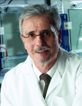 Yves Pommier合作论文数Developmental Therapeutics Branch and Laboratory of Molecular Pharmacology, National Cancer Institute, National Institutes of Health67
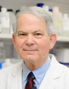 James H. Doroshow合作论文数Center for Cancer Research, National Cancer Institute46
 Wu Yongzhong合作论文数45Lu Jiamo合作论文数43
 Mark S. Cushman合作论文数Department of Medicinal Chemistry and Molecular Pharmacology, Purdue University41Jiang Guojian合作论文数40Meitzler Jennifer L合作论文数37Agnes Juhasz合作论文数36
 Krishnendu Roy合作论文数Vanderbilt Institute for Infection, Immunology and Inflammation, Vanderbilt University Medical Center;School of Engineering, Vanderbilt University24Liu Han合作论文数21Andrew Morrell合作论文数19Glenda Kohlhagen合作论文数18Agama Keli合作论文数17Nagarajan Muthukaman合作论文数16Mariam M Konaté合作论文数15
 Becky Diebold合作论文数12
 Zehong Miao (缪泽鸿)合作论文数Shanghai Institute of Materia Medica, Chinese Academy of Sciences;University of Chinese Academy of Sciences9Melinda G. Hollingshead合作论文数Healthy Pet Mobile Vet9Iris Dahan合作论文数9Xs Xiao合作论文数8
 Christophe H Marchand合作论文数8Mollie H. Wright合作论文数8
 Thomas S. Dexheimer合作论文数Center for Cancer Research, National Cancer Institute8Donna O. Butcher合作论文数8Kazutaka Takagi合作论文数7
 Olivier Sordet合作论文数7
 Lyuba Varticovski合作论文数St. Elizabeth’s Medical Center, Tufts University School of Medicine7Diana C Haines合作论文数6
 Muthusamy Jayaraman合作论文数6David J. Mallick合作论文数5
 Bart Staker合作论文数Seattle Struct Genom Ctr Infect Dis5Alexandra Ioanoviciu合作论文数5Zhiyong Liao合作论文数5
 Ana I. Robles合作论文数Diagnostics Evaluation Branch, Division of Cancer Treatment and Diagnosis, National Cancer Institute, National Institutes of Health;Office of Cancer Clinical Proteomics Research, National Cancer Institute, National Institutes of Health5
 Kurt W. Kohn合作论文数Center for Cancer Research, National Cancer Institute, National Institutes of Health5L Stewart合作论文数4Stephen L. Wang合作论文数4Hala R. Makhlouf合作论文数4K. Agama合作论文数4
 Iwona E Weidlich合作论文数3Yun Ge合作论文数3
 Dennis Curran合作论文数Department of Chemistry, Dietrich School of Arts and Sciences, University of Pittsburgh3Marc C. Nicklaus合作论文数3
 Gary S. Laco合作论文数3
 Linghua Meng (蒙凌华)合作论文数Research Center for Molecular Recognition and Synthesis, Department of Chemistry, Fudan University;Shanghai Institute of Materia Medica, Chinese Academy of Sciences3
 Michael Placzek合作论文数Bioorganic and Medicinal Chemistry Laboratories, Northeastern University3
 Andrew G Stephen合作论文数Protein Chemistry Laboratory, National Cancer Institute at Frederick3




