A requirement for effective chemotherapy is that therapeutic agents reach all cancer cells. However, only a small fraction of the dose accumulates in tumors and is taken up by cancer cells. Encapsulation of drugs into nanoparticles, exploiting the enhanced permeability and retention effect, has moderately improved tumor accumulation. The nanoparticles, being substantially larger than molecular drugs, encounter more severe obstacles in reaching the tumor cells resulting in heterogeneous tumor distribution. Delivery of drugs to the brain is even more challenging due to the blood-brain barrier. In this review, we describe the various barriers for successful delivery of nanoparticles, including the chaotic vasculature, heterogenous extravasation and dense extracellular matrix the nanoparticles need to penetrate. Ultrasound has been shown to enhance delivery of therapeutic agents across these barriers, and the various mechanisms for ultrasound-mediated drug delivery are discussed. The transport of nanoparticles is governed by diffusion and convection, depending on the concentration- and hydrostatic pressure gradient, respectively. Elevated interstitial fluid pressure within tumors eliminates the transcapillary pressure gradient except at the tumor periphery, and pressure gradients within the interstitial space are largely absent. Consequently, diffusion becomes the dominant transport mechanism, but diffusion of nanoparticles is inherently slow. Ultrasound combined with circulating microbubbles has been reported to improve the delivery of therapeutic agents, resulting in improved therapeutic outcomes. This has been shown both for tumors outside the brain and for tumors and other diseases in the brain. Ultrasound and microbubbles can enhance extravasation by increasing both paracellular and transcellular transport, which is particularly important for crossing the blood-brain barrier. Oscillations of the vascular wall induced by ultrasound and microbubbles can improve fluid flow through the extracellular matrix. Increased interstitial hydraulic conductivity and reduced solid stress, associated with enhanced delivery of nanoparticles, have been reported. In addition, ultrasound alone (no microbubbles) can enhance diffusion by releasing nanoparticles bound to extracellular matrix components. Understanding the underlying mechanisms of ultrasound–mediated drug delivery is essential for optimizing therapeutic outcomes. Integrating experimental data with computational modeling provides powerful tools for generating new mechanistic insights. Rakesh Jain and colleagues have been instrumental in advancing this integrated approach by developing sophisticated mathematical models and simulation frameworks alongside with advanced experimental techniques. Their research has provided new insights into key transport processes and parameters which are highly valuable for elucidating the fundamental mechanisms governing ultrasound-mediated transport.
Ageing is established as the most significant risk factor for disease. About 75% of people over 75 years have diabetes or pre-diabetes and/or hyperlipidaemia which are established risk factors for cardiovascular outcomes, and risk factors for age-related conditions such as dementia, sarcopenia, frailty and osteoporosis. Age-related changes in the liver microcirculation, in particular relating to the cells lining the blood vessels, the liver sinusoidal endothelial cells (LSEC), are a potential cause for dyslipidaemia and insulin resistance in old age. There is also loss of LSEC mediated waste clearance functions essential for homeostasis. Finding ways to reverse these age-related changes in the LSEC will fill a significant gap in therapeutic options available for the treatment of ageing disorders. Such therapies may also benefit patients with fibrotic livers, since LSEC changes in this disease resemble those seen in the ageing LSEC in many aspects. Nanoparticles that access systemic circulation frequently accumulate in the liver. This could be utilized as a promising strategy for targeted drug delivery to the liver. The present study assessed if poly(alkyl-cyanoacrylate) nanoparticles (PACA NPs) are a suitable vector for the targeted transport of such therapeutics to LSEC, to reverse age-related changes such as fenestration/porosity loss. Mice were co-injected with PACA NPs and formaldehyde denatured serum albumin (FSA) and their livers were then examined by microscopy. PACA and FSA co-localised to LSEC, including at the sub-cellular level in endocytic vesicles. Isolated LSEC were challenged with Nile Red (NR668) labelled PACA NPs, which were rapidly internalized. HEK293 cells overexpressing stabilin-2 internalized PACA NPs, suggesting that stabilin-2 mediates PACA uptake on LSEC. Cultured LSEC from aged mice were challenged with PACA NPs containing sildenafil and examined with scanning electron microscopy to determine effects on fenestrations. Sildenafil PACA reversed age-related changes LSEC fenestration frequency and porosity at 3-fold lower sildenafil concentrations than sildenafil alone. If sildenafil PACA induces similar changes in vivo, age-related reduction of LSEC porosity could be reversed by the targeted delivery of sildenafil via PACA NPs.
OBJECTIVE:Ultrasound in combination with microbubbles can enhance accumulation and improve the distribution of various therapeutic agents in tumor tissue, leading to improved efficacy. Understanding the impact of treatment on the tumor microenvironment, concurrently with how microenvironment attributes affect treatment outcome, will be important for selecting appropriate patient cohorts in future clinical trials. The main aim of this work was to investigate the influence of ultrasound and microbubbles on the functional vasculature of cancer tissue. METHODS:Four different tumor models in mice (bone, pancreatic, breast and colon cancer) were characterized with respect to vascular parameters using contrast-enhanced ultrasound imaging. The effect of treatment with microbubbles and ultrasound was then investigated using immunohistochemistry and confocal microscopy, quantifying the total amount of vasculature and fraction of functional vessels. Two different microbubbles were used, the clinical contrast agent SonoVue and the large bubbles generated by Acoustic Cluster Therapy (ACT), tailored for therapeutic purposes. RESULTS:The colon cancer model displayed slower flow but a higher vascular volume than the other models. The pancreatic model showed the fastest flow but also the lowest vascular volume. Ultrasound and SonoVue transiently reduced the amount of functional vasculature in breast and colon tumors immediately after treatment. No reduction was observed for ACT, likely due to shorter ultrasound pulses and lower pressures applied. CONCLUSION:Variation between tumor models due to tissue characteristics emphasizes the importance of evaluating treatment suitability in the specific tissue of interest, as altered perfusion could have a large impact on drug delivery and therapeutic outcome.
OBJECTIVE:The chicken chorioallantoic membrane (CAM) is used to study biological processes, including drug delivery, and provides an in ovo model with vasculature available for intravital microscopy. Ultrasound and microbubbles have been shown to be a promising strategy for improving drug delivery to solid tumors and across the blood-brain barrier. Acoustic cluster therapy (ACT) based on large microbubbles with a volume 50-100 times larger than regular contrast agent microbubbles is of particular interest. The objective of this study was to expand our understanding of the mechanisms underlying ACT and its role in drug delivery, particularly in blood flow and extravasation. METHODS:The CAMs were cultured ex ovo and used at embryonic day 13-14, with the vascular effects and extravasation of a co-injected macromolecule (2 MDa fluorescein isothiocyanate-dextran) recorded during ultrasound exposure (1 MHz, mechanical index 0.4 or 0.8, 10,000 cycles) in combination with the microbubble Sonazoid or ACT (activation 2.7 MHz, mechanical index 0.4 to form the large bubble, enhancement 0.5 MHz, mechanical index 0.2 to oscillate the large bubble). RESULTS:Extravasation of 2 MDa dextran demonstrated two different kinetics, rapid and slow, and were observed both for ACT and ultrasound and Sonazoid. ACT bubbles were easily visible in the vasculature, and vascular occlusion or opening and changing in the direction of blood flow were observed. CONCLUSION:Slow and fast extravasation kinetics could be due to diffusion or ultrasound-induced convection. This study demonstrates that the CAM model is highly useful for studying bioeffects induced by sonopermeation at a vascular level.
Lung cancer is one of the most common cancers and a leading cause of death, with poor prognosis and high unmet clinical need. Chemotherapy is a common part of the treatment, either alone or in combination with other treatment modalities, but with limited efficacy and severe side effects. Encapsulation of drugs into nanoparticles can enable a more targeted delivery with reduced off-target toxicity. Delivery to the lungs is however often insufficient due to various biological barriers in the body and in the tumor microenvironment. Here we demonstrate that by incorporating drug-loaded nanoparticles into air-filled microbubbles, a more effective targeting to the lungs can be achieved. Fluorescence imaging and mass spectrometry revealed that the microbubbles could significantly improve accumulation of drug in the lungs of mice, compared to injecting either the free drug by itself or only the drug-loaded nanoparticles. Therapeutic efficacy was verified in a preclinical mouse model with non-small cell lung cancer, monitoring tumor growth by luminescence.
The delivery of nanoparticles to tumors has been shown preclinically to be improved by microbubble-mediated ultrasound. However, the mechanisms and biological effects are not fully understood. In this study, we explored the influence of the tumor microenvironment on nanoparticle uptake and microdistribution both with and without ultrasound and microbubble treatment. Three murine tumor models, KPC (pancreatic ductal adenocarcinoma), 4T1 (triple negative mammary carcinoma) and CT26 (colon carcinoma), were characterized with respect to extracellular matrix composition, tumor stiffness and perfusion. KPC and 4T1 tumors presented higher levels of collagen and hyaluronic acid and were stiffer compared to CT26, whereas all three tumors had similar levels of sulfated glycosaminoglycans. Furthermore, the 4T1 tumors appeared poorly vascularized with a lower cell density compared to KPC and CT26. All three tumors presented similar nanoparticle uptake, but extravasated nanoparticles traveled significantly shorter in KPC tumors compared to 4T1 and CT26. The effect of ultrasound and microbubble treatment on the tumor uptake and penetration of polymer nanoparticles into the extracellular matrix were evaluated using a treatment protocol previously shown to increase nanoparticle delivery to tumors. Interestingly, we found a significant increase in nanoparticle uptake in the soft CT26 tumor, but no effect of the ultrasound treatment in the stiff KPC and 4T1 tumors, suggesting that tumor stiffness is an important parameter for treatment with ultrasound and microbubbles. Ultrasound treatment resulted in a modest but not statistically significant improvement in nanoparticle penetration through the extracellular matrix. In tumors demonstrating increased uptake of nanoparticles following ultrasound treatment, the uptake correlated positively with blood volume. These findings emphasize the importance of taking the tumor microenvironment into consideration when optimizing ultrasound parameters for delivery of nanomedicine.
OBJECTIVE:Ultrasound-triggered bubble-mediated local drug delivery has shown potential to increase therapeutic efficacy and reduce systemic side effects, by loading drugs into the microbubble shell and triggering delivery of the payload on demand using ultrasound. Understanding the behavior of the microbubbles in response to ultrasound is crucial for efficient and controlled release. METHODS:In this work, the response of microbubbles with a coating consisting of poly(2-ethyl-butyl cyanoacrylate) (PEBCA) nanoparticles and denatured casein was characterized. High-speed recordings were taken of single microbubbles, in both bright field and fluorescence. RESULTS:The nanoparticle-loaded microbubbles show resonance behavior, but with a large variation in response, revealing a substantial interbubble variation in mechanical shell properties. The probability of shell rupture and the probability of nanoparticle release were found to strongly depend on microbubble size, and the most effective size was inversely proportional to the driving frequency. The probabilities of both rupture and release increased with increasing driving pressure amplitude. Rupture of the microbubble shell occurred after fewer cycles of ultrasound as the driving pressure amplitude or driving frequency was increased. CONCLUSION:The results highlight the importance of careful selection of the driving frequency, driving pressure amplitude and duration of ultrasound to achieve the most efficient ultrasound-triggered shell rupture and nanoparticle release of protein-and-nanoparticle-stabilized microbubbles.
Optimising drug delivery to tumours remains an obstacle to effective cancer treatment. A prerequisite for successful chemotherapy is that the drugs reach all tumour cells. The vascular network of tumours, extravasation across the capillary wall and penetration throughout the extracellular matrix limit the delivery of drugs. Ultrasound combined with microbubbles has been shown to improve the therapeutic response in preclinical and clinical studies. Most studies apply microbubbles designed as ultrasound contrast agents. Acoustic Cluster Therapy (ACT®) is a novel approach based on ultrasound-activated microbubbles, which have a diameter 5–10 times larger than regular contrast agent microbubbles. An advantage of using such large microbubbles is that they are in contact with a larger part of the capillary wall, and the oscillating microbubbles exert more effective biomechanical effects on the vessel wall. In accordance with this, ACT® has shown promising therapeutic results in combination with various drugs and drug-loaded nanoparticles. Knowledge of the mechanism and behaviour of drugs and microbubbles is needed to optimise ACT®. Real-time intravital microscopy (IVM) is a useful tool for such studies. This paper presents the experimental setup design for visualising ACT® microbubbles within the vasculature of tumours implanted in dorsal window (DW) chambers. It presents ultrasound setups, the integration and alignment of the ultrasound field with the optical system in live animal experiments, and the methodologies for visualisation and analysing the recordings. Dextran was used as a fluorescent marker to visualise the blood vessels and to trace drug extravasation and penetration into the extracellular matrix. The results reveal that the experimental setup successfully recorded the kinetics of extravasation and penetration distances into the extracellular matrix, offering a deeper understanding of ACT’s mechanisms and potential in localised drug delivery.
Pancreatic ductal adenocarcinoma (PDAC) remains a challenging malignancy, mainly due to its resistance to chemotherapy and its complex tumour microenvironment characterised by stromal desmoplasia. There is a need for new strategies to improve the delivery of drugs and therapeutic response. Relevant preclinical tumour models are needed to test potential treatments. This paper compared orthotopic and subcutaneous PDAC tumour models and their suitability for drug delivery studies. A novel aspect was the broad range of tumour properties that were studied, including tumour growth, histopathology, functional vasculature, perfusion, immune cell infiltration, biomechanical characteristics, and especially the extensive analysis of the structure and the orientation of the collagen fibres in the two tumour models. The study unveiled new insights into how these factors impact the uptake of a fluorescent model drug, the macromolecule called 800CW. While the orthotopic model offered a more clinically relevant microenvironment, the subcutaneous model offered advantages for drug delivery studies, primarily due to its reproducibility, and it was characterised by a more efficient drug uptake facilitated by its collagen organisation and well-perfused vasculature. The tumour uptake seemed to be influenced mainly by the structural organisation and the alignment of the collagen fibres and perfusion. Recognising the diverse characteristics of these models and their multifaceted impacts on drug delivery is crucial for designing clinically relevant experiments and improving our understanding of pancreatic cancer biology.
Immunocompetent murine models are important tools for preclinical evaluation of immunotherapies. Here, six different immunocompetent tumor models based on four different cell lines were characterized, including metastatic lung cancer (CMT 167), triple-negative breast cancer (4T1), pancreatic cancer (KPCY), and colon cancer (MC38). The tumors were implanted subcutaneously or orthotopically before the animals were treated with anti-PD1 checkpoint inhibitor. A range of innate and adaptive immune cells were then quantified by flow cytometry of single-cell suspensions from the tumors. Furthermore, confocal laser scanning microscopy was used to quantify the density and distribution of T-cells in frozen sections. A model-dependent cellular immune landscape was observed, with variable responsiveness toward anti-PD1, ranging from the most responsive MC38 colon cancer model to the least responsive 4T1 breast cancer model. The study provides an overview of the immune landscape of these tumor models, and a foundation for further elucidation of pro-tumor and anti-tumor mechanisms behind heterogeneous responses towards immunotherapies.
Objective: Currently available cytotoxic treatments have limited effect on pancreatic ductal adenocarcinoma (PDAC) because desmoplastic stroma limits drug delivery. Efforts have been made to overcome these barriers by drug targeting the tumor microenvironment. Results so far are promising, but without clinical impact. Our aim was to investigate whether ultrasound and microbubbles could improve the uptake and therapeutic response of conventional chemotherapy.Methods: Orthotopic pancreatic tumors growing in mice were treated with commercially available FOLFIRINOX (fluorouracil, irinotecan, oxaliplatin and calcium folinate) and SonoVue microbubbles combined with focused ultrasound. Tumor uptake of platinum (Pt) was measured by inductively coupled plasma mass spectroscopy (ICP-MS), and tumor volumes were measured by ultrasound imaging.Discussion: Uptake of Pt, the active ingredient of oxaliplatin, was significantly increased after ultrasound treatment of orthotopic PDAC tumors. Multiple injections with FOLFIRONOX increased the amount of Pt in tumors. How-ever, the enhanced accumulation did not improve therapeutic response. Increased uptake of Pt confirms that ultrasound and microbubbles have potential in clinical practice with existing drugs.Conclusion: The lack of therapeutic response, despite increased uptake in tumor tissue, emphasizes the importance of studying how to overcome stromal barriers.
Objective: The blood-brain barrier (BBB) is an obstacle for cerebral drug delivery. Controlled permeabilization of the barrier by external stimuli can facilitate the delivery of drugs to the brain. Acoustic Cluster Therapy (ACT (R)) is a promising strategy for transiently and locally increasing the permeability of the BBB to macromolecules and nanoparticles. However, the mechanism underlying the induced permeability change and subsequent enhanced accumulation of co-injected molecules requires further elucidation. Methods: In this study, the behavior of ACT (R) bubbles in microcapillaries in the murine brain was observed using real-time intravital multiphoton microscopy. For this purpose, cranial windows aligned with a ring transducer centered around an objective were mounted to the skull of mice. Dextrans labeled with 2 MDa fluorescein isothio-cyanate (FITC) were injected to delineate the blood vessels and to visualize extravasation.Discussion: Activated ACT (R) bubbles were observed to alter the blood flow, inducing transient and local increases in the fluorescence intensity of 2 MDa FITC-dextran and subsequent extravasation in the form of vascular out-pouchings. The observations indicate that ACT (R) induced a transient vascular leakage without causing substantial damage to the vessels in the brain.Conclusion: The study gave novel insights into the mechanism underlying ACT (R)-induced enhanced BBB permeability which will be important considering treatment optimization for a safe and efficient clinical translation of ACT (R).
Colorectal and ovarian cancers frequently develop peritoneal metastases with few treatment options. Intraperitoneal chemotherapy has shown promising therapeutic effects, but is limited by rapid drug clearance and systemic toxicity. We therefore encapsulated the cabazitaxel taxane in poly(alkyl cyanoacrylate) (PACA) nanoparticles (NPs), designed to improve intraperitoneal delivery. Toxicity of free and encapsulated cabazitaxel was investigated in rats by monitoring clinical signs, organ weight and blood hematological and biochemical parameters. Pharmacokinetics, biodistribution and treatment response were evaluated in mice. Biodistribution was investigated by measuring both cabazitaxel and the 2-ethylbutanol NP degradation product. Drug encapsulation was shown to increase intraperitoneal drug retention, leading to prolonged intraperitoneal drug residence time and higher drug concentrations in peritoneal tumors. As a result, encapsulation of cabazitaxel improved the treatment response in two in vivo models bearing intraperitoneal tumors. Together, these observations indicate a strong therapeutic potential of NP-based cabazitaxel encapsulation as a novel treatment for peritoneal metastases.
Poly (alkyl cyanoacrylate) (PACA) polymeric nanoparticles (NPs) are promising drug carriers in drug delivery. However, the selection of commercially available alkyl cyanoacrylate (ACA) monomers is limited, because most monomers were designed for use in medical and industrial glues and later repurposed for drug encapsulation. This study therefore aimed to seek out novel ACA materials for use in NP systems using a toxicity led screening approach. A multistep strategy, including cytotoxicity screening of alcohols as degradation products of PACA (44 alcohols), NPs (14 polymers), and a final in vivo study (2 polymers) gave poly (2-ethylhexyl cyanoacrylate) PEHCA as a promising novel PACA candidate. For the first time, this work presents cytotoxicity data on several novel ACAs, PEHCA in vivo toxicity data, and miniemulsion polymerisation-based encapsulation of the cabazitaxel and NR688 in novel PACA candidates. Furthermore, several of the ACA candidates were compatible with a wider selection of lipophilic active pharmaceutical ingredients (APIs) versus commercially available controls. Combined, this work demonstrates the potential benefits of expanding the array of available ACA materials in drug delivery. Novel ACAs have the potential to encapsulate a wider range of APIs in miniemulsion polymerisation processes and may also broaden PACA applicability in other fields.
The aim of this study was to develop high load-capacity antibubbles that can be visualized using diagnostic ultrasound and the encapsulated drug can be released and delivered using clinically translatable ultrasound. The antibubbles were developed by optimising a silica nanoparticle stabilised double emulsion template. We produced an emulsion with a mean size diameter of 4.23 ± 1.63 µm where 38.9 ± 3.1% of the droplets contained a one or more cores. Following conversion to antibubbles, the mean size decreased to 2.96 ± 1.94 µm where 99% of antibubbles were <10 µm. The antibubbles had a peak attenuation of 4.8 dB/cm at 3.0 MHz at a concentration of 200 × 103 particles/mL and showed distinct attenuation spikes at frequencies between 5.5 and 13.5 MHz. No increase in subharmonic response was observed for the antibubbles in contrast to SonoVue®. High-speed imaging revealed that antibubbles can release their cores at MIs of 0.6. In vivo imaging indicated that the antibubbles have a long half-life of 68.49 s vs. 40.02 s for SonoVue®. The antibubbles could be visualised using diagnostic ultrasound and could be disrupted at MIs of ≥0.6. The in vitro drug delivery results showed that antibubbles can significantly improve drug delivery (p < 0.0001) and deliver the drug within the antibubbles. In conclusion antibubbles are a viable concept for ultrasound guided drug delivery.
Biodistribution of nanoencapsulated bioactive compounds is primarily determined by the size, shape, chemical composition and surface properties of the encapsulating nanoparticle, and, thus, less dependent on the physicochemical properties of the active pharmaceutical ingredient encapsulated. In the current work, we aimed to investigate the impact of formulation type on biodistribution profile for two clinically relevant nanoformulations. We performed a comparative study of biodistribution in healthy rats at several dose levels and durations up to 14-day post-injection. The studied nanoformulations were nanostructured lipid carriers incorporating the fluorescent dye IR780-oleyl, and polymeric nanoparticles containing the anticancer agent cabazitaxel. The biodistribution was approximated by quantification of the cargo in blood and relevant organs. Several clear and systematic differences in biodistribution were observed, with the most pronounced being a much higher (more than 50-fold) measured concentration ratio between cabazitaxel in all organs vs. blood, as compared to IR780-oleyl. Normalized dose linearity largely showed opposite trends between the two compounds after injection. Cabazitaxel showed a higher brain accumulation than IR780-oleyl with increasing dose injected. Interestingly, cabazitaxel showed a notable and prolonged accumulation in lung tissue compared to other organs. The latter observations could warrant further studies towards a possible therapeutic indication within lung and conceivably brain cancer for nanoformulations of this highly antineoplastic compound, for which off-target toxicity is currently dose-limiting in the clinic.
Systemic injections of chemotherapeutics often deliver only minor fractions of the administered dose to the targeted pathology, resulting in unwanted toxicity towards healthy tissue. In brain tissue, drug delivery is impaired by the highly selective nature of the blood brain barrier impeding the influx of most substances. Acoustic Cluster Therapy (ACT®) is a platform for targeted therapeutic enhancement, facilitating increased local drug transfer. The platform is comprised of ACT® clusters, a mix of microbubbles and microdroplets and low intensity (diagnostic) ultrasound insonation. Intravenously injected ACT® clusters circulate freely through the body before activation by localised insonation at the targeted pathology. In the ultrasound field, microbubbles transfer energy to microdroplets, which undergo a liquid-to-gas phase transition, forming larger ACT® bubbles that transiently deposit in the targeted microvasculature. Further exposure to ultrasound results in controlled volume oscillation of the ACT® bubbles, inducing a range of biomechanical effects. The effects lead to enhanced extravasation across the endothelial barrier, facilitating the transport of co-administered chemotherapeutics to the targeted tissue. Proof of concept studies showed an increased therapeutic efficacy of standard of care drugs, when combined with ACT® treatment. Furthermore, ACT® treatment induced a controlled and temporal opening of the blood brain barrier observed by the extravasation of fluorescent macromolecules into brain tissue.
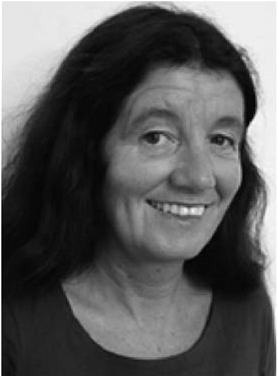 Catharina de Lange Davies合作论文数Department of Physics, Norwegian University of Science and Technology36
 Yrr Mørch合作论文数Dept. of Polymer Particles and Surface Chemistry, SINTEF Materials and Chemistry22
 Einar Sulheim合作论文数20
 Andreas Åslund合作论文数Department of Biotechnology and Nanomedicine, SINTEF Industry18
 Sigrid Berg合作论文数Dept. of Electron. & Telecommun., Norwegian Univ. of Sci. & Technol.;c;Dept. of Electron. & Telecommun., Norwegian Univ. of Sci. & Technol.15
 Rune Hansen合作论文数9
 Melina Mühlenpfordt合作论文数7
 Torp Sverre H合作论文数7
 Annemieke Van Wamel合作论文数7
 Ruth Schmid合作论文数SINTEF6
 Astrid Bjørkøy合作论文数6Astrid Hyldbakk合作论文数5
 Marieke Olsman合作论文数Dept Phys, Norwegian Univ Sci & Technol5
 Sven Even Borgos合作论文数Department of Biotechnology and Nanomedicine, SINTEF5
 Gunhild Mari Mælandsmo合作论文数Department of Tumor Biology, Institute for Cancer Research, Oslo University Hospital4
 Abhilash D. Pandya合作论文数Department of Tumor Biology, Institute for Cancer Research, Division of Cancer Medicine, Oslo University Hospital4Baghirov Habib合作论文数4
 Karianne Giller Fleten合作论文数4
 Kirsten Sandvig合作论文数Dept. of Molecular Cell Biology, Institute for Cancer Research, Oslo University Hospital;Institute for molecular Biosciences, University of Oslo;Institute for Cancer Research, The Norwegian Radium Hospital, Oslo4
 Tore-Geir Iversen合作论文数4Olav Engebraaten合作论文数Institute for Cancer Research, University of Oslo4
 Kjersti Flatmark合作论文数University of Oslo;Department of Tumor Biology, Norwegian Radium Hospital;Department of Tumor Biology, Institute for Cancer Research, Oslo University Hospital4Tore Skotland合作论文数4
 Wanja Kildal合作论文数4Caroline Einen合作论文数3Markus Fusser合作论文数3Krister Vikedal合作论文数NTNU, Norwegian Univ Sci & Technol3
 Anders Øverbye合作论文数3
 Hanne Askautrud合作论文数Division of Medicine, University of Oslo;Institute for Cancer Genetics and Informatics, Oslo University Hospital3
 Spiros Kotopoulis合作论文数Institute of Medical Engineering, Ruhr-Universität Bochum3Emma Bøe Olsen合作论文数2Matilde Maardalen合作论文数2Jana Kim合作论文数2Kristine Andreassen合作论文数2Margrete Haram合作论文数2
 Petros Tesfamichael Yemane合作论文数2
 Viktoria Sereti合作论文数Dept Hlth Technol, Tech Univ Denmark2
 Rasmus Eliasen合作论文数Dept Hlth Technol, Tech Univ Denmark2
 Twan Lammers合作论文数Helmholtz Institute of Biomedical Engineering, RWTH Aachen University;Faculty of Medicine and University Hospital, RWTH Aachen;University of Twente2
 Hak Sjoerd合作论文数2
 G.P.R. Lajoinie (Guillaume)合作论文数Physics of Fluids Group, University of Twente2
 Chrit Moonen合作论文数UK Focused Ultrasound Foundation2
 Eva Lovisa Christina von Haartman合作论文数2
 Fabian Kiessling合作论文数Institute for Experimental Molecular Imaging, RWTH Aachen University;Helmholtz Institute for Biomedical Engineering, RWTH Aachen University2
 Andrew Urquhart合作论文数2
 Segers Tim合作论文数Physics of Fluids Group and MESA+ Institute of Nanotechnology, P.O. Box 217, 7500 AE Enschede, The Netherlands. t.j.segers@utwente.nl.2
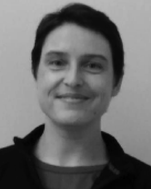 Lélu Sylvie合作论文数2
 Michel Versluis合作论文数Technical Medical Centre, University of Twente;MESA+ Research Institute for Nanotechnology, University of Twente2
 G. (Gerrit) Storm合作论文数Department of Pharmaceutics, Utrecht University;Ayuvant;Phospholipid Research Center;Liposoma B.V.;Universiteit Twente2




