Background and study aims:Endoscopic ultrasound-guided radiofrequency ablation (EUS-RFA) is a nonsurgical treatment option for managing pancreatic lesions. We sought to evaluate the safety and efficacy of EUS-RFA for large (≥4 cm) branch-duct intraductal papillary mucinous neoplasms (BD-IPMNs). Patients and methods:Patients with a definitive diagnosis of BD-IPMN who declined or were unfit for surgery underwent EUS-RFA in a single-arm prospective trial. Ablation was performed using a 19G EUS-RFA needle. RFA applications were delivered up to a maximum threshold of 45 seconds or 400 ohms impedance. Safety was assessed using AGREE guidelines. Potential for efficacy was assessed using cyst volume and cyst fluid KRAS GNAS mutations using next-generation sequencing (NGS). Adverse events (AEs) were analyzed per RFA session, while response was analyzed per BD-IPMN. Results:Thirty BD-IPMNs (mean diameter 4.6 ± 1.7 cm; 80% multilocular) in 25 participants (mean age 74.1 ± 8.3 years) underwent 41 EUS-RFA sessions. AEs occurred in 12.2% of procedures (5/41), the majority being AGREE Grade 3A (9.8%, 4/41). During a mean follow-up of 18 ± 5 months, 22 of 28 BD-IPMNs (78.6%) achieved ≥ 50% reduction in cyst volume, and 11 (39.3%) demonstrated complete (≥90%) response. Among 26 BD-IPMNs that revealed KRAS GNAS mutations, follow-up NGS was performed in 17, with 88.2% showing loss of detectable mutations. Conclusions:EUS-RFA in large, predominantly multilocular BD-IPMNs shows promising volumetric efficacy. Safety may be improved through refined energy delivery and technical advances. Molecular response remains exploratory and requires further validation. Long-term studies assessing progression-free outcomes are needed to define its role as an organ-preserving therapeutic option.
Background and Aims:Non-gastric subtype of branch duct (BD)-intraductal papillary mucinous neoplasms (IPMNs) are associated with high-grade dysplasia/invasive adenocarcinoma (HGD/IC) and disease progression. We evaluated preoperative prediction of gastric vs. non-gastric BD-IPMN subtypes and assessed a needle-based confocal laser endomicroscopy-guided artificial intelligence algorithm (nCLE-AI) for detecting HGD/IC in pathologist-reclassified BD-IPMNs. Methods:Participants with resected BD-IPMNs were enrolled from prospective studies (2015-2024). Phase 1: lesions were reclassified by subtype and dysplasia grade through blinded pathologist review, with discordant cases receiving MUC immunostaining and consensus review. Phase 2: using this reclassified pathology data, preoperative clinical and morphological features were analyzed to predict BD-IPMN subtypes. Phase 3: nCLE-AI performance in detecting HGD/IC within reclassified gastric and non-gastric BD-IPMNs was evaluated using preoperative endomicroscopy videos. Results:Among 63 resected BD-IPMNs (mean diameter=35.0±10.1 mm), 38% were classified as HGD/IC. Phase 1: The interobserver agreement among pathologists for subtype classification was moderate (k=0.52; 95%CI: 0.27-0.77). Phase 2: Multivariable analysis or preoperative variables revealed Kyoto high-risk stigmata (aOR=11.568, p=0.007), unifocal lesions (aOR=8.354, p=0.041), and lower BMI (aOR=1.37, p=0.04) predicted non-gastric subtype. Phase 3: The nCLE-AI algorithm using presurgical endomicroscopy imaging showed comparable sensitivity for detecting HGD/IC in non-gastric and gastric IPMN subtypes (83% vs. 82%, p=0.92), but significantly higher specificity (100% vs. 44%, p=0.06) and accuracy (87% vs. 53%, p<0.02) in the non-gastric subtype. Conclusion:Moderate interobserver variability in BD-IPMN subtype classification among pathologists highlights the need for immunohistochemistry and consensus review in challenging cases. Preoperative clinical variables can predict non-gastric subtype which is associated with less favorable prognosis. nCLE-AI shows improved performance in detecting HGD/IC in non-gastric BD-IPMNs, where accurate risk stratification is particularly important due to higher risk of progression.
BACKGROUND:The optimal timing for endoscopic retrograde cholangiopancreatography (ERCP) in managing uncomplicated acute biliary pancreatitis (ABP) remains unclear. This population-based study assessed the impact of ERCP timing on outcomes in hospitalized ABP patients without cholangitis or sepsis. METHODS:Using the Nationwide Readmission Database (2016 to 2020), patients with ABP who underwent ERCP were identified, excluding those with cholangitis/sepsis. ERCP timing was categorized as early (0 to 1 d) or delayed (2 to 7 d). Outcomes, including mortality, 30-day readmissions, and health care resource utilization were analyzed using multivariable logistic regression. RESULTS:Among 54,250 noninfectious ABP patients, 42.13% underwent ERCP. There was no significant difference in in-hospital mortality between early and delayed ERCP groups (0.26% vs. 0.24%, P =0.73). However, 30-day readmission rates rose with each day of delay in the delayed ERCP group ( P <0.001) and were higher overall (8.26% vs. 7.51%, P =0.03). Delayed ERCP was associated with increased 30-day readmissions (OR: 1.11, 95% CI: 1.00-1.22); using alternate categorical thresholds, ERCP on day 4 or later was also linked to higher odds of readmission (OR: 1.35, 95% CI: 1.18-1.54). Early ERCP was associated with reduced mean length of stay by 1.51 days (95% CI: -1.51 to -1.50; P <0.001) and hospital costs by $1536.45 (95% CI: -$1545.37 to -$1527.52; P <0.001) after multivariable adjustment. Compared with day 4 or later, ERCP on days 2 to 3 further reduced stay by 2.42 days and costs by $3756.95 (both P <0.001). CONCLUSION:Delayed ERCP was associated with increased 30-day readmission rates and greater health care resource utilization and ERCP performed within 4 days may improve hospital quality metrics in patients with noninfectious acute biliary pancreatitis.
Advancements in endoscopic ultrasound-guided tissue acquisition (EUS-TA) for solid pancreatic masses have prompted updates in society guideline recommendations. However, the extent to which these recommendations have been adopted in clinical practice remains unclear. Thus, we sought to characterize current practice patterns and beliefs regarding EUS-TA for solid pancreatic masses in a large, international cohort of endosonographers. We developed and distributed an electronic survey to 1,281 endosonographers worldwide between September and November 2024. The survey aimed to evaluate EUS-TA practices across six domains: A) needle selection, B) sampling technique, C) method guiding the number of passes, D) specimen handling, E) quality indicator (QI) monitoring, and F) factors influencing decision-making. The response rate was 30
Cavernous transformation of the portal vein is a rare condition that is often misdiagnosed or diagnosed late due to its insidious onset and nonspecific clinical presentation. It is most often associated with prothrombotic conditions, liver disease, malignancy, or local inflammation. We report a rare case involving a 65‑year‑old woman with HIV infection (CD4 count 96 cells/µL) who developed portal vein cavernous transformation leading to obstructive biliopathy. Cholangioscopy excluded intrinsic mucosal pathology or neoplasm, and additional testing ruled out other hypercoagulable disorders. Following placement of a biliary stent, her cholestatic symptoms resolved and her alkaline phosphatase and bilirubin levels declined. This case highlights HIV as an unusual precipitant of portal vein cavernous transformation and discusses imaging findings, underlying etiologies, and other management options.
BACKGROUND AND AIMS:EUS-guided gallbladder drainage (EUS-GBD) has been found to be a safe and effective treatment option for patients with cholecystitis. There are limited data regarding the use of EUS-GBD in patients with symptomatic cholelithiasis without cholecystitis. Therefore, the aim of this study was to examine the safety and efficacy of EUS-GBD for the management of gallstones in nonoperative candidates without cholecystitis. METHODS:This 2-center retrospective study examined patients with symptomatic cholelithiasis without cholecystitis treated with EUS-GBD via lumen-apposing metal stent (LAMS) placement who were deemed nonsurgical candidates. The primary outcome was complete gallstone clearance, and secondary outcomes included clinical success, technical success of LAMS placement, and adverse events. RESULTS:A total of 23 patients received EUS-GBD for symptomatic cholelithiasis in the absence of cholecystitis. LAMSs were successfully placed in all patients (technical success rate, 100%). Lithotripsy was performed in 9% of patients, and complete stone clearance was achieved in 100% of patients (87% achieved clearance within 1 treatment). Sixteen (70%) patients underwent follow-up endoscopy, during which 1 patient was noted to have small residual diminutive stones on cholecystoscopy via the LAMS. Interval imaging in all patients who did not undergo follow-up endoscopy revealed stone clearance and decompression of the gallbladder. There were no procedure-related adverse events noted, and no recurrence of symptoms was reported over a mean follow-up period of 9 months. CONCLUSIONS:This 2-center case series provides preliminary evidence that EUS-GBD may offer a safe and effective method for treating symptomatic cholelithiasis without cholecystitis in nonoperative candidates.
 Darwin L. Conwell合作论文数167Hart Phil A合作论文数Division of Gastroenterology, Hepatology, and Nutrition, The Ohio State University Wexner Medical Center135Hinton Alice合作论文数111
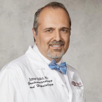 Samer Eldika合作论文数Medicine Department, Stanford University71Georgios I. Papachristou合作论文数Ohio State University Wexner Medical Center69Stacey Culp合作论文数57
 Peter Lee合作论文数Div Gastroenterol, Hosp Univ Penn48
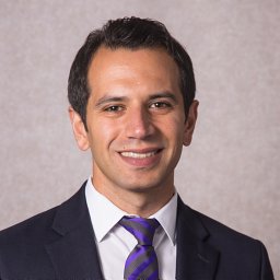 Hussan Hisham合作论文数Medical Center, The Ohio State University48
 Luis F. Lara合作论文数University of Texas Southwestern Medical School46Mitchell Ramsey合作论文数45
 Samuel Han合作论文数Sch Med, UC Denver45
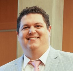 Peter P Stanich合作论文数Internal Medicine, Mayo Clinic45
 Wei Chen合作论文数Berkshire Medical Center45
 Kyle Porter合作论文数Center for Biostatistics, The Ohio State University42
 Zobeida Cruz-Monserrate合作论文数39Jordan Burlen合作论文数38
 Timothy M Pawlik合作论文数Department of Surgery, College of Medicine, The Ohio State University Wexner Medical Center36
 Mary Dillhoff合作论文数Department of Surgery, The Ohio State University34Rohan M. Modi合作论文数33William A. Ross合作论文数33Hamza Shah合作论文数31
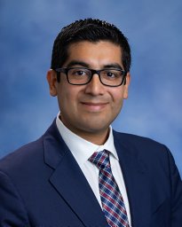 Machicado Jorge D合作论文数Internal Medicine Department, University of Texas Health Science Center at Houston30Anjuli Luthra合作论文数29
 Jeffrey H. Lee合作论文数Department of Gastroenterology Hepatology and Nutrition, Division of Internal Medicine, The University of Texas MD Anderson Cancer Center29Walker Jon P合作论文数28
 Manoop S. Bhutani合作论文数Department of Gastroenterology Hepatology and Nutrition, Division of Internal Medicine, The University of Texas MD Anderson Cancer Center;Department of Radiation Oncology, Division of Radiation Oncology, The University of Texas MD Anderson Cancer Center;Department of Endoscopic Research and Development, The University of Texas MD Anderson Cancer Center28
 Khalid Mumtaz合作论文数Department of Medicine, Aga Khan University Hospital27
 Zarine Ketul Shah合作论文数26Swanson Benjamin合作论文数25
 Andrei Manilchuk合作论文数Department of Surgery, Ohio State University School of Medicine and Public Health25Li Feng合作论文数23
 Raj Shah合作论文数Division of Gastroenterology and Hepatology, School of Medicine, University of Colorado Anschutz Medical Campus22Sean T. Mccarthy合作论文数22Jeffery R. Groce合作论文数21
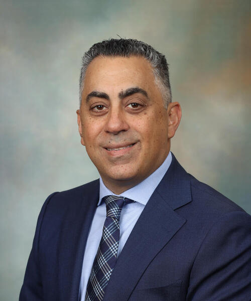 Tanios S. Bekaii-Saab合作论文数Mayo Clinic Cancer Center20Jordan M. Cloyd合作论文数The Ohio State University Wexner Medical Center20D. L. Conwell合作论文数19
 Schmidt Carl合作论文数Wexner Med Ctr, Ohio State Univ19
 Peter Muscarella合作论文数Montefiore Med Ctr, Dept Surg, 111 E 210th St, Bronx, NY 10467 USA18
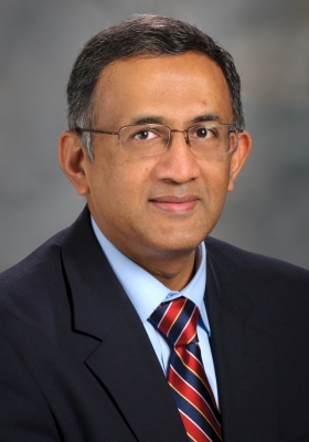 Gottumukkala S. Raju合作论文数Department of Gastroenterology Hepatology and Nutrition, Division of Internal Medicine, The University of Texas MD Anderson Cancer Center17
 Amrit Kamboj合作论文数16Groce Royce合作论文数16Andrew Kruger合作论文数16
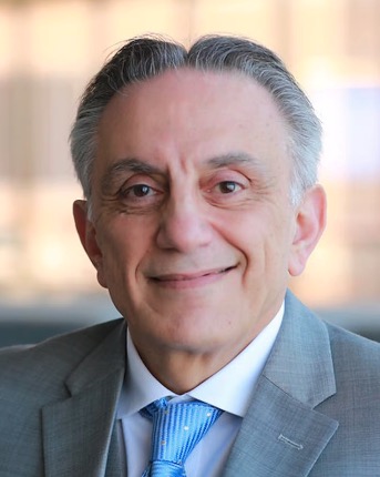 Elias Anaissie合作论文数CTI Clinical Trial and Consulting Services16
 Bart Barlogie合作论文数Icahn School of Medicine at Mount Sinai15Abhik Bhattacharya合作论文数14
 Emmanuel Ugbarugba合作论文数14Cheng Zhang合作论文数14Oza Veeral M合作论文数14




