Athletes are commonly imaged for focal swelling, persistent pain, or a new palpable abnormality after training or injury. These imaging findings are frequently not neoplastic, although several benign processes can appear sufficiently mass-like or aggressive to raise concern for sarcoma or an aggressive lesion. Such "pseudotumors" are more common in routine musculoskeletal practice than primary malignancy. This review approaches the problem as it is encountered in practice: first, the palpable abnormality that prompts imaging, and second, the incidental or symptomatic injury-related lesion that appears worrisome on radiographs, ultrasound, CT, or MRI. Radiographs should be obtained as a first line of imaging. Both radiographs and CT remain essential when evaluating mineralization, ossification, periosteal reaction, cortical injury, or stress fracture. Ultrasound is particularly useful for imaging evaluation of superficial lesions, fluid collections, vascular abnormalities, and dynamic processes such as muscle herniation. MRI is the principal problem-solving modality for deep or indeterminate lesions because it shows compartment of origin, tissue plane affected, internal architecture, marrow involvement, and enhancement patterns. In athletes, the most useful question is often whether the finding fits the story. A benign injury-related lesion should occur in a plausible location, involve the expected tissue plane, and change over time in a predictable way. Discordant features, including progressive enlargement, unexplained deep location, infiltrative margins, nodular internal enhancement, bone destruction, neurovascular encasement, or failure to evolve as expected, should prompt further imaging evaluation to exclude a more ominous diagnosis.
To describe a reproducible framework for bulk large language model (LLM)–based extraction of structured cartilage-lesion data from knee MRI reports and to benchmark seven LLM configurations against multiple radiologists using the modified Outerbridge classification. In this IRB-approved retrospective study, 100 non-contrast knee MRI reports (January 2019 to January 2025) were randomly selected from 66,479 eligible examinations and independently graded by five readers (four fellowship-trained musculoskeletal radiologists with 6–21 years of post-fellowship experience and one fourth-year resident) and seven LLM configurations, comprising six Azure OpenAI deployments (GPT-4.1, GPT-5.1-mini, GPT-5.3, GPT-5.4, GPT-5.4-mini, GPT-5.4-nano) and one locally hosted open-weight model (Qwen2.5-32B-Instruct), across a fixed 20-surface anatomic taxonomy. Pairwise Cohen quadratic-weighted κ was computed at compartment and surface levels. Bootstrap 95
To assess current trends and impacts of hybrid (HYB) and work from home (WFH) practice models on the musculoskeletal (MSK) radiology workforce and explore implications for recruitment and the future of the subspecialty. A 38-question voluntary, anonymous survey was distributed to the Society of Skeletal Radiology (SSR) membership (n = 1060). Descriptive statistics and chi-squared test were performed to analyze demographics, trends, and preferences for HYB-WFH practice models. Thematic analysis of open-ended responses identified perceptions and impacts of HYB-WFH. The survey response rate was 30
Objective:To evaluate explainable AI models for predicting Knee osteoarthritis (KOA) progression using quantitative MRI and clinical data, for two outcomes: a composite endpoint (radiographic plus pain progression) and a radiographic-only progression endpoint. Method:We analyzed 600 participants from the Foundation for the National Institutes of Health Osteoarthritis Biomarkers Consortium (FNIH), part of case-control cohort in the Osteoarthritis Initiative (OAI). The participants were grouped as composite progressors (n = 194), radiographic-only (n = 103), pain-only (n = 103), and non-progressors (n = 200). Input features included demographic data, Kellgren-Lawrence grade, joint space width, WOMAC pain score, and quantitative volume MRI measurements of KOA-related, including cartilage, bone marrow lesions, osteophytes, effusion-synovitis (ES), and Hoffa's synovitis (HS), at baseline and as 24-month change. Data were split into stratified 80% training and 20% held-out test sets, with 10-fold cross-validation used for model tuning within the training set. Five classifiers (random forest, XGBoost, logistic regression, decision tree, and multilayer perceptron). We applied multiple explainability methods, including Gini importance, SHAP values, regression coefficients, and permutation importance. Results:Radiographic progression was most accurately predicted using longitudinal change features (random forest AUC = 0.87). Baseline features alone also yielded strong performance (AUC = 0.80). Composite progression was more difficult to predict (AUCs = 0.66-0.70). Across models, key expandability factors included medial femoral cartilage loss, BMLs in the medial tibia and femur, medial osteophyte, and ES volumes. Conclusion:Explainable machine learning using quantitative MRI enable interpretable prediction of KOA progression. This is the first study in the FNIH/OAI cohort to integrate longitudinal quantitative MRI features with model-agnostic explanations across multiple classifiers.
Diversity, equity, and inclusion (DEI) is important for delivering high-quality, culturally competent care and ensuring equal access to resources and opportunities in healthcare. However, the implementation of DEI has been met with unique challenges and successes across the globe. The International Skeletal Society (ISS), a multidisciplinary musculoskeletal society, made a conscious effort to promote DEI. This article discusses advantages and controversies of DEI approaches, DEI initiatives implemented by the ISS, and experiences of the ISS DEI committee members from their respective continents. The ISS DEI committee implemented educational webinars with expert panel discussions, revising membership criteria and policies for enhancing inclusiveness, advising on programing and speakers for the annual meeting, and fostering mentorship. From a global perspective, in North America, DEI has improved health outcomes and patient care, but anti-DEI legislation has posed significant challenges. Europe relies on international recruitment but faces challenges in staff retention. South America's cultural diversity necessitates culturally sensitive approaches, but discussions about DEI are scarce, and gender inequalities persist in leadership. In Africa, DEI principles are underdeveloped, with limited engagement among stakeholders. In Asia, DEI is emerging, with more women being appointed to faculty positions and leadership roles in academic societies. The implementation of meaningful DEI initiatives requires long-term institutional buy-in and the global participation and commitment of employees and institutional leaders at all levels.
RATIONALE:The percentage of women in radiology has remained at 25 % for decades. Our institutions identified the need to recruit, support and retain women in our departments in order to change this status quo. METHODS:Our institutions created two different frameworks for Women in Radiology (WIR) programs in order to meet the needs of each distinct department. The organizational structure (trainee led vs faculty led and hybrid), the need for departmental chair support, and types of events and speakers are outlined with unique programs provided by each institution. Collaboration with established programs for women within the institutions and within the region was utilized to support the developing programs. RESULTS:Each of the (WIR) programs has been successful in creating community, impact and sustainable programming, including a peer and faculty mentoring programs for all genders, improvements in gender equity in the workplace, the implementation of trainee parental support programs, and an overall increase in the percentage of women trainees and faculty. Over the last 3 years, the integration of the two programs into a single Women in Radiology program, also encompassing the new enterprise radiology group, has led the way during the initiation of a large institutional merger from two institutions to one large institution. OUTCOMES:We have provided two frameworks for developing a successful women in radiology program as well as a roadmap for combining best practices in a time of change and institutional merger at a major academic institution.
Objective:To use software-based magnetic resonance imaging (MRI) measures of multiple features of knee osteoarthritis (KOA) to predict radiographic and pain progression in persons with KOA, and compare to a study that used primarily semi-quantitative (SQ) scoring. Design:Data from the Foundation for the National Institutes of Health Osteoarthritis Biomarkers Consortium (FNIH) nested case-control study (600 subjects divided into case and control groups based on knee pain and/or radiographic progression) were used. The MRI Osteoarthritis Software Scoring (MOSS) was used to quantitatively assess medial femoral cartilage, bone marrow lesions, osteophyte volume, effusion-synovitis volume, and a measure of Hoffa's synovitis at baseline and 24-months using readers with diverse levels of expertise. Association between baseline and baseline to 24-month change with progressor status was examined and discriminative ability assessed using the c-statistic (AUC) computed under 10-fold cross validation. Results:AUC values ranged from 0.690 to 0.726 to predict combined pain/radiographic progression and from 0.709 to 0.804 to predict radiographic progression alone. Bone marrow lesions and osteophyte volume played a role in all analyses. Medial femoral cartilage was significant for all but the cross-sectional analysis involving pain progression. Comparison to results from a separate publication showed that MOSS offered similar discrimination to a published model that primarily used SQ scoring. Conclusions:We found a high level of discrimination particularly for radiographic progression analysis. Use of fast automated software and readers with varied prior experience make MOSS a useful tool for enriching future clinical trials and for other large studies of KOA.
Objective The purposes of this study were to evaluate the prevalence of persistent postoperative synovitis three months after anterior cruciate ligament reconstruction (ACLR) using ultrasonographic superb microvascular imaging (SMI) and evaluate the intra-rater reliability of SMI synovitis and the level of agreement with other ultrasound synovitis assessment methods. Design Twenty-three individuals who had undergone primary ACLR took part in this prospective, IRB-approved study (11 females, 12 males; age=28.7±9.7 y; BMI=25.6±3.3 kg/m2). Three months after surgery, longitudinal and transverse ultrasound scans of the suprapatellar recess were performed at midline, medial to midline, and lateral to the midline. In addition to Power Doppler and B mode measures of synovitis and effusion, SMI was graded on two separate occasions at least two weeks apart. SMI intra-rater reliability was assessed with weighted Kappa analyses, and Kappa analyses were also used to assess the agreement between SMI synovitis and Power Doppler synovitis and B mode effusion. Results Three months following ACLR, 2 (8.7%) participants had no SMI synovitis, 9 (39.1%) participants had mild synovitis, and 12 (52.2%) had moderate synovitis. SMI synovitis grades demonstrated excellent intra-rater reliability (Kappa=0.93 [95%CI: 0.80, 1.06]) but demonstrated little agreement with Power Doppler synovitis grades (Kappa=0.29) or B mode effusion grades (Kappa=0.14). Conclusions Persistent postoperative synovitis was common with half of patients demonstrating moderate synovitis. SMI synovitis grading was reliable, but little agreement was noted between SMI and other ultrasound grades of synovitis or effusion suggesting that the different techniques are identifying distinct features of postoperative inflammation.
ObjectiveErosive hand osteoarthritis (eHOA) is a subtype of hand osteoarthritis (OA) that develops in finger joints with pre-existing OA and is differentiated by clinical characteristics (hand pain/disability, inflammation, and erosions) that suggest inflammatory or metabolic processes.MethodThis was a longitudinal nested case-cohort design among Osteoarthritis Initiative participants who had hand radiographs at baseline and 48-months, and biospecimens collected at baseline. We classified incident radiographic eHOA in individuals with ≥1 joint with Kellgren-Lawrence ≥2 and a central erosion present at 48-months but not at baseline. We used a random representative sample (n = 1282) for comparison. We measured serum biomarkers of inflammation, insulin resistance and dysglycemia, and adipokines using immunoassays and enzymatic colorimetric procedures, blinded to case status.ResultsEighty-six participants developed incident radiographic eHOA. In the multivariate analyses adjusted for age, gender, race, smoking, and body mass index, and after adjustment for multiple analyses, incident radiographic eHOA was associated with elevated levels of interleukin-7 (risk ratio (RR) per SD = 1.30 [95% confidence interval (CI) 1.09, 1.55] p trend 0.01).ConclusionThis exploratory study suggests an association of elevated interleukin-7, an inflammatory cytokine, with incident eHOA, while other cytokines or biomarkers of metabolic inflammation were not associated. Interleukin-7 may mediate inflammation and tissue damage in susceptible osteoarthritic finger joints and participate in erosive progression.
Purpose: This study aimed to evaluate the degree of gender disparity in leadership positions at the top 25 medical schools in North America compared to their affiliated radiology departments. Methods: The academic rank and leadership appointment of medical school and radiology faculty were obtained from publicly available official websites between June-November 2022. Gender was determined using selfidentified pronouns on website biographies. Alternatively, gender API software was used. Finally, SCOPUS Elsevier was used to extract research output metrics including publication counts, citations, and h-indices. Statistical analysis was conducted using the IBM SPSS Statistics version 25 software. Results: 2216 individuals across 25 medical schools were included in this study. 1301 (58.7 %) were part of the medical school and 915 (41.3 %) were part of the affiliated radiology departments. Additionally, 1575 (71.1 %) were identified as men and 641 (28.9 %) as women. Rank biserial correlations showed a significant association between higher academic rank and male gender (rpb = 0.143, p <0.001) regardless of affiliation (medical school leadership versus radiology faculty); this disparity was largest at the highest academic ranks. Male gender was associated with higher research productivity relative to female gender regardless of affiliation (p < 0.001). There were minimal statistical differences in leadership positions between genders, however the proportion of men holding the position of dean was two times higher than women. Conclusion: The underrepresentation of women in academic medicine is prevalent in the top-ranking medical institutions in North America and disproportionately involves senior academic ranks and leadership positions.
OBJECTIVE:To evaluate gender differences in the association between metacarpal cortical thickness (Tcort)-a surrogate for bone density-and severity of radiographic hand osteoarthritis (HOA) in a longitudinal observational study. METHOD:Hand radiographs of 3575 participants (2039 F/1536 M) from the Osteoarthritis Initiative were assessed at baseline and 48 months. A reader used a semi-automated software tool to calculate Tcort, a measurement of the cortical thickness, for metacarpals 2-4. Average Tcort at baseline and change in Tcort from baseline to 48 months was determined and stratified by gender and age for 7 5-year age groups. Spearman's rank correlation coefficients were calculated for the association of baseline Tcort and 2 measures of baseline HOA severity: the sum of Kellgren-Lawrence (KL) grade and total number of joints with radiographic HOA. Longitudinally, logistic regression was used to assess the relationship of Tcort loss to new finger joint radiographic HOA, increase in KL grades, and incident hand pain. RESULTS:Male Tcort was higher than females. Significant correlations between Tcort and radiographic severity were noted for women but not men, with stronger associations among women >60 years (rho = -0.25; 95% confidence interval (CI) = -0.31 to -0.19). Statistically significant associations were seen between Tcort change and radiographic osteoarthritis change among women but not men, with substantial gender differences for Tcort change, particularly ages 50 to 70 years (p < 0.01; e.g., Tcort change ages 55 to <60: males = -0.182 (0.118), females = -0.219 (0.124)). CONCLUSION:We found significant HOA-related gender differences in Tcort, suggesting the involvement of female bone loss during and after menopause.
Among people with or without hand osteoarthritis, we aimed to identify characteristics of people (e.g., age and gender) with marginal erosions (MEs). We also examined changes in MEs during 48 months. We described radiographic severity and progression among joints with MEs, changes in MEs, or central erosions (CEs). We studied participants from the Osteoarthritis Initiative with baseline and 48-month hand radiographs. A radiologist and rheumatologist evaluated the radiographs for disease severity (Kellgren-Lawrence grades) and erosions (central or marginal), respectively. We used descriptive statistics to characterize participants and calculated frequencies at the joint level. Of the 3558 participants, 89 had a ME at baseline. People with MEs were more often male, older, and ever (former and current) smokers than those without a ME. There was no difference in inflammatory biomarkers or the presence of hand pain between individuals with and without a baseline ME. Almost all hands had only one ME (80
ObjectiveRadiographs are frequently obtained for patients with knee osteoarthritis (KOA), with magnetic resonance imaging (MRI) reserved for those with complex KOA. There are few data on how often subsequent MRI reveals clinically actionable but unanticipated findings. The purpose of this study is to estimate the prevalence of these findings on MRI for patients managed nonoperatively for suspected meniscal tears.MethodsThe Treatment of Meniscal Problems and Osteoarthritis (TeMPO) study enrolled patients aged 45 to 85 years with knee pain, osteoarthritis (Kellgren-Lawrence [KL] grades 0-3), and suspected meniscal tear. We reviewed baseline MRI and recorded notable findings, including subchondral insufficiency fractures of the knee (SIFKs), avascular necrosis (AVN), tumors, and nonsubchondral fractures. Other baseline data included demographic characteristics, Knee Injury and Osteoarthritis Outcome Score, duration of knee symptoms, and KL grade.ResultsStudy-ordered MRI was performed on 760 patients, with 61 concerning findings identified (8.03%, 95% confidence interval 6.09%-9.96%). A total of 25 participants had SIFKs, 10 had nonsubchondral fractures, 4 had AVN, 8 had benign tumors, and 14 had other clinically relevant findings.ConclusionWe estimated the prevalence of clinically relevant incidental findings on MRI to be 8.03% in middle-aged adults with mild to moderate KOA and suspected meniscal tear. These data may prompt clinicians to be more aware of the range of findings that can underlie knee symptoms, some of which could change management but may require different modalities of imaging to detect. Future research is needed to pinpoint factors associated with these concerning findings so that patients who are at risk can be identified and referred for advanced imaging.
The objective of this study is to assess the status and perception of work from home (WFH) in three academic musculoskeletal radiology divisions. A 17-item anonymous online survey related to WFH was administered to all musculoskeletal (MSK) faculty (n = 27) of three academic radiology divisions between April and July 2023. Survey items included demographics, clinical work environment, presence of a WFH policy, number of days WFH and desired WFH days, commute, perceived benefits and drawbacks of WFH, and symptoms of burnout. Statistical analysis included the Cochrane-Armitage trend test and Fisher exact test. The study was exempt from IRB approval. The survey response rate was 85
INTRODUCTION Metacarpal cortical thickness (MCT), a surrogate for bone density, has been well studied in people with rheumatoid arthritis but much less so for hand OA (HOA). OBJECTIVE To investigate the association of MCT with radiographic HOA severity. METHODS We performed a software measurement of MTC on the dominant hand radiograph of 3,575 participants from the OAI at the baseline and 48-month visits. Spearman's rank correlation coefficients (rho) were calculated for the association of baseline MTC and 2 measures of baseline HOA severity: the sum of Kellgren and Lawrence (KL) grade and total number of joints with radiographic HOA. Longitudinally, logistic regression with odds ratios were used to assess the relationship of MTC loss to new finger joint radiographic OA and an increase in KL grades. The results were stratified by gender and into two age groups: 45-60 years and > 60 years. RESULTS The baseline results are in Table 1, and the longitudinal results are in Table 2. For women, we found a weak correlation between baseline MTC and ROA for ages 45-60 years; the correlation was higher for the > 60 years age group. For MTC change, we found higher odds ratios in women for the 45-60 year group than for the > 60 year group. No significant correlations were seen between MCT and HOA for men either cross-sectionally or longitudinally. CONCLUSION We found significant associations between MCT and ROA status in women for both baseline and 48-month change but not for men. The consideration of differences between men and women may have implications for understanding the structural nature of HOA. It may be important in developing targeted interventions to manage symptoms and improve outcomes for affected individuals.
The diagnosis and management of chronic nonspinal osteomyelitis can be challenging, and guidelines regarding the appropriateness of performing percutaneous image-guided biopsies to acquire bone samples for microbiological analysis remain limited. An expert panel convened by the Society of Academic Bone Radiologists developed and endorsed consensus statements on the various indications for percutaneous image-guided biopsies to standardize care and eliminate inconsistencies across institutions. The issued statements pertain to several commonly encountered clinical presentations of chronic osteomyelitis and were supported by a literature review. For most patients, MRI can help guide management and effectively rule out osteomyelitis when performed soon after presentation. Additionally, in the appropriate clinical setting, open wounds such as sinus tracts and ulcers, as well as joint fluid aspirates, can be used for microbiological culture to determine the causative microorganism. If MRI findings are positive, surgery is not needed, and alternative sites for microbiological culture are not available, then percutaneous image-guided biopsies can be performed. The expert panel recommends that antibiotics be avoided or discontinued for an optimal period of 2 weeks prior to a biopsy whenever possible. Patients with extensive necrotic decubitus ulcers or other surgical emergencies should not undergo percutaneous image-guided biopsies but rather should be admitted for surgical debridement and intraoperative cultures. Multidisciplinary discussion and approach are crucial to ensure optimal diagnosis and care of patients diagnosed with chronic osteomyelitis.
Imaging plays a pivotal role in osteoarthritis research, particularly in epidemiological and clinical trials of knee osteoarthritis (KOA), with the ultimate goal being the development of an effective drug treatment for future prevention or cessation of disease. Imaging assessment methods can be semi-quantitative, quantitative, or a combination, with quantitative methods usually relying on software to assist. The software generally attempts image segmentation (outlining of relevant structures). New techniques using artificial intelligence (AI) or deep learning (DL) are currently a frequent topic of research. This review article provides an overview of the literature to date, focusing primarily on the current status of quantitative software-based assessment techniques of KOA using magnetic resonance (MR) imaging. We will concentrate on the imaging evaluation of three specific structural imaging biomarkers: bone marrow lesions (BMLs), meniscus, and synovitis consisting of effusion synovitis (ES) and Hoffa's synovitis (HS). A brief clinical and imaging background review of osteoarthritis evaluation, particularly relating to these three structural markers, is provided as well as a general summary of the software methods. A summary of the literature with respect to each KOA assessment method will be presented overall as well as with respect to each specific biomarker individually. Novel techniques, as well as future goals and directions using quantitative imaging assessment, will be discussed.
 Jeffrey Duryea合作论文数23Lena F Schaefer合作论文数15
 Timothy E. McAlindon合作论文数Graduate School of Biomedical Sciences, Tufts University;School of Medicine, Tufts University14
 Driban Jeffrey B合作论文数Tufts Med Ctr, Boston, MA USA14Roberts Mary B合作论文数14
 Jacob C. Mandell合作论文数Brigham and Women's Hospital, Harvard Medical School13
 Charles B. Eaton合作论文数School of Public Health, Brown University;Center for Primary Care and Prevention, Kent County Hospital12
 Haugen Ida K合作论文数Dept Rheumatol, Diakonhjemmet Hosp11Shah Nehal合作论文数11
 Bharti Khurana合作论文数8Kransdorf Mark J合作论文数7
 Kenneth S Lee合作论文数Department of Radiology, School of Medicine and Public Health, University of Wisconsin-Madison6
 Kathleen Vanni合作论文数Division of Rheumatology, Inflammation, and Immunity, Brigham and Women's Hospital, 60 Fenwood Road, Boston, MA, 02115, USA6
 Mihra S Taljanovic合作论文数6
 Guenette Jeffrey P合作论文数Warren Alpert School of Medicine, Brown University6
 Daniel Hal Solomon合作论文数Orthopaedic and Arthritis Center, Brigham and Women's Hospital;Harvard Medical School6
 Neena Kapoor合作论文数5Lu Bing合作论文数5
 Adam Singer合作论文数Department of Diagnostic Radiology, Jackson Memorial Hospital5
 Collins Jamie E合作论文数Brigham & Womens Hosp5Ali Guermazi合作论文数5Catherine H. Phillips合作论文数4
 Tetyana Gorbachova合作论文数4Meera Sury合作论文数4
 Darel E Heitkamp合作论文数Department of Radiology, Indiana University School of Medicine4Alice S Ha合作论文数4
 Angelisa Paladin合作论文数4Ali Sayed合作论文数4
 Antonia Chen合作论文数Department of Orthopaedic Surgery, UT Southwestern Medical Center4
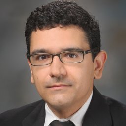 Behrang Amini合作论文数Division of Diagnostic Imaging, Department of Musculoskeletal Imaging, The University of Texas MD Anderson Cancer Center4
 Miriam A. Bredella合作论文数Department of Radiology, Harvard Medical School, Massachusetts General Hospital4Ramin Khorasani合作论文数4
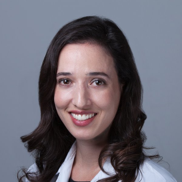 Sara K. Tedeschi合作论文数Orthopaedic and Arthritis Center, Brigham and Women's Hospital;Harvard Medical School4Anna Rozenshtein合作论文数4Leon Lenchik合作论文数4Jennifer K. Bussell合作论文数3Taryn Hodgdon合作论文数3Alan Klitzke合作论文数3Anarosa Campos合作论文数3Omar Musbahi合作论文数3Alyssa Wohlfahrt合作论文数3Shanna A Matalon合作论文数3Penny Wang合作论文数3Seoyoung C Kim合作论文数3
 Anthony Paul Trace合作论文数Eastern Virginia Medical School3
 G. Morgan Jones合作论文数3
 Garg Rajesh K合作论文数3
 Jeffrey N. Katz合作论文数Department of Epidemiology and Environmental Health, Harvard T.H. Chan School of Public Health, Harvard University;Brigham and Women's Hospital3Laur Olga合作论文数3




