Mitochondrial ATP-dependent proteases are essential for maintaining protein homeostasis through degradation of damaged or misfolded proteins. Among these, the ClpXP protease complex locates in mitochondrial matrix and contributes to mitochondrial quality control under physiological and stress conditions. This work demonstrates a quantitative fluorescence microscopy workflow to assess mitochondrial targeting of the fluorescent peptidyl inhibitor FAM-FAPAL-CMK and evaluate mitochondrial morphological changes associated with ClpXP inhibition in mammalian cells. HeLa cells were treated with FAM-FAPAL-CMK and analyzed using confocal microscopy combined with immunofluorescence staining of mitochondrial markers and quantitative image analysis. Colocalization analysis using Costes thresholding and Manders' overlap coefficients demonstrated mitochondrial enrichments of the inhibitor signal. As a consequence, inhibition of ClpP altered mitochondrial morphology. Immunoblot analysis showed no significant change in ClpP protein abundance upon inhibitor treatment. Taken together, this work describes a reproducible imaging-based workflow that will enable interrogation of mitochondrial ClpXP functions in intact cells in response to perturbations of homeostasis, such as oxidative stress.
The bacteriophage T4 replisome is a complex molecular machine responsible for DNA replication in the T4 phage. It consists of multiple proteins that work together to ensure efficient and accurate replication of the phage genome. The replisome comprises DNA polymerases, helicase, primase, and other accessory proteins, which coordinate leading- and lagging-strand synthesis. Extensive research over the years, including protein analysis, enzyme kinetics, and structural investigations, has provided insights into the organization and function of these proteins, along with their dynamics and coordination at the replication fork. The T4 replisome serves as a useful model system for understanding molecular fidelity, enzymatic interplay, and fundamental principles of DNA replication.
Cancer cells have the unique capability to upregulate the de novo nucleotide biosynthesis supporting cell survival under nucleotide deprivation. We probe the role of metabolic channeling and membrane-less metabolic compartmentalization by mitochondria-proximal dynamic de novo pyrimidine and purine biosynthesis metabolons, the pyrimidinosome and the purinosome, respectively. We designed in-cell stable isotope label incorporation assays (13C6 glucose, 15N2 glutamine) for detection of metabolic channeling, revealing the function and enzymatic composition of these complexes. Moreover, we discovered that the mitochondrially compartmentalized GOT2 dependent generation of aspartic acid feeds the channeled nucleotide synthesis instead of the bulk cytosolic pool or the GOT1 activity. While a low flux diffusive pathway generates the pathway intermediates in an accumulative process, it's the channeled pathway that successfully generates the end product nucleotides. Our results demonstrate how metabolic channeling and efficient de novo nucleotide biosynthesis is fueled by coordination of mitochondrially compartmentalized metabolic events with cytosolic metabolons in cancer cells.
Purines are the building blocks of DNA/RNA and hence essential metabolites. While the contributions of external purine salvage as well as the de novo purine biosynthesis (DNPB) have been widely studied, the contribution of lysosome mediated DNA/RNA digestion and external reabsorption into the cytosol remains unknown. Here, we address that question as well as the role of lysosome-mediated purine recycling and its coordination with DNPB in maintaining total purine pools in human cancer cell lines. By combining in-cell stable isotope incorporation assay with quantitative metabolomics we show: cellular uptake of external purines and their internal generation are equivalent; an upregulation in lysosome biogenesis that functions in response to purine deficiency caused by methotrexate (MTX) and lometrexol (LTX) treatment. This leads to increased RNA digestion as visualized by a newly developed intracellular RNA-FRET oligo assay. Interestingly, downregulation of lysosomal RNase activity through knockdown of RNAseT2 increased RNA accumulation and a compensating increase in DNPB.
We describe peptide mapping through Split Antibiotic Resistance Complementation (SpARC-map), a method to identify the probable interface between two interacting proteins. Our method is based on in vivo affinity selection inside a bacterial host and uses high-throughput DNA sequencing to infer probable protein-protein interaction (PPI) interfaces. SpARC-map uses only routine microbiology techniques, with no reliance on specialized instrumentation, dedicated reagents, or reconstituting protein complexes in vitro. SpARC-map can be tuned to detect PPIs over a broad range of affinities, multiplexed to probe multiple PPIs in parallel, and its nonspecific background can be precisely measured, enabling the sensitive detection ofweak PPIs. Using SpARC-map, we recover known PPI interfaces in the p21-PCNA, p53-MDM2, and MYC-MAX complexes. We also use SpARC-map to probe the purinosome, the weakly bound complex of six purine biosynthetic enzymes, where no PPI interfaces are known. There, we identify interfaces that satisfy structural requirements for substrate channeling, as well as protein surfaces that participate in multiple distinct interactions, which we validate using site-specific photocrosslinking in live human cells. Finally, we show that SpARC-map results can impose stringent constraints on machine learning-based structure prediction.
Extrachromosomal DNA (ecDNA) presents a major challenge for cancer patients. ecDNA renders tumours treatment resistant by facilitating massive oncogene transcription and rapid genome evolution, contributing to poor patient survival1-7. At present, there are no ecDNA-specific treatments. Here we show that enhancing transcription-replication conflict enables targeted elimination of ecDNA-containing cancers. Stepwise analyses of ecDNA transcription reveal pervasive RNA transcription and associated single-stranded DNA, leading to excessive transcription-replication conflicts and replication stress compared with chromosomal loci. Nucleotide incorporation on ecDNA is markedly slower, and replication stress is significantly higher in ecDNA-containing tumours regardless of cancer type or oncogene cargo. pRPA2-S33, a mediator of DNA damage repair that binds single-stranded DNA, shows elevated localization on ecDNA in a transcription-dependent manner, along with increased DNA double strand breaks, and activation of the S-phase checkpoint kinase, CHK1. Genetic or pharmacological CHK1 inhibition causes extensive and preferential tumour cell death in ecDNA-containing tumours. We advance a highly selective, potent and bioavailable oral CHK1 inhibitor, BBI-2779, that preferentially kills ecDNA-containing tumour cells. In a gastric cancer model containing FGFR2 amplified on ecDNA, BBI-2779 suppresses tumour growth and prevents ecDNA-mediated acquired resistance to the pan-FGFR inhibitor infigratinib, resulting in potent and sustained tumour regression in mice. Transcription-replication conflict emerges as a target for ecDNA-directed therapy, exploiting a synthetic lethality of excess to treat cancer.
In this study, we advance our understanding of the spatial relationship between the purinosome, a liquid condensate consisting of six enzymes involved in de novo purine biosynthesis, and mitochondria. Previous research has shown that purinosomes move along tubulin toward mitochondria, suggesting a direct uptake of glycine from mitochondria. Here, we propose that the purinosome is located proximally to the mitochondrial transporters SLC25A13 and SLC25A38, facilitating the uptake of glycine, aspartate, and glutamate, essential factors for purine synthesis. We utilized the proximity ligation assay and APEX proximity labeling to investigate the association between purinosome proteins and mitochondrial transporters. Our results indicate that purinosome assembly occurs close to the mitochondrial membrane under purine-deficient conditions, with the transporters migrating to be adjacent to the purinosome. Furthermore, both targeted and non-targeted analyses suggest that the SLC25A13-APEX2-V5 probe accurately reflects endogenous cellular status. These findings provide insights into the spatial organization of purine biosynthesis and lay the groundwork for further investigations into additional proteins involved in this pathway.
Extrachromosomal DNA (ecDNA) presents a major challenge for precision medicine, contributing to poor survival for patients with oncogene-amplified tumours. EcDNA renders tumours resistant to targeted treatments by facilitating massive transcription of oncogenes and rapid genome evolution. At present, there are no ecDNA- specific treatments. Here we show that enhancing transcription replication conflict enables targeted elimination of ecDNA-containing cancers, exposing an actionable vulnerability. Stepwise analyses of ecDNA transcription reveal landscapes of pervasive RNA transcription and associated single-stranded DNA, leading to excessive transcription replication conflicts and replication stress (RS) compared to chromosomal loci. Nucleotide incorporation onto growing DNA strands is markedly slower on ecDNA, and RS is significantly higher in ecDNA-containing tumours regardless of cancer type or oncogene cargo. Replication Protein A2 phosphorylated on serine 33, a mediator of DNA damage repair that binds single-stranded DNA, shows elevated localization on ecDNA in a transcription dependent manner, along with increased DNA double strand breaks, and activation of the S-phase checkpoint kinase, CHK1. Genetic or pharmacological CHK1 inhibition abrogates the DNA replication check point, causing extensive and preferential tumour cell death in ecDNA-containing tumours as they enter S-phase. To exploit this vulnerability, we develop a highly selective, potent, and bioavailable oral CHK1 inhibitor, BBI-2779, and demonstrate that it preferentially kills ecDNA-containing tumour cells. In a gastric cancer model containing FGFR2 on ecDNA, BBI-2779, suppresses tumour growth and prevents ecDNA-mediated acquired resistance to the pan-FGFR inhibitor infigratinib, resulting in potent and sustained tumour regression in mice. These results reveal transcription-replication conflict as an ecDNA-generated vulnerability that can be targeted as an ecDNA-directed therapy and suggest that synthetic lethality of excess can be exploited as a strategy for treating cancer.### Competing Interest StatementH.Y.C. is a co-founder of Accent Therapeutics, Boundless Bio, Cartography Biosciences, Orbital Therapeutics, and an advisor of 10x Genomics, Arsenal Biosciences, Chroma Medicine, Exai Bio, and Spring Discovery. P.S.M. is a co-founder and advisor of Boundless Bio. S.C, E.T., R.H., J.P., A.S., S.M. T.M, T. P. S.K., and C.A.H. are employees of Boundless Bio. V.B. is a co-founder, consultant, SAB member and has equity interest in Boundless Bio and Abterra, and the terms of this arrangement have been reviewed and approved by the University of California, San Diego in accordance with its conflict-of-interest policies. The remaining authors declare no competing interests.
The T4 bacteriophage gp41 helicase and gp61 primase assemble into a primosome to couple DNA unwinding with RNA primer synthesis for DNA replication. How the primosome is assembled and how the primer length is defined are unclear. Here we report a series of cryo-EM structures of T4 primosome assembly intermediates. We show that gp41 alone is an open spiral, and ssDNA binding triggers a large-scale scissor-like conformational change that drives the ring closure and activates the helicase. Helicase activation exposes a cryptic hydrophobic surface to recruit the gp61 primase. The primase binds the helicase in a bipartite mode in which the N-terminal Zn-binding domain and the C-terminal RNA polymerase domain each contain a helicase-interacting motif that bind to separate gp41 N-terminal hairpin dimers, leading to the assembly of one primase on the helicase hexamer. Our study reveals the T4 primosome assembly process and sheds light on the RNA primer synthesis mechanism. Here the authors present a methodical cryo-EM analysis to show how a T4 bacteriophage gp41 helicase hexamer and a gp61 primase assemble with a template-primer into an active primosome.
Over the past fifteen years, we have unveiled a new mechanism by which cells achieve greater efficiency in de novo purine biosynthesis. This mechanism relies on the compartmentalization of de novo purine biosynthetic enzymes into a dynamic complex called the purinosome. In this review, we highlight our current understanding of the purinosome with emphasis on its biophysical properties and function and on the cellular mechanisms that regulate its assembly. We propose a model for functional purinosomes in which they consist of at least ten enzymes that localize near mitochondria and carry out de novo purine biosynthesis by metabolic channeling. We conclude by discussing challenges and opportunities associated with studying the purinosome and analogous metabolons.
There is growing evidence that mammalian cells deploy a mitochondria-associated metabolon called the purinosome to perform channeled de novo purine biosynthesis (DNPB). However, the molecular mechanisms of this substrate-channeling pathway are not well defined. Here, we present molecular evidence of protein-protein interactions (PPIs) between the human bifunctional phosphoribosylaminoimidazole carboxylase/succinocarboxamide synthetase (PAICS) and other known DNPB enzymes. We employed two orthogonal approaches: bimolecular fluorescence complementation, to probe PPIs inside live, intact cells, and co-immunoprecipitation using StrepTag-labeled PAICS that was reintegrated into the genome of PAICS-knockout HeLa cells (crPAICS). With the exception of amidophosphoribosyltransferase, the first enzyme of the DNPB pathway, we discovered PAICS interacts with all other known DNPB enzymes and with MTHFD1, an enzyme which supplies the 10-formyltetrahydrofolate cofactor essential for DNPB. We show these interactions are present in cells grown in both purine-depleted and purine-rich conditions, suggesting at least a partial assembly of these enzymes may be present regardless of the activity of the DNPB pathway. We also demonstrate that tagging of PAICS on its C terminus disrupts these interactions and that this disruption is correlated with disturbed DNPB activity. Finally, we show that crPAICS cells with reintegrated N-terminally tagged PAICS regained effective DNPB with metabolic signatures of channeled synthesis, whereas crPAICS cells that reintegrated C-terminally tagged PAICS exhibit reduced DNPB intermediate pools and a perturbed partitioning of inosine monophosphate into AMP and GMP. Our results provide molecular evidence in support of purinosomes and suggest perturbing PPIs between DNPB enzymes negatively impact metabolite flux through this important pathway.
Enzymes within the de novo purine biosynthetic pathway spatially organize into dynamic intracellular assemblies called purinosomes. The formation of purinosomes has been correlated with growth conditions resulting in high purine demand, and therefore, the cellular advantage of complexation has been hypothesized to enhance metabolite flux through the pathway. However, the properties of this cellular structure are unclear. Here, we define the purinosome in a transient expression system as a biomolecular condensate using fluorescence microscopy. We show that purinosomes, as denoted by formylglycinamidine ribonucleotide synthase granules in purine-depleted HeLa cells, are spherical and appear to coalesce when two come into contact, all liquid-like characteristics that are consistent with previously reported condensates. We further explored the biophysical and biochemical means that drive the liquid-liquid phase separation of these structures. We found that the process of enzyme condensation into purinosomes is likely driven by the oligomeric state of the pathway enzymes and not a result of intrinsic disorder, the presence of low-complexity domains, the assistance of RNA scaffolds, or changes in intracellular pH. Finally, we demonstrate that the heat shock protein 90 KDa helps to regulate the physical properties of the condensate and maintain their liquid-like state inside HeLa cells. We show that disruption of heat shock protein 90 KDa activity induced the transformation of formylglycinamidine ribonucleotide synthase clusters into more irregularly shaped condensates, suggesting that its chaperone activity is essential for purinosomes to retain their liquid-like properties. This refined view of the purinosome nize into dynamic condensates within human cells.
With the elucidation of myriad anabolic and catabolic enzyme-catalyzed cellular pathways crisscrossing each other, an obvious question arose: how could these networks operate with maximal catalytic efficiency and minimal interference? A logical answer was the postulate of metabolic channeling, which in its simplest embodiment assumes that the product generated by one enzyme passes directly to a second without diffusion into the surrounding medium. This tight coupling of activities might increase a pathway's metabolic flux and/or serve to sequester unstable/toxic/reactive intermediates as well as prevent their access to other networks. Here, we present evidence for this concept, commencing with enzymes that feature a physical molecular tunnel, to multi-enzyme complexes that retain pathway substrates through electrostatics or enclosures, and finally to metabolons that feature collections of enzymes assembled into clusters with variable stoichiometric composition. Lastly, we discuss the advantages of reversibly assembled metabolons in the context of the purinosome, the purine biosynthesis metabolon.
I endeavor to share how various choices—some deliberate, some unconscious—and the unmistakable influence of many others shaped my scientific pursuits. I am fascinated by how two long-term, major streams of my research, DNA replication and purine biosynthesis, have merged with unexpected interconnections. If I have imparted to many of the talented individuals who have passed through my lab a degree of my passion for uncloaking the mysteries hidden in scientific research and an understanding of the honesty and rigor it demands and its impact on the world community, then my mentorship has been successful.
Abstract Paintings often consist of highly complex layered structures that contain a mixture of organic and inorganic materials at each layer depending upon the artist’s technique. A comprehensive analysis of the chemical composition could provide critical information on the sequence of events that led to an artwork’s current state. In this investigation, we have employed a novel imaging technique known as Secondary Ion Mass Spectrometry (SIMS) to chemically map the cross-section of a paint film sampled from Leonardo da Vinci’s Adoration of the Magi at a submicron resolution using a C60+ ion source. A wide range of materials were found to be spatially localized at the different layers such as: protein, carbon, silicates, fatty acids, salts and lead associated compounds. An interaction of chloride salts with the lead white was observed in the priming layer. The chloride salts disrupted most of the priming layer suggesting that a cleaning process took place that removed most of the priming layer before the monochrome brown repaints were added on top at a later time.
The purinosome is a dynamic metabolic complex composed of enzymes responsible for de novo purine biosynthesis, whose formation has been associated with elevated purine demand. However, the physiological conditions that govern purinosome formation in cells remain unknown. Here, we report that purinosome formation is up-regulated in cells in response to a low-oxygen microenvironment (hypoxia). We demonstrate that increased purinosome assembly in hypoxic human cells requires the activation of hypoxia inducible factor 1 (HIF-1) and not HIF-2. Hypoxia-driven purinosome assembly was inhibited in cells lacking 5-aminoimidazole-4-carboxamide ribonucleotide formyltransferase/IMP cyclohydrolase (ATIC), a single enzyme in de novo purine biosynthesis, and in cells treated with a small molecule inhibitor of ATIC homodimerization. However, despite the increase in purinosome assembly in hypoxia, we observed no associated increase in de novo purine biosynthesis in cells. Our results indicate that this was likely due to a reduction in mitochondrial one-carbon metabolism, resulting in reduced mitochondrion-derived one-carbon units needed for de novo purine biosynthesis. The findings of our study further clarify and deepen our understanding of purinosome formation by revealing that this process does not solely depend on cellular purine demand.
 Patricia A. Benkovic合作论文数52Michelle M. Spiering合作论文数36
 Richard Lerner合作论文数The Scripps Research Institute27
 Dale L. Boger合作论文数Department of Chemistry, The Scripps Research Institute20
 Ali Tavassoli合作论文数School of Chemistry, Faculty of Engineering and Physical Sciences, University of Southampton19
 Ian Wilson合作论文数Department of Integrative Structural and Computational Biology, The Scripps Research Institute;The Skaggs Institute for Chemical Biology, The Scripps Research Institute;Salk Institute for Biological Studies19Stephen J. Baker合作论文数16
 Anthony J Berdis合作论文数15
 Sharon Hammes-Schiffer合作论文数Department of Chemistry, Princeton University15
 Anthony M. Pedley合作论文数Chemistry, The Pennsylvania State University;Chemistry, The Pennsylvania State University14
 Carol A. Fierke合作论文数Department of Biochemistry, Brandeis University14
 Keith Schray合作论文数14Victoria A. Roberts合作论文数14
 Jon D. Stewart合作论文数Department of Chemistry, University of Florida13
 Jarrod B. French合作论文数Dept Chem, SUNY Stony Brook13
 Kim Janda合作论文数Department of Chemistry, The Scripps Research Institute;The Skaggs Institute for Chemical Biology, The Scripps Research Institute;Worm Institute For Research & Medicine, The Scripps Research Institute;Skaggs Graduate School of Chemical and Biological Sciences13MA Trakselis合作论文数12C. Tony Liu合作论文数12
 Marc Ostermeier合作论文数Department of Chemical and Biomolecular Engineering, Whiting School of Engineering, Johns Hopkins University;Ostermeier LAB, Johns Hopkins University12Ishmael Faoud T合作论文数12
 Stefan Lutz合作论文数Department of Chemistry, Emory University12
 Gordon G. Hammes合作论文数Department of Biochemistry, Duke University12
 Elizabeth D. Getzoff (Elizabeth Getzoff)合作论文数Department of Integrative Structural and Computational Biology, Scripps Research Institute12Senthil K. Perumal合作论文数Department of Chemistry, Wesleyan University12Mark Hedglin合作论文数Department of Chemistry, Ithaca College12Robert A. Lazarus合作论文数12
 Peter Wright合作论文数Department of Integrative Structural and Computational Biology, The Scripps Research Institute12
 Jae Hoon Shim合作论文数Department of Chemistry, Pennsylvania State University12Maria Manosas合作论文数LPS-ENS-CNRS12
 James Inglese合作论文数Division of Preclinical Innovation, Assay Development and Screening Technology Laboratory, National Center for Advancing Translational Sciences, National Institutes of Health11Frank G. Salinas合作论文数11
 Kenneth Johnson合作论文数Department of Molecular Biosciences, College of Natural Sciences, University of Texas at Austin11
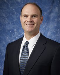 John W. Tomsho合作论文数11Scott W. Nelson合作论文数10CA CAPERELLI合作论文数10Hong Zhao合作论文数Department of Chemistry, The Pennsylvania State University10
 Tony Jun Huang合作论文数Department of Mechanical Engineering and Materials Science, Pratt School of Engineering, Duke University10Kazunari Taira合作论文数10Mark S. Warren合作论文数10Vincent Croquette合作论文数10
 Lin Wang合作论文数Wuhan University of Technology Wuhan University of Technology9
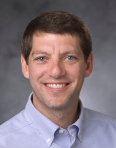 Andrew B. Nixon合作论文数School of MedicineDepartment of Medicine, Duke University9Barbara Fenn Kaboord合作论文数9WT MUELLER合作论文数9
 Songon An合作论文数9
 Zhiquan Zhang合作论文数8
 Chan Chung Yu合作论文数Department of Engineering Science and Mechanics, The Pennsylvania State University8
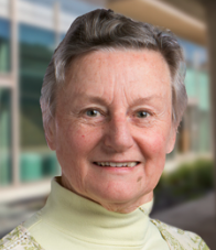 Jane Dyson合作论文数Department of Integrative Structural and Computational Biology, The Scripps Research Institute8
 Carston R. Wagner合作论文数Department of Medicinal Chemistry, College of Pharmacy, University of Minnesota8




