The vibration properties of materials play a role in their conduction of electric charges. Ionic conductors such as electrodes and solid electrolytes are also relevant in this respect. The vibration properties are typically assessed with infrared and Raman spectroscopy, and inelastic neutron scattering, which all allow for the derivation of the phonon density of states (PDOS) in part of a full portion of the Brioullin zone. Nuclear resonant vibration spectroscopy (NRVS) is a novel method that produces the element-specific PDOS from Mössbauer-active isotopes in a compound. We employed NRVS operando on a pouch cell battery containing a Li57FePO4 electrode, and thus could derive the PDOS of the 57Fe in the electrode during charging and discharging. The spectra reveal reversible vibrational changes associated with the two-phase conversion between LiFePO4 and FePO4, as well as signatures of metastable intermediate states. We demonstrate how the NRVS data can be used to tune the atomistic simulations to accurately reconstruct the full vibration structures of the battery materials in operando conditions. Unlike optical techniques, NRVS provides bulk-sensitive, element-specific access to the full phonon spectrum under realistic operando conditions. These results establish NRVS as a powerful method to probe lattice dynamics in working batteries and to advance the understanding of ion transport and phase transformation mechanisms in electrode materials.
Extremophiles are organisms that live and grow under extreme conditions. Such conditions include extreme cold, extreme heat, and extreme pressure. Understanding the viability of life under these stressors has applications for human health, biotechnology, bioremediation, and the search for life beyond our planet. Understanding the dynamics of proteins in extremophiles is key to understanding their functions and their broader applications. One proposed framework for understanding extremophile protein dynamics is the corresponding states hypothesis. This is the idea that the flexibility of homologous proteins from different extremophilic organisms will be comparable at their respective growth temperatures. However, the hypothesis is not universally accepted as evidence is contradictory. This conflicted body of evidence calls for a comprehensive experimental review using a variety of methods. Our study aims to contribute to this body of evidence by including X-ray diffraction, small-angle X-ray scattering, and molecular dynamics measurements at a range of temperatures on multiple extremophile rubredoxins. We utilized rubredoxin samples of two hyperthermophiles, Pyrococcus furiosus and Pyrococcus yayanosii, and two psychrophiles, Polaromonas glacialis and Polaromonas glacialis Eur3.
Iron-sulfur clusters fulfill numerous roles throughout biology. The reduced [2Fe-2S] + cluster offers unique electronic and magnetic properties due to its mixed-valent nature and can serve as an essential model for understanding electron transfer, electron delocalization, and accessible spin states not only in mixed-valent dimers, but potentially larger iron sulfur clusters. Recently a series of mixed-valent diiron dichalcogenide complexes [L 2 Fe 2 Q 2 ] − (Q = S ( 1 ), Se ( 2 ), Te ( 3 ), L = 2,6-diisopropylphenyl β-diketiminate ligand) were synthesized and characterized, where complex 1 showed a typical S = 1/2 spin state, while complexes 2 and 3 exhibited intermediate S = 3/2 spin states, potentially enabled by the minimization of vibronic coupling. Here we studied the vibrational dynamics of the Fe and Te centers in these complexes using 57 Fe and 125 Te nuclear resonance vibrational spectroscopy (NRVS), coupled with DFT calculations. The findings suggest that heavy character of larger chalcogen atoms results in decreased vibronic coupling. The observation of an intermediate spin state is shown to be unattainable for lighter Fe 2 Q 2 cores. This highlights the crucial role of vibronic coupling in modulating the electronic structure of mixed-valence systems and should enhance understanding of the electronic structure in more complex biological Fe-S clusters.
We herein present a comprehensive analysis of the oxidation-state-dependence of the 57Fe partial vibrational density of states (PVDOS), measured using nuclear resonance vibrational spectroscopy (NRVS), across a complete oxidation state series of iron-sulfur cubane (Fe4S4) clusters, which covers the [Fe4S4]0/1+/2+/3+/4+ redox levels. It is thereby revealed that the electronic/magnetic (and the thereof resulting geometric) symmetry of the respective complexes imprints onto their vibrational architecture, providing a rare experimental insight into the elusive relationship between these fundamental attributes. Modeling of our results at the broken-symmetry (BS) density functional theory (DFT) level allowed rationalizing the observed trends and supports the empirical correlations.
In this letter, the first nuclear resonant vibrational spectroscopy (NRVS) measurement on Na-Fe3O4/HZSM-5 multifunction catalyst for CO2 -> gasoline reactions is reported and discussed. A series of model or standard samples were also measured to provide comparisons. The Na-Fe3O4/HZSM-5 at the end of the catalytic reaction is identified via NRVS as a pure form of Fe5C2 without Fe3O4 or Fe(0). In the future, the information regarding to various catalytic intermediates can also be investigated via NRVS either on the extracted samples or with an in-situ measurement on the reaction cells. The advantages of NRVS can provide benefits for these researches.
Iron‑sulfur clusters are the primordial prosthetic groups for living systems, and they have even been proposed as partly responsible for the origin of life. They play a role in essential biological processes such as electron transfer, enzyme catalysis, DNA replication and repair, small molecule sensing, iron homeostasis, apoptosis, and human health and disease. They have frequently been studied by resonance Raman, electron paramagnetic resonance, and Mössbauer spectroscopies. Over the past two decades, we have used a synchrotron method called Nuclear Resonance Vibrational Spectroscopy (NRVS) to examine the vibrational dynamics of a wide variety of FeS clusters in model systems and native proteins, ranging in complexity from single Fe sites in small rubredoxins to the [7Fe-9S-C-Mo-R-homocitrate] cluster in nitrogenases.
Among all enzymatic metallocofactors, those found in nitrogenases, the P and L or M clusters, stand out for their intricate structures. They are assembled by proteins of the Nif gene cluster from Fe2S2 rhombs—the smallest building blocks in FeS cluster chemistry—through a sequence of reactions constructing a Fe8S8 precursor. To advance our understanding of how enzymes selectively build such elaborate inorganic molecules, here we parallel the biosynthetic pathway by reporting the rational stepwise assembly of [Fe8S8]m+ (m = 2, 4, 6) clusters from [Fe2S2]2+ rhombs within an extensive cyclic synthetic network. A [Fe8S8]4+ cluster of unique topology is identified, for which we coin the term ‘interlocked’ double cubane. As a molecular analogue of the NifB K cluster, a proposed precursor to both the P and L or M clusters, its preparation and the characterization of all related intermediates, offers fundamental insights into the molecular mechanisms governing the assembly of both biogenic and synthetic FeS clusters. Elucidating the nature of the metallocofactors in nitrogenase enzymes, and preparing synthetic analogues of these clusters, is a classic target for bioinorganic chemists. Now the transformation of [Fe2S2]2+ rhombs to [Fe8S8]n+ clusters has been achieved through a series of redox- and ligand-substitution reactions.
Cubane-type iron-sulfur clusters (Fe4S4) are some of the most versatile metallocofactors and, as such, among multiple functions, primarily responsible for mediating challenging electron transfers (ETs). Their efficient ET chemistry is enabled by a conflated interplay of cofactor-protein interactions, which can be categorized into the covalent first (1°) sphere ones and the noncovalent second (2°) sphere ones. The latter have remained particularly elusive, as they are difficult to observe and assess directly and independently. Accordingly, our understanding of these effects is hampered by their entangled nature. To address this, we herein leverage a systematic series of synthetic Fe4S4 complexes, which allows spectroscopically investigating 2° sphere electrostatic interactions and covalent 1° sphere interactions separately from one another. We expand the study of 1° sphere interactions with a histidine-type ligand in [Fe4S4]1+ complexes to the [Fe4S4]2+ and [Fe4S4]3+ oxidation states, supporting the notion that 1° sphere interactions "fine-tune" the electronic/magnetic structure of these systems in a manner that persists at ambient temperatures. In contrast, scrutinizing the 2° sphere electric dipolar interactions in [Fe4S4]1+,2+,3+ complexes revealed that although similar effects are observable at extremely low temperatures, no significant alteration of the clusters' gross electronic/magnetic structure persists at the temperatures relevant to enzyme function. These results thus not only systematically catalogue the influence of 1° sphere covalent and 2° sphere electrostatic interactions on the observables and properties of Fe4S4 complexes, but also establish a clear energetic distinction between the two. As such, they will facilitate identifying the elusive 2° sphere interactions in biological systems, while also strengthening our biophysical understanding of structure-function relationships in Fe4S4 cofactors.
Nitrogenase (N2ase) is a critical enzyme which catalyzes the reaction of N2 → NH3 in nature. Studies on the spectroscopy and photochemistry of trans-[FeII(DMeOPrPE)2(N2)H][BPh4] (1) and its isotopologues (2-6) provide a possible first step to evaluate the geometries and properties of the real N2ase-N2 structure(s). In this article, we have used FT-IR, FT-Raman, synchrotron-based nuclear resonant vibrational spectroscopy (NRVS) and DFT calculations to examine and assign the normal modes of these complexes. In addition, we have monitored their wavelength dependent photochemistry using mid-IR, near-IR, NRVS, and Mössbauer spectroscopies. Two distinct photolysis pathways are observed with mid-IR at (nominal) 4 K - (1) the cleavage of Fe-N2 bond in UV or visible light photolyses, which presents a unipolar disappearance of the N2 peak at 2094 cm-1 and is recombinable; (2) the ejection of trans hydrogen atom with UV irradiation, which has a pair of bipolar peaks with the disappearance of N2 at 2094 cm-1 and the appearance of a new species at 2056 cm-1 and is non-recombinable. The latter peak is well aligned with the N2 peak in an FeI reference complex (7). The combination of mid IR monitored photolysis/recombination and NRVS monitored photolysis form the central evidence for the conclusions in this article. In particular, the FeII-N2 and FeI-H·dissociations are in competition with each other in UV or UV-inclusive photolyses of this dinitrogen hydride complex. In addition, near-IR and Mössbauer also provide consistent evidence about FeI. This wavelength dependent photochemical work is the first one on a reaction active N2ase-N2 model complex and it also demonstrates the competition ejection between two axial ligands (H· and N2). It offers valuable information for future studies on real N2ase-N2 and its photolysis products.
In this publication, the potential non-gadolinium magnetic resonant imaging agent—nanoparticulate K2Mn[Fe(CN)6]—its comparison sample KFe[Co(CN)6], as well as their reference samples were measured and analyzed using Mn, Co and Fe L-edge X-ray absorption spectroscopy (L XAS). From the information obtained, we conclude that K2Mn[Fe (CN)6] has a high spin (hs)-Mn(II) and a low spin (ls)-Fe(II), while KFe[Co(CN)6] has an hs-Fe(II) and an ls-Co(III). In these Prussian blue (PB) analog structures, the L XAS analysis also led to the conclusion that the hs-Mn(II) in K2Mn[Fe(CN)6] or the hs-Fe(II) in KFe[Co(CN)6] bonds to the N in the [M(CN)6]4−/3− ions (where M = Fe(II) or Co(III)), while the ls-Fe(II) in K2Mn[Fe(CN)6] or the ls-Co(III) in KFe[Co(CN)6] bonds to the C in the [M(CN)6]4−/3− ion, suggesting the complexed metalloligand [Mn(II) or Fe(II)] occupies the N-bound site in PB. Then, nuclear resonant vibrational spectroscopy (NRVS) was used to confirm the results from the L XAS measurements: the Mn(II), Eu(III), Gd(III), Fe(II) cations complexed by [M(CN)6]n−-metalloligand all take the N-bound site in PB-like structures. Our NRVS studies also prove that iron in the K2Mn[Fe(CN)6] compound has a 2+ oxidation state and is surrounded by the C donor atoms in the [M(CN)6]n− ions.
An azadithiolate bridged CN- bound pentacarbonyl bis-iron complex, mimicking the active site of [Fe-Fe] H2ase is synthesized. The geometric and electronic structure of this complex is elucidated using a combination of EXAFS analysis, infrared and Mössbauer spectroscopy and DFT calculations. The electrochemical investigations show that complex 1 effectively reduces H+ to H2 between pH 0-3 at diffusion-controlled rates (1011 M-1 s-1) i.e. 108 s-1 at pH 3 with an overpotential of 140 mV. Electrochemical analysis and DFT calculations suggests that a CN- ligand increases the pKa of the cluster enabling hydrogen production from its Fe(i)-Fe(0) state at pHs much higher and overpotential much lower than its precursor bis-iron hexacarbonyl model which is active in its Fe(0)-Fe(0) state. The formation of a terminal Fe-H species, evidenced by spectroelectrochemistry in organic solvent, via a rate determining proton coupled electron transfer step and protonation of the adjacent azadithiolate, lowers the kinetic barrier leading to diffusion controlled rates of H2 evolution. The stereo-electronic factors enhance its catalytic rate by 3 order of magnitude relative to a bis-iron hexacarbonyl precursor at the same pH and potential.
Homocitrate is an organic component in the active site of nitrogenases (N2ase), where their cofactors are in the forms of [Δ-MoFe7S9C(R-Hhomocit)(N-his)(S-cys)], [Δ-VFe7S8C(CO3)(R-Hhomocit/homocit)(N-his)(S-cys)], and [Δ-FeFe7S9C(R-Hhomocit /homocit)(N-his)(S-cys)] (FeMo/V/Fe-cos) respectively, which are based on high-resolution crystallography, XES (XES = X-ray Emission Spectroscopy), ESEEM (ESEEM = Electron Spin Echo Envelope Modulation), and cryogenic electron microscopy. The protonated form of FeMo-co is set up on IR (Infra-red), VCD (Vibrational Circular Dichroism) spectroscopies and the comparisons of model complexes with protein structures in different oxidation states. Homocitrate ligand coordinates with molybdenum through its α-alkoxido and vicinal carboxyl groups, which is served as a possible proton source during the N2 reduction. Here a brief review has been given on the isolations, spectroscopies and structural characterizations of molybdenum and vanadium homocitrates and their homologs, including citrates, malates, tartrates, lactates and glycolates. The structures and configurations of Mo/V-homocitrato complexes are compared with the coordination environments of molybdenum, vanadium and iron atoms in FeMo/V/Fe-cos. Early theoretical calculations for the protonation of α-alkoxyl group in homocitrate of FeMo-co were also reviewed. A delivery pathway of hydrogen is suggested for the protonation and deprotonation of homocitrate in FeMo/V-cos.
Extremophile organisms are known that can metabolize at temperatures down to − 25 °C (psychrophiles) and up to 122 °C (hyperthermophiles). Understanding viability under extreme conditions is relevant for human health, biotechnological applications, and our search for life elsewhere in the universe. Information about the stability and dynamics of proteins under environmental extremes is an important factor in this regard. Here we compare the dynamics of small Fe-S proteins – rubredoxins – from psychrophilic and hyperthermophilic microorganisms, using three different nuclear techniques as well as molecular dynamics calculations to quantify motion at the Fe site. The theory of ‘corresponding states’ posits that homologous proteins from different extremophiles have comparable flexibilities at the optimum growth temperatures of their respective organisms. Although ‘corresponding states’ would predict greater flexibility for rubredoxins that operate at low temperatures, we find that from 4 to 300 K, the dynamics of the Fe sites in these homologous proteins are essentially equivalent.
[FeFe] hydrogenases are enzymes that have acquired a unique capacity to synthesize or consume molecular hydrogen (H2). This function relies on a complex catalytic mechanism involving the active site and two distinct electron and proton transfer networks working in concert. By an analysis based on terahertz vibrations of [FeFe] hydrogenase structure, we are able to predict and identify the existence of rate-promoting vibrations at the catalytic site and the coupling with functional residues involved in reported electron and proton transfer networks. Our findings suggest that the positioning of the cluster is influenced by the response of the scaffold to thermal fluctuations, which in turn drives the formation of networks for electron transfer through phonon-assisted mechanisms. Thus, we address the problem of linking the molecular structure to the catalytic function through picosecond dynamics, while raising the functional gain brought by the cofactors or clusters, using the concept of fold-encoded localized vibrations.
CO-bound forms of nitrogenase are N2-reduction inhibited and likely intermediates in Fischer-Tropsch chemistry. Visible-light photolysis at 7 K was used to interrogate all three known CO-related EPR-active forms as exhibited by the α-H195Q variant of Azotobacter vinelandii nitrogenase MoFe protein. The hi(5)-CO EPR signal converted to the hi-CO EPR signal, which reverted at 10 K. FT-IR monitoring revealed an exquisitely light-sensitive "Hi-2" species with bands at 1932 and 1866 cm-1 that yielded "Hi-1" with bands at 1969 and 1692 cm-1, which reverted to "Hi-2". The similarities of photochemical behavior and recombination kinetics showed, for the first time, that hi-CO EPR and "Hi-1" IR signals arise from one chemical species. hi(5)-CO EPR and "Hi-2" IR signals are from a second species, and lo-CO EPR and "Lo-2" IR signals, formed after prolonged illumination, are from a third species. Comparing FT-IR data with CO-inhibited MoFe-protein crystal structures allowed assignment of CO-bonding geometries in these species.
DNA primase is a DNA-dependent RNA polymerase that synthesizes short oligonucleotide primers required for processive DNA replication. Like many enzymes involved in DNA replication and repair, human DNA primase contains a redox-active, high potential [4Fe4S] cluster whose associated redox state serves as an on/off “switch” for DNA binding. The structural basis for this “switch,” however, is poorly understood, given the 25 Å distance between the [4Fe4S] cluster and DNA binding region. Examination of available crystallographic data suggests the [4Fe4S] cluster may undergo a change in structure upon substrate binding, but this information alone is not sufficient to form conclusions about allostery.
The nitrogenase (N2ase) enzyme family is responsible for the conversion of dinitrogen into biologically accessible ammonia, a critical step in the global nitrogen cycle. Carbon monoxide (CO) has long been known as an inhibitor of dinitrogen reduction, but it can also be reduced to hydrocarbons catalyzed by all three N2ases, namely the wild-type Mo enzyme and select variants and the V and Fe nitrogenases, both of which are orders of magnitude more effective. CO interactions with N2ases are thus relevant to both dinitrogen fixation and Fischer-Tropsch-like chemistry. Here, we investigated the interaction of CO with the α-R277H variant of the Azotobacter vinelandii N2ase MoFe protein, in which the α-subunit 277Arg residue is replaced by His and results in production of only the S = 3/2 EPR signal (denoted as hi(5)-CO). Fourier-transform infrared (FT-IR) spectroscopy was used to follow the photolysis of CO bound to the α-R277H variant under cryogenic conditions. Multiple EPR-silent species were observed with FT-IR spectroscopic signatures previously assigned to CO-inhibited forms of the α-H195Q and α-H195N N2ase variants. The distribution of these CO-inhibited forms varied dramatically with pH over the range of pH 6.5 to pH 8.5, indicating protonation/deprotonation involvement.
AbstractZur Untersuchung von Metalloenzymen haben wir einen Aufbau für die Präparation katalytischer Intermediate und deren anschließende Charakterisierung mit spektroskopischen Techniken entwickelt. Mit diesem können Redoxreaktionen in Enzymen in Form von Lyophilisat, gelöst oder als Kristall in einem großen Temperaturbereich IR‐spektroskopisch in situ verfolgt werden. Zwei sauerstofftolerante [NiFe]‐Hydrogenasen wurden als Modellenzyme untersucht. Zunächst wurde der Aufbau zur Herstellung von komprimiertem Lyophilisat einer Hydrogenase in einem paramagnetischen Zustand mit verbrückendem Hydrid genutzt. Dies erleichterte die Charakterisierung durch57Fe‐kernresonante inelastische Streuung und erlaubte es, in Kombination mit DFT, die schwingungsspektroskopischen Merkmale dieses katalytischen Intermediats zu detektieren. Der In‐situ‐IR‐Aufbau lieferte zusammen mit Resonanz‐Raman‐Untersuchungen auch Einblicke in die Redoxchemie von Proteinkristallen. Eine Ergänzung röntgenkristallographischer Daten durch komplementäre spektroskopische Analysen ist daher essentiell.
 Hongxin Wang合作论文数75
 Uwe Bergmann合作论文数Department of Materials Science and Engineering, University of Wisconsin-Madison/Department of Physics, University of Wisconsin-Madison/SLAC National Accelerator Laboratory55
 Yoshitaka Yoda合作论文数52
 Pieter Glatzel合作论文数European Synchrotron Radiation Facility38Vladimir Pelmenschikov合作论文数Department of Physics;Stockholm Uni Versity;Department of Physics, Stockholm Uni Versity35
 Simon J George合作论文数CSIRO Petroleum34
 Simon J. George合作论文数School of Chemical Sciences, University of East Anglia29Weiwei Gu合作论文数27
 Leland Bruce Gee合作论文数Department of Chemistry, University of California, Davis26Yuming Xiao合作论文数Geophysical Laboratory;Carnegie Institution of Washington;Geophysical Laboratory, Carnegie Institution of Washington24J Vanelp合作论文数23Stephan Friedrich合作论文数Lawrence Livermore National Laboratory23
 Alex Guo合作论文数Department of Chemistry, Mellon College of Science, Carnegie Mellon University23
 Graham George合作论文数Department of Geological Sciences, University of Saskatchewan21
 Xianhong Wang (王献红)合作论文数Key Laboratory of Polymer Ecomaterials, Chinese Academy of Sciences;Changchun Institute of Applied Chemistry, Chinese Academy of Sciences;University of Chinese Academy of Sciences19Gang Peng合作论文数19
 Michael Adams合作论文数Department of Biochemistry and Molecular Biology, Franklin College of Arts and Sciences, University of Georgia19
 Thomas B. Rauchfuss合作论文数Department of Chemistry, College of Liberal Arts & Sciences, University of Illinois at Urbana-Champaign16
 Oliver C. Mullins合作论文数Mullins Reservoir Solutions;Schlumberger Companies16Jason Christiansen合作论文数Department of Biochemistry, Fralin Biotechnology Center16William E. Newton合作论文数15
 Vittal K. Yachandra合作论文数Lawrence Berkeley National Laboratory14Kenji Tamasaku合作论文数14
 Frank de Groot合作论文数Department of Chemistry, Utrecht University;X-ray Spectroscopy in the Debye Institute, Utrecht University;Stanford University14
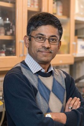 Aniruddha Deb合作论文数Department of Chemistry, College of Literature, Science, and the Arts, University of Michigan-Ann Arbor13
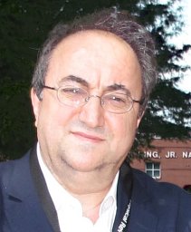 E. E. Alp合作论文数Argonne National Laboratory Advanced Photon Source Argon13
 Jiyong Zhao合作论文数U.S. Department of Energy’s Advanced Photon Source, Argonne National Laboratory13Tobias Funk合作论文数13
 Ralston Corie Y合作论文数12
 Stephen Ragsdale合作论文数Department of Biological Chemistry, University of Michigan Medical School12
 Aubrey D Scott合作论文数12
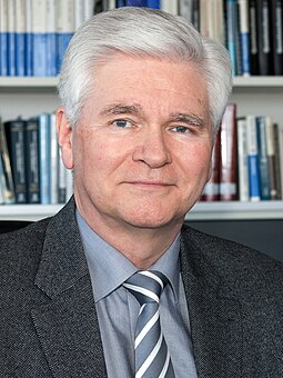 Wolfgang Lubitz合作论文数Max Planck Institute for Chemical Energy Conversion12
 Robert Scott合作论文数Department of Chemistry, Franklin College of Arts and Sciences, University of Georgia12
 Hendrik G Visser合作论文数Department of Chemistry;University of the Orange Free State;Department of Chemistry, University of the Orange Free State12Ko Hodgson合作论文数11Mm Grush合作论文数11
 Kenneth Sauer合作论文数Department of Chemistry, University of California, Berkeley;Physical Biosciences Division, Lawrence Berkeley National Laboratory, University of California11Matt C. Smith合作论文数Department of Biological and Agricultural Engineering, The University of Georgia11Francis E. Jenney合作论文数Georgia Campus - Philadelphia College of Osteopathic Medicine, Suwanee, United States11
 Cindy C Pham合作论文数Lawrence Berkeley Natl Lab, Mol Biophys & Integrated Bioimaging Div, Berkeley, CA 94720 USA10
 Elton J. Cairns合作论文数Department of Chemical and Biomolecular Engineering, College of Chemistry, University of California, Berkeley10
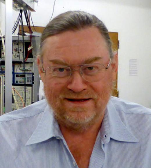 R. David Britt合作论文数Department of Chemistry, University of California, Davis10
 Harry B. Gray合作论文数Department of Applied Physics and Materials Science, Division of Engineering and Applied Science, California Institute of Technology;Beckman Institute Laser Resource Center, California Institute of Technology10
 James Swartz合作论文数Department of Chemical Engineering, Stanford University;Department of Bioengineering, Stanford University;Swartz Biotechnology Laboratory, School of Engineering, Stanford University10Christie H. Dapper合作论文数10JH Robblee合作论文数10Li-Fen Yan合作论文数9
 Saeed Kamali-Moghaddam合作论文数9
 Lars Lauterbach合作论文数9




