There have been several methods established for immunohistochemical labeling of the PGP 9.5 antigen in human tissue for the assessment of epidermal nerve fiber density, none of which uses neutral-buffered formalin as the preferred fixative for paraffin-embedded tissue. The literature maintains that formalin-fixed paraffin embedded tissues are unable to be used for this purpose and that other fixatives must be used due to the cross-linkages caused by formalin fixation. This study was undertaken to develop a standardized method for the use of formalin-fixed paraffin embedded tissues for immunohistochemical labeling and assessment of epidermal nerve fiber density. Formalin-fixed paraffin embedded tissues from the punch biopsies of patients suspected to have small fiber neuropathy were prepared for immunohistochemical labeling using heat-induced epitope retrieval for one hour at 92 degrees C. The tissues were then stained with a polyclonal rabbit anti-human PGP 9.5 primary antibody. The resulting stains were then evaluated by a licensed pathologist who counted the number of epidermal nerve fibers stained and recorded the epidermal length in millimeters. Human foreskin was used as the tissue control in these studies. Satisfactory immunohistochemical labeling of epidermal nerve fibers was achieved from formalin-fixed paraffin embedded tissues through the use of heat-induced epitope retrieval. The authors of this paper have concluded that formalin-fixed paraffin embedded tissues may be used to achieve satisfactory immunohistochemical labeling for the assessment of epidermal nerve fiber density.
Abstract Introduction Deep partial thickness burns generally require more than 2-3 weeks to heal often resulting in hypertrophic scarring and patient morbidity. Because the skin of pigs most closely resembles that of humans, the pig is often used as an experimental burn wound model. We describe the development of a reproducible burn wound model in Yorkshire pigs. Methods We created standardized 5 x 5 cm burns on the backs and flanks of anesthetized domestic pigs (30 kg) using a radiant heating device set at 600 degrees C and applied to the animal’s skin for 5, 10, or 15 seconds. Full thickness 4 mm punch biopsies were obtained from the burns at 4 and 24 hours for burn depth evaluation. The tissue sections were stained with Hematoxylin and Eosin, HMGB-1 (a marker of necrosis) and activated cleaved caspase 3 (a marker of apoptosis). The tissue sections were viewed by a board-certified dermatopathologist blinded to duration of exposure. The primary outcome was the deepest level of injury to the blood vessels or epithelial appendices (hair follicles or apocrine glands). Results Exposure of the pigs’ skin to the heating device for periods of 5, 10, and 15 seconds resulted in superficial partial, deep partial and full thickness burns respectively. Average depth of dermal injury 24 hours after exposure was 0.4 mm, 1.3 mm, and 2.4 mm for exposure times of 5, 10 and 15 seconds respectively. Conclusions We describe a standardized porcine model for creating reproducible superficial partial, deep partial and full thickness burns. Applicability of Research to Practice This model is now being used to investigate novel therapies for deep burns.
There have been several studies on the use of the Verhoeff van Gieson staining method to demonstrate thermal effects on tissues. However, this method has rarely been used for the analysis of periodontal tissues. This study was undertaken to compare the quality and effectiveness of the Verhoeff van Gieson (VVG) staining method with conventional hematoxylin & eosin (H&E) in measuring the thermal effects in gingival tissues. Periodontal tissues around bovine mandibular teeth were treated using different surgical lasers (wavelengths of 10,600 nm, 970 nm, and 445 nm) at 2 W power setting. Measurements of the depth of the coagulation zone were recorded for all treatment groups in sample tissues stained with H&E as well as the VVG-staining method. Measures were interpreted by a trained pathologist. A statistical analysis was performed using the Wilcoxon signed-rank test to determine if there was a statistically significant difference between values recorded for the light penetration depth on tissues stained with each of the two staining methods. It was determined that there was no significant difference in the recorded values (P = 0.23). We have concluded that the VVG-stained tissues were better able to visualize the depth of thermal damage and thus may make it easier for someone not well trained to interpret the depth of light penetration in these tissues.
Surgical excision and grafting of deep partial-thickness (DPT) and full-thickness (FT) burns is a cornerstone of wound care. The use of commercially available topical enzymatic agents has been limited due to slower and less complete eschar removal than surgical excision. Using a porcine model of DPT and FT burns, we compared the eschar removal efficacy of a bromelain-enriched enzymatic agent derived from the stems of pineapple plants and a commercially available collagenase. We created 40 DPT and 40 FT burns on four anesthetized Yorkshire pigs. Eschar removal was initiated 24 hours later. Two pigs each were randomly assigned to collagenase or the bromelain-enriched agent. The bromelain-enriched agent was applied topically once for 4 hours followed by a 2-hour soaking. The collagenase was applied topically daily until complete removal of eschar or for up to 14 days. All bromelain-enriched treated FT burns underwent complete removal of the eschar after a single application while none of the collagenase-treated FT burns underwent complete removal of the eschar even after 14 days of treatment. All bromelain-enriched treated DPT burns had complete eschar removal after the single application. None of the collagenase-treated DPT burns experienced complete removal of eschar after 10 days; by day 14, 35% had complete eschar removal, 30% had >50% eschar removed, and 35% had <50% eschar removed. We conclude that eschar removal is quicker and more complete with the bromelain-enriched compared with collagenase debriding agent.
Background: This study aimed to evaluate the penetration depth of 445 and 970 nm diode lasers using both initiated and non-initiated fibers on bovine gingiva in an ex vivo model. Methods: In this in vitro experiment, 445 nm and 970 nm diode lasers were compared in pulsed (35 Hz, duty cycle: 50%) and continuous mode with both initiated and non-initiated tips. All experimental groups had laser output settings of 2 Watts with a 320 μm thick glass fiber utilized for delivery. Two posterior teeth were randomly selected from four bovine mandibles for each group. The teeth were irradiated for 30 s buccal and 30 s lingual before the gingiva was harvested for histological evaluation. Samples were fixed in 10% formalin and stained via elastin Van Gieson. Sections were examined microscopically to evaluate collagen denaturation due to thermal effects, which were measured and compared using a mixed-effect model with Tukey adjustment. Results: The 445 nm wavelength groups displayed significantly higher penetration depths when compared to the 970 nm wavelength groups. The initiated tip groups tended to display a lower penetration depth than non-initiated groups; however, these values were similar (p > 0.05) for the 445 nm pulsed lasers (initiated vs. non-initiated). Conclusions: We can conclude that the 445 nm laser wavelength displayed greater thermal denaturation in bovine gingiva ex vivo when compared to the 970 nm wavelength. Additionally, the pulsed settings displayed less thermal penetration depth when compared to the continuous emission modes of the same power output. However, additional studies are necessary to better compare initiated and non-initiated tips using the novel blue laser light.
Background: With laser irradiation emerging as an adjunctive treatment utilized in nonsurgical periodontal therapy, it is important to understand the variance of penetration depth among the different laser wavelengths.Purpose: To evaluate the thermal penetration depth, as a photothermal effect, of carbon dioxide (CO2)-, erbium: doped yttrium-aluminum-garnett (Er:YAG)-, and erbium, chromium: yttrium-scandium-gallium-garnett (Er,Cr:YSGG)-lasers on the bovine gingiva in an in vitro model.Methods: Four mandibles from freshly slaughtered cows were utilized in this study. Buccal and lingual root debridement was provided using three different laser wavelengths, all in pulsed settings. A CO2- (10,600 nm), Er:YAG- (2940 nm), and Er,Cr:YSGG- (2780 nm) were utilized to irradiate pockets of two mandibular posterior teeth in each group. Laser power output settings were set to 2 W. The posterior teeth were irradiated for 30 sec buccal and 30 sec lingual of each tooth for all selected treatment test groups. Instrumentation with curettes was performed as a control group. Gingival flaps, including the entire gingiva, were fixed in 10% formalin and stained via Elastin van Gieson. Sections were examined microscopically to evaluate thermal damage and statistically compared using mixed effect model with Tukey adjustment.Results: The CO2-laser irradiation presented a statistically significant lower mean compared to Er,Cr:YSGG-laser (p < 0.0001). Er,Cr:YSGG-laser had a higher penetration depth compared to Er:YAG-laser (p < 0.0001). There was no statistically significant difference found in penetration depth between CO2- and Er:YAG-laser irradiation.Conclusions: It can be concluded that all tested pulsed lasers had minimal penetration depth into the gingiva. However, the pulsed CO2- and Er:YAG-lasers presented lower thermal effects compared to Er,Cr:YSGG-laser in vitro.
PURPOSE:CMC 2.24, a chemically modified curcumin, was developed as a novel, pleiotropic MMP-inhibitor to treat various inflammatory/collagenolytic diseases including periodontitis. To date, this compound has shown efficacy in vitro, in cell culture, and in vivo (oral administration) in mice, rats and dogs. In preparation for possible Phase I human clinical trials, the current study describes the maximum-tolerated-dose (MTD), pharmacokinetics (PK), and toxicology of CMC 2.24 in the rat model.METHODS:For the MTD study, 30 Sprague-Dawley rats were randomly distributed into 5 groups (3M/3F per group): Placebo (vehicle; carboxymethylcellulose) and CMC 2.24 at various doses (50, 100, 500, 1000 mg/kg/day), were administered once daily by oral gavage for 5 days. For the PK study, 24 rats were administered either Placebo or CMC 2.24 (100mg/kg/day) once daily for 28 days or only once (500 or 1000 mg/kg). Analysis of this test compound was done using LC/MS/MS for PK evaluation on blood samples drawn from rats at multiple time points. The animals were sacrificed after 5 or 28 days of treatment, and blood chemistry and serology were analyzed. Major organs (heart, lung, liver, kidney, spleen, intestine, brain) were histologically examined at necropsy.RESULTS:Orally administered, CMC 2.24 did not produce significant changes in body weight, food consumption or adverse events in the MTD and toxicology studies. Moreover, no obvious pathologic changes were observed based on histology, hematology, serum biochemistry, or necropsy compared to placebo-treated controls. The PK study demonstrated a peak-blood concentration (Cmax) at 45 mins after oral administration of 2.24 and a serum half-life of 10 hours.CONCLUSION:In conclusion, CMC 2.24, orally administered to rats once a day, appears to be safe and effective at a wide range of doses, consistent with efficacy previously demonstrated in studies on animal models of various collagenolytic diseases, such as periodontitis, diabetes and cancer.
During the 2020 coronavirus (SARS-CoV-2) pandemic, several cutaneous lesions were identified, including pseudo-chilblain, vesicular, urticarial, maculopapular, and livedo/necrosis. A 59-year-old obese man with probable COVID-19 developed painful cyanosis with histopathologic capillary thrombosis of toes, and the cyanosis persisted for nearly 22 months. Shortly after initial exposure to family members with documented SARS-CoV-2, he developed upper respiratory symptoms, yet his anti-SARS-CoV-2 antibody and nasal swab RT-PCR tests were repeatedly negative. Two family members were hospitalized and one of them succumbed with documented SARS-CoV-2 pneumonia within 10 days of exposure. Biopsy specimen of the distal toe 16 weeks after initial exposure showed papillary dermal capillary thrombosis with endothelial swelling, telangiectasia, and peri-eccrine lymphocytic infiltrates resembling pernio. Overall, this is the first case of biopsy specimen of "long COVID toe" following presumed SARS-CoV-2 exposure, with a demonstration of thrombotic vasculopathy, toe cyanosis, and pernio-like pathology.
Whether the depth and healing of scalds and contact burns are similar is controversial. Due to water's greater heat capacity, we hypothesized that when exposed to similar temperatures and durations of exposure, burns caused by hot water would be deeper than those caused by contact with hot metal. Forty standardized burns were created in two anesthetized female domestic pigs using a brass bar or circulating heated water. In one pig, the temperature was kept constant (95°C) while the duration of exposure varied (5, 10, 15 seconds) In the second pig, the exposure time was kept constant (10 seconds) while the temperature of exposure varied (70°C, 80°C, 98°C). Periodic punch biopsies were taken to determine burn depth immediately after injury, percentage burns reepithelialized within 21 days, and depth of scar at 28 days. The analysis was performed using analysis of variance. When the temperature was held constant, duration of exposure (5, 10, and 15 seconds) was associated with scar depth (2.1 vs 3.8 vs 5.0 mm, respectively, P = 0.001) but not with burn depth (2.0 vs 2.2 vs 2.3 mm, respectively, P = 0.10). When exposure duration was held constant, temperature (70°C, 80°C, 98°C) was associated with scar depth (0.6 vs 1.7 vs 3.6, P < 0.001) but not with burn depth (1.2 vs 1.5 vs 1.7 mm, respectively, P = 0.21). Burn depths were greater for scald than contact burns although not significantly greater. After controlling for temperature, the difference in scar depth between scalds and contact burns was statistically significant (marginal means 3.0 for contact burns, 4.3 for scalds, P = 0.008). We conclude that burns created in swine with circulating hot water result in deeper scars than those created by contact with a brass bar when controlling for temperature and duration of exposure.
Abstract Introduction Whether the depth and healing of scalds and contact burns are similar is controversial. Prior studies have not compared scalds and contact burns using identical temperatures and durations of exposure limiting direct comparison. We hypothesized that when exposed to similar temepratures and durations of exposure, burns caused by hot water and contact with hot metal would be similar. Methods Multiple 2.5 or 5 cm diameter standardized cutaneous burns were created in two anesthetized female domestic pigs (~45kg) using a brass bar or circulating heated water in replicates. The temperatures were controlled and kept constant using thermosensors. In one pig, the temperature was kept constant (95 degrees C) while the duration of exposure varied (5, 10, 15 sec.) In the second pig, the exposure time was kept constant (10 sec.) while the temperature of exposure varied (70, 80, 98 degrees C). Burns were covered with a topical antibiotic ointment and frequent dressing changes were performed for 4 weeks. Periodic punch biospies were taken to determine burn depth immediately after injury, percentage burns reepithelialized within 21 days, and depth of scar at 28 days. Analysis was performed using analysis of variance. Results When temperature was held constant, duration of exposure (5, 10, and 15 sec.) was associated with scar depth (2.1 vs 3.8 vs 5.0 mm respectively, P=0.001) and there was a trend for an association with burn depth (2.0 vs 2.2 vs 2.3 mm respectively, P=0.10). When exposure duration was held constant, temperature (70, 80, 98 degrees C) was associated with scar depth (0.6 vs 1.7 vs 3.6, P< 0.001) but not significantly associated with burn depth (1.2 vs 1.5 vs 1.7 mm respectively, P=0.21). For each pig a comparison of burn and scar depth by device generally showed that depths were greater for scald than contact burns although not significantly greater. Only scar depth in the first pig showed a difference with a significance of less than 0.10 (2.9 for contact vs 4.1 mm for scalds, P=0.08). After controlling for temperature the difference between scalds and contact burns was statistically significant (marginal means 3.0 for contact burns, 4.3 for scalds, P=0.008). When combining data from both pigs and controlling for temperatures and duration, scar depths for scalds (2.7 mm) vs contacts (1.9 mm) showed a significant difference (P=0.02) while burn depth was not quite significant (1.7 vs 1.5 mm, P=0.07). Almost all burns were reepithelialized by day 21. Conclusions Burns created in swine with circulating hot water tend to be deeper and result in deeper scars than those created by contact with a brass bar when controlling for temperature and duration of exposure. Applicability of Research to Practice Scalds of similar temperature and exposure times result in deeper scars than contact burns.
Transplantation of human xenografts onto immunocompromised mice is a powerful research tool for studying wound healing. However, differences in healing between humans and mice and their small size limit this model. We determined whether human cadaver skin xenografts transplanted onto pigs with severe combined immune deficiency (SCID) would survive and not be rejected. Meshed (1:1.5), cryopreserved human cadaver skin was transplanted onto 10 partial thickness dermatome wounds in each of two normal domestic pigs and two SCID pigs. Autografts (n = 2/animal) from the four animals were used as controls. In normal pigs, all autografts were engrafted and healed with a minimal, if any, inflammation and scarring. All human xenografts were rejected by the normal pigs within 5–11 days and associated with an intense T‐cell inflammatory response. In contrast, both autografts and xenografts were engrafted and survived the 28‐day study in the SCID pigs with a minimal inflammation and no gross scarring.
Burns are dynamic injuries that tend to progress in depth over several days. Currently, there are no FDA-approved therapies aimed at reducing burn injury progression. We have previously demonstrated that tadalafil (a phosphodiesterase-5 inhibitor used as a vasodilator to treat erectile dysfunction) reduces necrosis in the unburned interspaces compared with naproxen, NAC, or their combination in a rat comb burn model. In this study we tested the hypothesis that tadalafil would speed reepithelialization and reduce scarring of deep partial thickness burns in a validated vertical progression porcine burn model. A validated animal burn model was used in which 60 deep partial thickness burns were created on the backs and flanks of anesthetized domestic pigs (25–30 kg) using an aluminum bar preheated to 80 degrees C for 20 seconds. After removing the necrotic epidermis to simulate blister formation and rupture, the animals were randomly treated with oral tadalafil 2.5 mg or a placebo immediately after injury and once daily thereafter for seven days. All wounds were treated with a topical triple antibiotic ointment every other day. Periodic imaging and full thickness biopsies were conducted to monitor healing over 28 days. The mean percentages wound reepithelialization at days 8, 11, 15, 18, 21, and 28 for wounds treated with tadalafil and placebo were 14±2% vs. 3±4%, 28±39% vs. 1±2%, 88±30% vs. 21±33%, 95±19 vs. 72±40, and 98±13% vs. 97±8% respectively (p<0.01 for all except day 28). The mean time to complete wound reepithelialization was significantly faster in burns treated with tadalafil than control burns (15.3±1.9 vs. 21.9±5.4 days, p<0.001). Scar depth was significantly less in burns treated with tadalafil (2.6±1.6 mm vs. 5.2±1.5 mm, p<0.001). Wounds treated with tadalafil were less erythematous than control wounds. There were no wound infections or systemic adverse events in any of the wounds or animals respectively. Once daily oral tadalafil speeds reepithelialization and reduces scarring and erythema in porcine deep partial thickness burns. Further studies are required to determine if tadalafil reduces improves healing in humans.
Hypertrophic scarring after burns is common resulting in significant disfigurement and dysfunction. Further advances in understanding the pathobiology of scarring and the development of novel therapies aimed at reducing scarring are hindered by the lack of appropriate large animal models. Transplantation of human xenografts onto immune compromised mice is a powerful research tool for studying wound healing. However, differences in healing between humans and mice and their small size limits this model. Recently, a severe combined immune deficiency (SCID) pig was accidentally discovered. We determined whether human cadaver skin xenografts transplanted onto SCID pigs would survive and not be rejected. Split thickness, meshed (1:1.5), cryopreserved human cadaver skin obtained from a skin bank was transplanted onto 10 partial thickness dermatome wounds in each of two normal domestic pigs and two SCID pigs. Autografts (n=2/animal) from the 4 animals were used as controls. Animals were followed for 4 weeks and periodic digital images and full thickness biopsies were obtained to monitor healing using H&E stains as well as T-cell specific CD3-antibodies. Human specific HLA antibodies were used to determine the origin of the transplanted skin in SCID pigs. In normal pigs, all autografts were engrafted and healed with minimal if any inflammation and scarring. All human xenografts were rejected by the normal pigs within 5-11 days and associated with an intense T-cell inflammatory response. In contrast, both autografts and xenografts were engrafted and survived the 28-day study in the SCID pigs with minimal inflammation in only 1/20 xenografts and no gross scarring in any wounds. Human specific antibodies (HLA-ABC) confirmed the human source of the healed xenografts. This study serves as proof-of-concept that human cadaver skin survives on SCID pigs for at least 28 days. Further development of this model is ongoing to determine if human hypertrophic scars or keloids can be transplanted onto SCID pigs. We believe that a humanized scar model in pigs will be helpful in the development of novel therapies aimed at preventing or reducing hypertrophic scarring after burns.
Burn conversion from second to third degree is common leading to delayed healing and scarring. We hypothesized that tadalafil, a phosphodiesterase 5 inhibitor (PDE5I) that results in vasodilation, would reduce burn conversion leading to faster reepithelialization and less scarring of partial thickness porcine burns. We conducted a prospective, randomized, controlled, animal experiment using six female pigs (25-30 kg). We created 20 standardized partial thickness burns on each of the animals with an aluminum bar preheated to 80 °C and applied for 20 seconds to the pigs' dorsum. Three animals each were randomized to oral tadalafil 2.5 mg or control vehicle once daily for 1 week. Main outcomes were time to reepithelialization and depth of scarring at 28 days. A sample of 60 burns in each treatment group had 80% power to detect a 2-day difference in time to reepithelialization. Mean (95% CI) time to reepithelialization in burns treated with tadalafil and control were 14.9 (14.1-15.7) vs. 19.7 (18.2-21.3) days, respectively; mean difference 4.8 (3.1-6.6) days. After controlling for pig and within pig differences, mean time to reepithelialization was 6.5 (3.7-9.3) days shorter in burns treated with tadalafil compared with controls. Mean (95% CI) scar depth in burns treated with tadalafil and control were 2.7 (2.3-3.1) vs. 3.7 (3.1-4.2) mm. respectively, mean difference 1 (0.3-1.7) mm. After controlling for pig and within pig differences, scar depth in tadalafil-treated burns was 1.5 (0.7-2.3) mm lower compared with controls. We conclude that once daily oral tadalafil shortened time to reepithelialization and reduced scarring in a partial thickness porcine burns model.
Peters plus syndrome (MIM #261540 PTRPLS), characterized by defects in eye development, prominent forehead, hypertelorism, short stature and brachydactyly, is caused by mutations in the β3-glucosyltransferase (B3GLCT) gene. Protein O-fucosyltransferase 2 (POFUT2) and B3GLCT work sequentially to add an O-linked glucose β1-3fucose disaccharide to properly folded thrombospondin type 1 repeats (TSRs). Forty-nine proteins are predicted to be modified by POFUT2, and nearly half are members of the ADAMTS superfamily. Previous studies suggested that O-linked fucose is essential for folding and secretion of POFUT2-modified proteins and that B3GLCT-mediated extension to the disaccharide is essential for only a subset of targets. To test this hypothesis and gain insight into the origin of PTRPLS developmental defects, we developed and characterized two mouse B3glct knockout alleles. Using these models, we tested the role of B3GLCT in enabling function of ADAMTS9 and ADAMTS20, two highly conserved targets whose functions are well characterized in mouse development. The mouse B3glct mutants developed craniofacial and skeletal abnormalities comparable to PTRPLS. In addition, we observed highly penetrant hydrocephalus, white spotting and soft tissue syndactyly. We provide strong genetic and biochemical evidence that hydrocephalus and white spotting in B3glct mutants resulted from loss of ADAMTS20, eye abnormalities from partial reduction of ADAMTS9 and cleft palate from loss of ADAMTS20 and partially reduced ADAMTS9 function. Combined, these results provide compelling evidence that ADAMTS9 and ADAMTS20 were differentially sensitive to B3GLCT inactivation and suggest that the developmental defects in PTRPLS result from disruption of a subset of highly sensitive POFUT2/B3GLCT targets such as ADAMTS20.
Introduction: Platelet rich plasma (PRP) is rich in growth factors and has been shown to improve healing in a variety of wounds. We determined the effects of PRP on healing and scarring in full thickness porcine burns with and without tangential excision and grafting (TEG). Methods: Standardized full thickness 5cm by 5cm burns were created on each of the backs and flanks of 10 anesthetized female pigs (25 kg) using a validated model. The burns were created with a heating device that emits heat at a temperature of 400 degrees C for a period of 30 s. The burns were randomized to one of six treatments: no TEG or PRP, no TEG+PRP, early (day 2) TEG and no PRP, early TEG+PRP, late (day 14) TEG and no PRP, and late TEG+PRP. Tangential excision was performed down to viable tissue and autografts were 0.2mm thick. When used, a thin layer of autologous PRP was applied below the graft. All wounds were then treated with a topical antibiotic ointment 3 times weekly for 42 days. Digital images and full thickness biopsies were taken at 9, 11, 14, 18, 21, 28, 35 and 42 days after injury to determine percentage reepithelialization, scar depth, and scar contraction. Tissue sections were stained with H&E and viewed by a dermatopathologist masked to treatment assignment. Results: There was no reduction in platelet and white blood cell concentrations in PRP and blood samples for the first 14 days after-full thickness burns. A total of 120 burns were created on 10 animals evenly distributed between the six treatment groups. Burns undergoing early TEG reepithelialized fastest and with the thinnest scars followed by late TEG. Burns that did not undergo TEG had the slowest reepithelialization and greatest amount of scarring. Application of PRP had no additional effects on reepithelialization, scar depth, or scar contraction in any of the treatment groups. Conclusions: Addition of PRP had similar effects on reepithelialization and scarring of full thickness porcine burns as standard topical antibiotic ointment regardless of whether the burns underwent excision or grafting or the timing of excision and grafting. (C) 2018 Elsevier Ltd and ISBI. All rights reserved.
Objectives: There are no well accepted animal models of chronic wounds, limiting advances in understanding and treatment of chronic ulcers. We developed a porcine wound model which combines multiple factors involved in chronic wounds to create a contaminated necrotic eschar and evaluated the debriding efficacy of a novel bromelain based enzymatic debriding agent (EscharEx). Methods: Contaminated ischemic wounds were created on the flanks of domestic pigs by 'sandwiching' the skin between 2 'O' rings (1 placed on the surface of the skin and the other underneath the skin) for 24h prior to dermatomal excision of the necrotic eschar and its contamination with Staphylococcus aureus and Candida albicans. After confirming the development of infected eschars, additional animals were used to compare the effects of daily application of topical EscharEx or its hydrating vehicle on eschar debridement as a control. Results: In all cases, application of the 'O' rings resulted in full thickness necrotic ecshars with invasive infections, which did not reepithelialize and sloughed off spontaneously within 14-21 days. All wounds reepithelialized within 28-42 days forming contracted scars. All EscharEx treated eschars were completely debrided within 7-9 days, while no debridement was evident in eschars treated with the control gel. Conclusions: Our model simulates the initial phase of chronic wounds characterized by a contaminated necrotic eschar allowing evaluation of wound debriding agents, and that a bromelain-based debriding agent completely debrides the contaminated necrotic eschars within one week in this model. (C) 2017 Elsevier Ltd and ISBI. All rights reserved.
Burns are dynamic injuries characterized by progressive tissue death and continuous severe pain over the course of several days. The extent of burn injury progression determines the ultimate patient outcome. Initial burns result in a central zone of necrosis surrounded by a potentially viable zone of ischemia. Several mechanisms have been proposed to explain injury progression, including oxidant and cytokine stress resulting from either ischemia/reperfusion and/or inflammation, but no proven therapy has emerged. To address the unmet need to limit burn injury progression, the root cause of this process must be delineated. For this reason, we have recently focused on post-burn blood vessel occlusion, currently ascribed to microthrombi. We have found that blood vessel occlusion is initially, mainly and persistently caused by erythrocyte aggregation. Although thermal-induced cell necrosis is the immediate cause of cell death, apoptotic cells from persistent ischemia/anoxia, admixed with inflammatory cells, form a band between viable and nonviable tissue 24hours later. The delayed cell death by apoptosis appears to be the main attractant for inflammatory cells. Finally, we posit that fibrinogen elevation arising from inflammation provides stimulus for additional erythrocyte aggregation, further extending blood vessel occlusion. In our view this persistent occlusion with resultant prolonged tissue ischemia/anoxia, not ischemia/reperfusion, is the root cause of burn injury progression concomitant with associated severe and persistent pain. Epiviosamines, a new class of peptides, appear to selectively dilate microvasculature, and may provide therapy for burn injury progression.
Objectives: A major goal of burn management is to reduce the progression of necrosis in the zone of ischemia surrounding the central zone of necrosis. A rat comb burn model is often used to assess the progression of necrosis in the zone of ischemia. We compared various combinations of naproxen [NPX], N-acetyl cysteine [NAG], and tadalafil [TD] (a phosphodiesterase-5 inhibitor used as a vasodilator to treat erectile dysfunction) in a rat comb burn model to determine their effects on injury progression. Methods: We created two comb burns on the backs of 40 anesthetized Sprague-Dawley ratsu sing a brass comb with four rectangular prongs preheated in boiling water and applied for 30 s, resulting in four rectangular 10 x 20mm full-thickness burns separated by three 5 x 20mm unburned interspaces, representing the ischemic zones. We randomized five animals each to daily oral gavage with TD (1 mg/kg), NPX (10mg/kg), NAC (500mg/kg), NAC+NPX, TD+NPX, TD+NAC, TD+NPX+NAC, or normal saline [NS]. Wounds were observed daily for gross evidence of necrosis in the unburned interspaces and full-thickness biopsies from the interspaces were evaluated with Hematoxylin & Eosin seven days after injury for histological evidence of necrosis. Results: The percentages of interspaces with histological evidence of necrosis at day seven were TD-40%, NPX-93%, NAC-97%, NS-87%, TD + NPX-50%, TD + NAC-40%, TD + NPX+ NAC33%, and NPX +NAC-60% (P <0.001). Repeated measures ANOVA demonstrated reduced gross percentage of interspace area undergoing necrosis in all groups that included TD, compared with all groups not including TD (P <0.001). There were no differences among the various treatments within the groups that did or did not include TD. Conclusions: Daily oral therapy with tadalafil reduces necrosis in the unburned interspaces compared with naproxen, NAC, or their combination in a rat comb burn model. Addition of naproxen or NAC to tadalafil does not further reduce injury progression. (C) 2018 Elsevier Ltd and ISBI. All rights reserved.
Platelet-rich plasma (PRP) derived from autologous peripheral blood is rich in platelets that release growth factors and cytokines. We determined the effects of topically applied autologous PRP in a partial thickness porcine burn model. Partial thickness burns were created on the backs and flanks of six domestic pigs (24 burns each) using an aluminium bar preheated to 80° C for 20 seconds. After removing the necrotic epidermis, the burns were randomly treated with a topical antibiotic ointment or a single (day 2), double (days 2 and 7), or triple (days 2, 7, and 14) topical application of PRP that was prepared freshly before application. Periodic imaging and full thickness biopsies were conducted to monitor healing over 28 days. The percentage wound reepithelialization at days 11, 14, 18 and 21 did not differ significantly among the groups. By day 28 all wounds were completely (>95%) reepithelialized, and there were no differences among the groups. Time to complete healing (presented as mean, [SD]) did not differ among the groups (antibiotics, 17.1 [3.5]; single PRP, 17.6 [4.0]; double PRP, 18.4 [3.9]; and triple PRP, 17.7 [3.3] days; ANOVA P=0.43). Scar depth (presented as mean, [SD]) in mm at day 28 by treatment group was: antibiotic 5.0 [1.0], single PRP 5.5 [1.1], double PRP 5.4 [1.1], and triple PRP 5.5 [0.6], ANOVA P=0.026. We conclude that PRP results in similar rates of reepithelialization and scar depth to standard topical antibiotics in a partial thickness porcine burn model.
 Adam J. Singer合作论文数Department of Emergency Medicine55
 Richard A. Clark合作论文数20
 Breena R. Taira合作论文数Stony Brook University Medical Center14Lin Fubao合作论文数9Lior Rosenberg合作论文数9Toussaint Jimmy D合作论文数Department of Biomedical Engineering, Stony Brook University8Jean Rooney合作论文数6
 Alexander Dagum合作论文数6Won Taek Chung合作论文数5Raut Vivek P合作论文数Department of Bioengineering and Department of Chemistry, Clemson University5Tom Zimmerman合作论文数5Marcia Simon合作论文数4Ryon Anderson合作论文数4Hirth Douglas合作论文数4
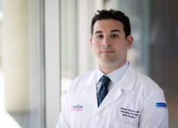 Fourman Mitchell S合作论文数Department of Orthopaedic Surgery, University of Pittsburgh Medical Center4
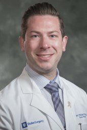 Brett Phillips合作论文数Division of Plastic, Maxillofacial, &amp;Oral Surgery, Duke University Medical Center;Division of Plastic, Maxillofacial, &amp; Oral Surgery, Duke University Medical Center4Crawford Laurie合作论文数4
 Steven Sandoval合作论文数Jet Propulsion Laboratory4Nathan E Estrin合作论文数3Nigel Zhang合作论文数3Thomas Zimmerman合作论文数3Lorne Golub合作论文数Department of Oral Biology and Pathology, School of Dental Medicine, Stony Brook University3
 Georgios E. Romanos合作论文数Department of Periodontolog, School of Dental Medicine, Stony Brook University;Eastman Institute for Oral Health, University of Rochester Medical Center3
 Francis Johnson合作论文数Department of Pharmacological Sciences, Renaissance School of Medicine, Stony Brook University3Wei Hou合作论文数Department of Epidemiology and Health Policy Research, University of Florida3
 Alexander Romanov合作论文数Arrhythmia Department, State Research Institute of Circulation Pathology3
 Mark M Melendez合作论文数3
 Mark Steven Wolff合作论文数2Evyatar Baer合作论文数2Agata Lesniewski合作论文数2Sami U. Khan合作论文数2hsiming j lee合作论文数2Joseph A Esposito合作论文数2Marcia G. Tonnesen合作论文数2N S Ramamurthy合作论文数2
 Juin Zhou合作论文数THz Biophoton Lab, SUNY Stony Brook2
 Mahmoud Ebrahimkhani合作论文数2
 Zachery B. Harris合作论文数Department of Biomedical Engineering, Stony Brook University2
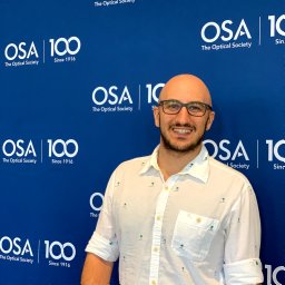 Omar Bassam Osman合作论文数Department of Biomedical Engineering, Stony Brook University2Yu Zhang合作论文数2Henry C. Thode合作论文数Department of Emergency Medicine, Renaissance School of Medicine, Stony Brook University2
 Lim Tae Ho合作论文数Department of Emergency Medicine, Hanyang University College of Medicine2
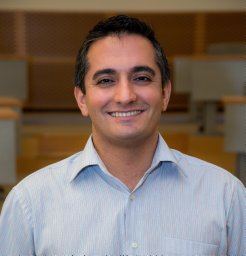 M Hassan Arbab合作论文数Appl. Phys. Lab., Univ. of Washington;c;Appl. Phys. Lab., Univ. of Washington2
 Muna S Elburki合作论文数School of Dental Medicine, Stony Brook University2Abraham Ariel B合作论文数2Laura M Rosenberg合作论文数2Xiaoti Xu合作论文数2
 Christopher K Tuggle合作论文数Department of Animal Science, College of Agriculture and Life Sciences, Iowa State University2Nicole Steinhauff合作论文数2




