In biological tissue, MR transverse relaxation stems from mechanisms spanning multiple scales, from molecular dipole-dipole interactions to mesoscopic field variations driven by tissue microstructure. While mesoscopic relaxation reflects cellular organization, its dynamics in white matter, specifically its dependence on axonal orientation and echo time, remain less investigated. This study employs theoretical frameworks and Monte-Carlo simulations using 3D EM-based white matter (WM) geometries to investigate how compartmentalization and realistic morphology drive these effects. Specifically, we simulate intra-axonal relaxation driven by magnetic fields induced by realistic axonal myelin sheaths and intra-axonal spheres as a model of iron-containing mitochondria. Our results confirm that orientation dependence of mesoscopic relaxation in WM is detectable and agrees with experimental observations. The time-dependence aligns with 1-dimensional short-range structural disorder, but at clinical echo times, this signature may be masked by dominant molecular relaxation. This work moves beyond idealized models to aid the development of more specific biophysical models of mesoscopic relaxation to achieve better biomarkers for neurodegenerative disease.
PURPOSE:To develop a generalized physiologically informed compartmental model for dynamic hyperpolarized [1-13C]pyruvate MRI that explicitly integrates arterial input, capillary transit, and transmembrane extraction to enhance accuracy and interpretability of metabolic rate quantification across diverse conditions. METHODS:We proposed a combined HEmodynamic and MEtabolic eXchange (HEMEX) model for this purpose. The model delineates vascular and tissue spaces, employing an arterial input function and a residue function to characterize capillary transit time distribution drawn from well-established tracer kinetics. A membrane transport term models pyruvate extraction, while intracellular conversion to lactate is described by conventional exchange kinetics. The model was hypothesized to more accurately describe the underlying biology compared to previous ratio-metric or compartmental models employed in the hyperpolarization MRI field. Monte Carlo simulations were used to evaluate Error-Propagation of model fitting, and various metabolic and hemodynamic scenarios were demonstrated in situ. Furthermore, the model was employed on a pig kidney dataset (n = 3) and fitted hemodynamic parameters were compared to that obtained from DCE imaging. RESULTS:Monte Carlo simulations revealed that model fitting was satisfactory at typical SNR-levels observed in vivo. It was also shown that HEMEX accurately models k PL $$ {k}_{PL} $$ across extraction-limited scenarios where ratio-metric methods exhibit biases. In vivo analyses revealed a moderate correlation (R2 = 0.48) between HEMEX-derived mean transit times and those derived from DCE modeling. CONCLUSION:HEMEX decouples delivery and metabolic kinetics, offering robust, physiologically interpretable quantification of HP [1-13C]pyruvate MRI data. Future work will expand the model to additional downstream metabolites and validate across diverse organs and pathologies.
Background and purpose:Diffusion tensor imaging (DTI) has been proposed to guide the anisotropic expansion from gross tumor volume to clinical target volume (CTV), aiming to integrate known tumor spread patterns into the CTV. This study investigate the potential of using a DTI atlas as an alternative to patient-specific DTI for generating anisotropic CTVs. Materials and Methods:The dataset consisted of twenty-eight newly diagnosed glioblastoma patients from a Danish national DTI protocol with post-operative T1-contrast and DTI imaging. Three different DTI atlases, spatially aligned to the patient images using deformable image registration, were considered as alternatives. Anisotropic CTVs were constructed to match the volume of a 15 mm isotropic expansion by generating 3D distance maps using either patient- or atlas-DTI as input to the shortest path solver. The degree of CTV anisotropy was controlled by the migration ratio, modeling tumor cell migration along the dominant white matter fiber direction extracted from DTI. The similarity between patient- and atlas-DTI CTVs was analyzed using the Dice Similarity Coefficient (DSC), with significance testing according to a Wilcoxon test. Results:The median (range) DSC between anisotropic CTVs generated using patient-specific and atlas-based DTI was 0.96 (0.93-0.97), 0.96 (0.93-0.97), and 0.95 (0.93-0.97) for the three atlases, respectively (p > 0.01), for a migration ratio of 10. The results remained consistent over the range of studied migration ratios (2 to 100). Conclusion:The high degree of similarity between all anisotropic CTVs indicates that atlas-DTI is a viable replacement for patient-specific DTI for incorporating fiber direction into the CTV.
Glioblastomas are aggressive brain tumors with dismal prognosis. One of the main bottlenecks for developing more effective therapies for glioblastoma stems from their histologic and molecular heterogeneity, leading to distinct tumor microenvironments and disease phenotypes. Effectively characterizing these features would improve the clinical management of glioblastoma. Glucose flux rates through glycolysis and mitochondrial oxidation have been recently shown to quantitatively depict glioblastoma proliferation in mouse models (GL261 and CT2A tumors) using dynamic glucose-enhanced (DGE) deuterium spectroscopy. However, the spatial features of tumor microenvironment phenotypes remain hitherto unresolved. Here, we develop a DGE Deuterium Metabolic Imaging (DMI) approach for profiling tumor microenvironments through glucose conversion kinetics. Using a multimodal combination of tumor mouse models, novel strategies for spectroscopic imaging and noise attenuation, and histopathological correlations, we show that tumor lactate turnover mirrors phenotype differences between GL261 and CT2A mouse glioblastoma, whereas recycling of the peritumoral glutamate-glutamine pool is a potential marker of invasion capacity in pooled cohorts, linked to secondary brain lesions. These findings were validated by histopathological characterization of each tumor, including cell density and proliferation, peritumoral invasion and distant migration, and immune cell infiltration. Our study bodes well for precision neuro-oncology, highlighting the importance of mapping glucose flux rates to better understand the metabolic heterogeneity of glioblastoma and its links to disease phenotypes.
Magnetic susceptibility MRI offers potential insights into the chemical composition and microstructural organization of tissue. However, estimating magnetic susceptibility in white matter is challenging due to anisotropic subvoxel Larmor frequency shifts caused by axonal microstructure relative to the B0 field orientation. Recent biophysical models have analytically described how axonal microstructure influences the Larmor frequency shifts, relating these shifts to a mesoscopically averaged magnetic field that depends on the axons' fiber orientation distribution function (fODF), typically estimated using diffusion MRI. This study is aimed at validating the use of MRI to estimate mesoscopic magnetic fields and determining whether diffusion MRI can faithfully estimate the orientation dependence of the Larmor frequency shift in realistic axonal microstructure. To achieve this, we developed a framework for performing Monte Carlo simulations of MRI signals in mesoscopically sized white matter axon substrates segmented with electron microscopy. Our simulations demonstrated that with careful experimental design, it is feasible to estimate mesoscopic magnetic fields. Additionally, the fODF estimated by the standard model of diffusion in white matter could predict the orientation dependence of the mesoscopic Larmor frequency shift. We also found that incorporating the intra-axonal axial kurtosis into the standard model could explain a significant amount of signal variance, thereby improving the estimation of the Larmor frequency shift. This factor should not be neglected when fitting the standard model.
Evaluating tissue microstructure and membrane integrity in the living human brain through diffusion-water exchange imaging is challenging due to requirements for a high signal-to-noise ratio and short diffusion times dictated by relatively fast exchange processes. The goal of this work was to demonstrate the feasibility of in vivo imaging of tissue micro-geometries and water exchange within the brain gray matter using the state-of-the-art Connectome 2.0 scanner equipped with an ultra-high-performance gradient system (maximum gradient strength=500 mT/m, maximum slew rate=600 T/m/s). We performed diffusion MRI measurements in 15 healthy volunteers at multiple diffusion times (13-30 ms) and b -values up to 17.5 ms/μm2. The anisotropic Kärger model was applied to estimate the exchange time between intra-neurite and extracellular water in gray matter. The estimated exchange time across the cortical ribbon was around (median±interquartile range) 13±8 ms on Connectome 2.0, substantially faster than that measured using an imaging protocol compatible with Connectome 1.0-alike systems on the same cohort. Our investigation suggested that the NEXI exchange time estimation using a Connectome 1.0 compatible protocol was more prone to residual noise floor biases due to the small time-dependent signal contrasts across diffusion times when the exchange is fast (≤20 ms). Furthermore, spatial variation of exchange time was observed across the cortex, where the motor cortex, somatosensory cortex and visual cortex exhibit longer exchange times compared to other cortical regions. Non-linear fitting for the anisotropic Kärger model was accelerated 100 times using a GPU-based pipeline compared to the conventional CPU-based approach. This study highlighted the importance of the chosen diffusion times and measures to address Rician noise in dMRI data, which can have a substantial impact on the estimated NEXI exchange time and require extra attention when comparing NEXI results between various hardware setups.
IntroductionApproximately one-third of all persons with multiple sclerosis (pwMS) are older, i.e., having an age ≥60 years. Whilst ageing and MS separately elicit deteriorating effects on brain morphology, neuromuscular function, and physical function, the combination of ageing and MS may pose a particular challenge. To counteract such detrimental changes, power training (i.e., a type of resistance exercise focusing on moderate-to-high loading at maximal intended movement velocity) presents itself as a viable and highly effective solution. Power training is known to positively impact physical function, neuromuscular function, as well as brain morphology. Existing evidence is promising but limited to young and middle-aged pwMS, with the effects of power training remaining to be elucidated in older pwMS.MethodsThe presented ‘Power Training in Older MS patients (PoTOMS)’ trial is a national, multi-center, parallel-group, randomized controlled trial. The trial compares 24 weeks of usual care(n = 30) to 24 weeks of usual care and power training (n = 30). The primary outcome is whole brain atrophy rate. The secondary outcomes include changes in brain micro and macro structures, neuromuscular function, physical function, cognitive function, bone health, and patient-reported outcomes.Ethics and disseminationThe presented study is approved by The Regional Ethics Committee (reference number 1-10-72-222-20) and registered at the Danish Data Protection Agency (reference number 2016-051-000001). All study findings will be published in scientific peer-reviewed journals and presented at relevant scientific conferences independent of the results. The www.clinicaltrials.gov identifier is NCT04762342.
This study aims to validate if MRI can measure anisotropic mesoscopic Larmor frequency shifts from white matter axonal microstructure relative to the B0 direction and if dMRI can estimate this anisotropy. Recent models describe how mesoscopic Larmor frequency shifts depend on induced magnetic fields by axons, described by an orientation distribution function. Using Monte-Carlo simulations of MRI signals in mesoscopic white matter axon substrates, we show MRI can estimate this mesoscopic frequency shift and dMRI can estimate its orientation dependence.
PurposeThe purpose of this study is to develop a method for selecting uniform wave vectors for double diffusion encoding (DDE) to improve the accuracy and reliability of diffusion measurements.MethodsThe method relies on identifying orthogonal wave vectors with rotations, and representing these rotations as points on a three-dimensional sphere in four dimensions using quaternions. This enables an electrostatic repulsion algorithm to achieve a uniform distribution of these points. The optimal points are then converted back into orthogonal wave vectors (or rotations).ResultsThe method was validated by comparing the distribution of directions to those generated by uniform sampling and by evaluating the error in the powder-averaged signal for various models. Our results demonstrate that the electrostatic repulsion approach effectively achieves a uniform distribution of wave vectors.ConclusionThe proposed method provides a systematic way to generate uniform diffusion directions suitable, for example, for DDE, enhancing the precision of diffusion measurements and reducing potential bias in experimental results. The method is also capable of generating uniform sets of B-tensors, and is thus applicable for general free waveform encoding.
Magnetic susceptibility imaging may provide valuable information about chemical composition and microstructural organization of tissue. However, its estimation from the MRI signal phase is particularly difficult as it is sensitive to magnetic tissue properties ranging from the molecular to macroscopic scale. The MRI Larmor frequency shift measured in white matter (WM) tissue depends on the myelinated axons and other magnetizable sources such as iron-filled ferritin. We have previously derived the Larmor frequency shift arising from a dense media of cylinders with scalar susceptibility and arbitrary orientation dispersion. Here we extend our model to include microscopic WM susceptibility anisotropy as well as spherical inclusions with scalar susceptibility to represent subcellular structures, biologically stored iron etc. We validate our analytical results with computer simulations and investigate the feasibility of estimating susceptibility using simple iterative linear least squares without regularization or preconditioning. This is done in a digital brain phantom synthesized from diffusion MRI (dMRI) measurements of an ex vivo mouse brain at ultra-high field.
MP-PCA denoising has become the method of choice for denoising MRI data since it provides an objective threshold to separate the signal components from unwanted thermal noise components. In rodents, thermal noise in the coils is an important source of noise that can reduce the accuracy of activation mapping in fMRI. Further confounding this problem, vendor data often contains zero-filling and other post-processing steps that may violate MP-PCA assumptions. Here, we develop an approach to denoise vendor data and assess activation “spreading” caused by MP-PCA denoising in rodent task-based fMRI data. Data was obtained from N = 3 mice using conventional multislice and ultrafast fMRI acquisitions (1 s and 50 ms temporal resolution, respectively), using a visual stimulation paradigm. MP-PCA denoising produced SNR gains of 64% and 39%, and Fourier Spectral Amplitude (FSA) increases in BOLD maps of 9% and 7% for multislice and ultrafast data, respectively, when using a small [2 2] denoising window. Larger windows provided higher SNR and FSA gains with increased spatial extent of activation that may or may not represent real activation. Simulations showed that MP-PCA denoising can incur activation “spreading” with increased false positive rate and smoother functional maps due to local “bleeding” of principal components, and that the optimal denoising window for improved specificity of functional mapping, based on Dice score calculations, depends on the data's tSNR and functional CNR. This “spreading” effect applies also to another recently proposed low-rank denoising method (NORDIC), although to a lesser degree. Our results bode well for enhancing spatial and/or temporal resolution in future fMRI work, while taking into account the sensitivity/specificity trade-offs of low-rank denoising methods.
The potential effect of compartmental water exchange and somas on the diffusion MRI signal in grey matter tissue are currently open questions with considerable implications for biophysical modelling of grey matter at (pre-)clinical diffusion time scales. Recent studies have begun to tackle these questions and found evidence that compartmental water exchange dominates the diffusion time dependence. Here, we extend the application of the Standard Model with exchange between the neurites and extracellular water (SMEX) to in vivo rat brain and again find evidence for exchange-dominated time dependence with very short neurite residence time on the order of 2 ms.
Marčenko-Pastur PCA (MPPCA) denoising is emerging as an effective means for noise suppression in MR imaging (MRI) acquisitions with redundant dimensions. However, MPPCA performance can be severely compromised by spatially correlated noise-an issue typically affecting most modern MRI acquisitions-almost to the point of returning the original images with little or no noise removal. In this study, we explore different threshold criteria for principal component analysis (PCA) component classification that enable efficient and robust denoising of MRI data even when noise exhibits high spatial correlations, especially in cases where data are acquired with Partial Fourier and when only magnitude data are available. We show that efficient denoising can be achieved by incorporating a-priori information about the noise variance into PCA denoising thresholding. Based on this, two denoising strategies developed here are: 1) General PCA (GPCA) denoising that uses a-priori noise variance estimates without assuming specific noise distributions; and 2) Threshold PCA (TPCA) denoising which removes noise components with a threshold computed from a-priori estimated noise variance to determine the upper bound of the Marčenko-Pastur (MP) distribution. These strategies were tested in simulations with known ground truth and applied for denoising diffusion MRI data acquired using pre-clinical (16.4T) and clinical (3T) MRI scanners. In synthetic phantoms, MPPCA denoising failed to denoise spatially correlated data, while GPCA and TPCA better classified components as dominated by signal/noise. In cases where the noise variance was not accurately estimated (as can be the case in many practical scenarios), TPCA still provides excellent denoising performance. Our experiments in pre-clinical diffusion data with highly corrupted by spatial correlated noise revealed that both GPCA and TPCA robustly denoised the data while MPPCA denoising failed. In in vivo diffusion MRI data acquired on a clinical scanner in healthy subjects, MPPCA weakly removed noised, while TPCA was found to have the best performance, likely due to misestimations of the noise variance. Thus, our work shows that these novel denoising approaches can strongly benefit future pre-clinical and clinical MRI applications.
PURPOSE:X-nuclei (also called non-proton MRI) MRI and spectroscopy are limited by the intrinsic low SNR as compared to conventional proton imaging. Clinical translation of x-nuclei examination warrants the need of a robust and versatile tool improving image quality for diagnostic use. In this work, we compare a novel denoising method with fewer inputs to the current state-of-the-art denoising method. METHODS:Denoising approaches were compared on human acquisitions of sodium (23 Na) brain, deuterium (2 H) brain, carbon (13 C) heart and brain, and simulated dynamic hyperpolarized 13 C brain scans, with and without additional noise. The current state-of-the-art denoising method Global-local higher order singular value decomposition (GL-HOSVD) was compared to the few-input method tensor Marchenko-Pastur principal component analysis (tMPPCA). Noise-removal was quantified by residual distributions, and statistical analyses evaluated the differences in mean-square-error and Bland-Altman analysis to quantify agreement between original and denoised results of noise-added data. RESULTS:GL-HOSVD and tMPPCA showed similar performance for the variety of x-nuclei data analyzed in this work, with tMPPCA removing ˜5% more noise on average over GL-HOSVD. The mean ratio between noise-added and denoising reproducibility coefficients of the Bland-Altman analysis when compared to the original are also similar for the two methods with 3.09 ± 1.03 and 2.83 ± 0.79 for GL-HOSVD and tMPPCA, respectively. CONCLUSION:The strength of tMPPCA lies in the few-input approach, which generalizes well to different data sources. This makes the use of tMPPCA denoising a robust and versatile tool in x-nuclei imaging improvements and the preferred denoising method.
PURPOSE:Estimating magnetic susceptibility using MRI depends on inverting a forward relationship between the susceptibility and measured Larmor frequency. However, an often-overlooked constraint in susceptibility fitting is that the Larmor frequency is only measured inside the sample, and after successful background field removal, susceptibility sources should only reside inside the same sample. Here, we test the impact of accounting for these constraints in susceptibility fitting.THEORY AND METHODS:Two different digital brain phantoms with scalar susceptibility were examined. We used the MEDI phantom, a simple phantom with no background fields, to examine the effect of the imposed constraints for various levels of SNR. Next, we considered the QSM reconstruction challenge 2.0 phantom with and without background fields. We estimated the parameter accuracy of openly-available QSM algorithms by comparing fitting results to the ground truth. Next, we implemented the mentioned constraints and compared to the standard approach.RESULTS:Including the spatial distribution of frequencies and susceptibility sources decreased the RMS-error compared to standard QSM on both brain phantoms when background fields were absent. When background field removal was unsuccessful, as is presumably the case in most in vivo conditions, it is better to allow sources outside the brain.CONCLUSION:Informing QSM algorithms about the location of susceptibility sources and where Larmor frequency was measured improves susceptibility fitting for realistic SNR levels and efficient background field removal. However, the latter remains the bottleneck of the algorithm. Allowing for external sources regularizes unsuccessful background field removal and is currently the best strategy in vivo.
Purpose To develop a denoising strategy leveraging redundancy in high-dimensional data. Theory and Methods The SNR fundamentally limits the information accessible by MRI. This limitation has been addressed by a host of denoising techniques, recently including the so-called MPPCA: principal component analysis of the signal followed by automated rank estimation, exploiting the Marchenko-Pastur distribution of noise singular values. Operating on matrices comprised of data patches, this popular approach objectively identifies noise components and, ideally, allows noise to be removed without introducing artifacts such as image blurring, or nonlocal averaging. The MPPCA rank estimation, however, relies on a large number of noise singular values relative to the number of signal components to avoid such ill effects. This condition is unlikely to be met when data patches and therefore matrices are small, for example due to spatially varying noise. Here, we introduce tensor MPPCA (tMPPCA) for the purpose of denoising multidimensional data, such as from multicontrast acquisitions. Rather than combining dimensions in matrices, tMPPCA uses each dimension of the multidimensional data's inherent tensor-structure to better characterize noise, and to recursively estimate signal components. Results Relative to matrix-based MPPCA, tMPPCA requires no additional assumptions, and comparing the two in a numerical phantom and a multi-TE diffusion MRI data set, tMPPCA dramatically improves denoising performance. This is particularly true for small data patches, suggesting that tMPPCA can be especially beneficial in such cases. Conclusions The MPPCA denoising technique can be extended to high-dimensional data with improved performance for smaller patch sizes.
Accurate estimation of microscopic magnetic field variations induced in biological tissue can be valuable for mapping tissue composition in health and disease. Here, we present an extension to Quantitative susceptibility mapping (QSM) to account for local white matter (WM) magnetic microstructure by using our previously presented model for solid cylinders with arbitrary orientations to describe axons in terms of concentric cylinders. Through computer simulations, we find that our model improves susceptibility estimation compared to QSM, and Susceptibility Tensor Imaging (STI) are substantially biased by the unaccounted-for structural anisotropy due to the mesoscopic frequency contribution, indicating the observed STI tensor might not represent susceptibility anisotropy as expected. Experiments in mouse brains acquired at ultrahigh field shows the mesoscopic contribution due to WM microstructure to be substantial. This in turn changed estimated susceptibility values up to 25 root-mean-squared-difference in WM compared to standard QSM. Our work underscores how microstructural field effects impact susceptibility estimates, and should not be neglected when imaging anisotropic tissue such as brain WM.
The signal to noise ratio (SNR) fundamentally limits the information accessible by magnetic resonance imaging (MRI). This limitation has been addressed by a host of denoising techniques, recently including so-called MPPCA: Principal component analysis (PCA) of the signal followed by automated rank estimation, exploiting the Marchenko-Pastur (MP) distribution of noise singular values. Operating on matrices comprised by data-patches, this popular approach objectively identifies noise components and, ideally, allows noise to be removed without introducing artifacts such as image blurring or non-local averaging. The MPPCA rank estimation, however, relies on a large number of noise singular values relative to the number of signal components to avoid such ill effects. This condition is unlikely to be met when data-patches and therefore matrices are small, for example due to spatially varying noise. Here, we introduce tensor MPPCA (tMPPCA) for the purpose of denoising multidimensional data, for example from multi-contrast acquisitions. Rather than combining dimensions in matrices, tMPPCA utilizes each dimension of the multidimensional data's inherent tensor-structure to better characterize noise, and to recursively estimate signal components. Relative to matrix-based MPPCA, tMPPCA requires no additional assumptions, and comparing the two in a numerical phantom and a multi-TE diffusion MRI dataset, tMPPCA dramatically improves denoising performance. This is particularly true for small data-patches, which we believe will improve denoising in cases of spatially varying noise.
The lentil isolectins, CMLcH A and CMLcH B, and pea isolectins, CMPSA A and CMPSA B, are compared in terms of their spectroscopic and saccharide binding properties. The paramagnetic contribution to the solvent proton magnetic relaxation dispersion profiles of solutions of the isolectins of each protein are found to be essentially identical. Electron paramagnetic resonance spectra suggest a high degree of octahedral symmetry at the Mn2+ site for both pairs of isolectins. The near-ultraviolet absorption spectra of CMLcH A and CMLcH B are identical, as are the spectra of CMPSA A and CMPSA B. Carbohydrate binding activities of the isolectins of each protein are compared using hemagglutination, precipitation, and precipitation-inhibition assays, and are found to be identical, although the activities of CMLcH and CMPSA differ somewhat. These results demonstrate that the spectroscopic and saccharide binding properties of the isolectins of CMLcH are essentially identical, as are those of the isolectins of CMPSA, and suggest that native mixtures of the isolectins may be treated as single proteins in further studies.
Characterizing neural tissue microstructure is a critical goal for future neuroimaging. Diffusion MRI (dMRI) provides contrasts that reflect diffusing spins’ interactions with myriad microstructural features of biological systems. However, the specificity of dMRI remains limited due to the ambiguity of its signals vis-à-vis the underlying microstructure. To improve specificity, biophysical models of white matter (WM) typically express dMRI signals according to the Standard Model (SM) and have more recently in gray matter (GM) taken spherical compartments into account (the SANDI model) in attempts to represent cell soma. The validity of the assumptions underlying these models, however, remains largely undetermined, especially in GM. To validate these assumptions experimentally, observing their unique, functional properties, such as the b−1/2 power-law associated with one-dimensional diffusion, has emerged as a fruitful strategy. The absence of this signature in GM, in turn, has been explained by neurite water exchange, non-linear morphology, and/or by obscuring soma signal contributions. Here, we present diffusion simulations in realistic neurons demonstrating that curvature and branching does not destroy the stick power-law behavior in impermeable neurites, but also that their signal is drowned by the soma signal under typical experimental conditions. Nevertheless, by studying the GM dMRI signal's behavior as a function of diffusion weighting as well as time, we identify an attainable experimental regime in which the neurite signal dominates. Furthermore, we find that exchange-driven time dependence produces a signal behavior opposite to that which would be expected from restricted diffusion, thereby providing a functional signature that disambiguates the two effects. We present data from dMRI experiments in ex vivo rat brain at ultrahigh field of 16.4T and observe a time dependence that is consistent with substantial exchange but also with a GM stick power-law. The first finding suggests significant water exchange between neurites and the extracellular space while the second suggests a small sub-population of impermeable neurites. To quantify these observations, we harness the Kärger exchange model and incorporate the corresponding signal time dependence in the SM and SANDI models.
 Leif Østergaard合作论文数46Brian Hansen合作论文数31
 Noam Shemesh合作论文数Champalimaud Centre for the Unknown, Champalimaud Foundation28Kim Mouridsen合作论文数22
 Simon Fristed Eskildsen合作论文数Center of Functionally Integrative Neuroscience, Department of Clinical Medicine, Aarhus University16
 Jonas Lynge Olesen合作论文数Dept Clin Med, Aarhus Univ13
 Khan Ahmad Raza合作论文数NMR Research Centre, Institute of Nuclear Medicine and Allied Sciences13Rafael Neto Henriques合作论文数Cognition and Brain Sciences Unit, Medical Research Council11Nagenthiraja Kartheeban合作论文数11
 Christopher D. Kroenke合作论文数Oregon National Primate Research Center, Oregon Health & Science University;Departments of Biomedical Engineering, Oregon Health & Science University11
 Mikkel B. Hansen合作论文数Center for Oxygen Microscopy and Imaging and Lundbeck Foundation Center for Theoretical Chemistry, University of Arhus10Valerij G. Kiselev合作论文数10
 Torben E Lund合作论文数Center of Functionally Integrative Neuroscience, Aarhus University9
 Andrey Chuhutin合作论文数Ctr Funct Integrat Neurosci, Aarhus Univ Hosp8
 Andrada Ianus合作论文数School of Biomedical Engineering and Imaging Sciences, Faculty of Life Sciences & Medicine, King's College London8Carsten Gyldensted合作论文数8Nina K. Iversen合作论文数8Louise Gyldensted合作论文数8
 Anders Rodell合作论文数Center for Functionally Integrative Neuroscience, Aarhus University Hospital8
 Anders Dyhr Sandgaard合作论文数Dept Clin Med, Aarhus Univ7
 Jesper Kallehauge合作论文数Department of Clinical Medicine, Aarhus University;Danish Centre for Particle Therapy, Aarhus University Hospital7Jakob Udby Blicher合作论文数7Angleys Hugo合作论文数7Hans Braendgaard合作论文数Department of Neurology, Aarhus University Hospital7Ryan Sangill合作论文数7
 Ove Wiborg合作论文数7Tietze Anna合作论文数6
 Irene Klærke Mikkelsen合作论文数Department of Clinical Medicine, Aarhus University;Center of Functionally Integrative Neuroscience, Aarhus University6Jesper Frandsen合作论文数6Jens Randel Nyengaard合作论文数6
 Kim Ryun Drasbek合作论文数6
 Lindegaard Jacob Christian合作论文数Department of Oncology, Aarhus University Hospital5
 Haack Søren合作论文数Department of Clinical Engineering, Aarhus University Hospital5Næss-Schmidt Erhard合作论文数Hammel Neurorehabilitation and Research Centre, Aarhus University Hospital5
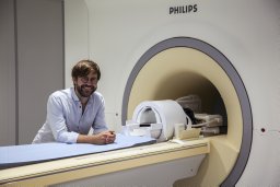 Henrik Lundell合作论文数5
 Erik Morre Pedersen合作论文数Department of Cardiology and Department Cardiothoracic and Vascular Surgery, Aarhus University Hospital5B Hansen合作论文数4
 Dmitry Novikov合作论文数Department of Radiology, Center for Biomedical Imaging, New York University School of Medicine4
 Tim Dyrby合作论文数Department of Applied Mathematics and Computer Science, Technical University of Denmark;Danish Research Centre for Magnetic Resonance, Copenhagen University Hospital4
 Jørgen Feldbæk Nielsen合作论文数4Leigland Lindsey A合作论文数4
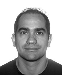 Gutiérrez-Jiménez Eugenio合作论文数4Alexander Blumen合作论文数Physikalisches Institut, Albert-Ludwigs-Universität Freiburg4
 Rikke B Dalby合作论文数4Mille Møller Thastum合作论文数3
 Santiago Coelho合作论文数Univ Leeds, Ctr Computat Imaging & Simulat Technol Biomed CIS, Sch Comp, Leeds, W Yorkshire, England3K. Tanderup合作论文数3Svendsen Susanne Wulff合作论文数3
 Andreas Schroder合作论文数3




