Breakdown of every transmembrane protein trafficked to lysosomes requires proteolysis of their hydrophobic helical transmembrane domains. Combining lysosomal proteomics with functional genomic datasets, we identified lysosomal leucine aminopeptidase (LyLAP; formerly phospholipase B domain-containing 1) as the hydrolase most tightly associated with elevated endocytosis. Untargeted metabolomics and biochemical reconstitution demonstrated that LyLAP is a processive monoaminopeptidase with preference for amino-terminal leucine. This activity was necessary and sufficient for the breakdown of hydrophobic transmembrane domains. LyLAP was up-regulated in pancreatic ductal adenocarcinoma (PDA), which relies on macropinocytosis for nutrient uptake. In PDA cells, LyLAP ablation led to the buildup of undigested hydrophobic peptides, lysosomal membrane damage, and growth inhibition. Thus, LyLAP enables lysosomal degradation of membrane proteins and protects lysosomal integrity in highly endocytic cancer cells.
Proteolysis of hydrophobic helices is required for complete breakdown of every transmembrane protein trafficked to the lysosome and sustains high rates of endocytosis. However, the lysosomal mechanisms for degrading hydrophobic domains remain unknown. Combining lysosomal proteomics with functional genomic data mining, we identify Lysosomal Leucine Aminopeptidase (LyLAP; formerly Phospholipase B Domain-Containing 1) as the hydrolase most tightly associated with elevated endocytic activity. Untargeted metabolomics and biochemical reconstitution demonstrate that LyLAP is not a phospholipase, but a processive monoaminopeptidase with strong preference for N-terminal leucine - an activity necessary and sufficient for breakdown of hydrophobic transmembrane domains. LyLAP is upregulated in pancreatic ductal adenocarcinoma (PDA), which relies on macropinocytosis for nutrient uptake, and its ablation led to buildup of undigested hydrophobic peptides, which compromised lysosomal membrane integrity and inhibited PDA cell growth. Thus, LyLAP enables lysosomal degradation of membrane proteins, and may represent a vulnerability in highly endocytic cancer cells. One sentence summary:LyLAP degrades transmembrane proteins to sustain high endocytosis and lysosomal membrane stability in pancreatic cancer.
Delivery of therapeutic agents in pancreatic cancer (PC) is impaired due to its hypovascular and desmoplastic tumor microenvironment. The Endothelin (ET)-axis is the major regulator of vasomotor tone under physiological conditions and is highly upregulated in multiple cancers. We investigated the effect of dual endothelin receptor antagonist bosentan on perfusion and macromolecular transport in a PC cell-fibroblast co-implantation tumor model using Dynamic Contrast-Enhanced Magnetic Resonance Imaging (DCE-MRI). Following bosentan treatment, the contrast enhancement ratio and wash-in rates in tumors were two- and nine times higher, respectively, compared to the controls, whereas the time to peak was significantly shorter (7.29 ± 1.29 min v/s 22.08 ± 5.88 min; p = 0.04). Importantly, these effects were tumor selective as the magnitudes of change for these parameters were much lower in muscles. Bosentan treatment also reduced desmoplasia and improved intratumoral distribution of high molecular weight FITC-dextran. Overall, these findings support that targeting the ET-axis can serve as a potential strategy to selectively enhance tumor perfusion and improve the delivery of therapeutic agents in pancreatic tumors.
Background The small molecule CCX559 is a novel, highly potent inhibitor of human PD-L1 (hPD-L1) in development as an oral treatment for cancer patients.1,2In vitro studies showed that CCX559 inhibits PD-L1 binding to PD-1 and induces PD-L1 internalization from the cell surface. To investigate the mechanism of action in vivo, we examined the effect of CCX559 distribution and clearance on tumor cell PD-L1 dynamics and anti-tumor activity. Methods CCX559 was administered orally at the clinically relevant dose of 30 mg/kg once daily for 7 days to human PD-L1 knock-in mice bearing MC38 tumors (average volume 60-100 mm3). The MC38 cells were engineered to express hPD-L1, as CCX559 does not cross-react with mouse PD-L1. CCX559 levels in tumors and organs, including lung, liver, kidney, spleen, heart, and brain, were quantitated by mass-spectrum analysis at 1, 5 and 12 days after the last dose. Cell surface and intracellular PD-L1 were detected using flow cytometry and immunohistochemistry. Results Dosing CCX559 for 7 days significantly reduced hPD-L1-MC38 tumor growth compared to vehicle control, and the reduction in tumor volume persisted post dosing until the end of study. The average CCX559 level on day 1 post dose was significantly higher in tumors than plasma and other organs (27.9 µg/g tissue vs. 0.007 – 1.4 µg/g). In tumors CCX559 induced hPD-L1 internalization into intracellular vesicles and reduced the cell surface level by over 90% compared to vehicle control. CCX559 levels dropped 98% in plasma, tumor and tissues by day 5 post dose, but the level in the tumors was still above the IC90 for inhibiting PD-L1; consistent with this, intracellular PD-L1 was also observed. The drug was completely cleared by day 12 post dose, but tumor cell surface hPD-L1 in the CCX559 arm was only partially recovered compared to the vehicle control. Conclusions In a preclinical model, higher levels of CCX559 were observed in tumors than in plasma and other organs. Cessation of dosing led to clearance of CCX559 within days, but recovery of tumor cell surface PD-L1 levels was delayed, perhaps as a result of higher CCX559 levels in the tumor. The in vivo properties of CCX559 suggest that PD-L1 inhibition in tumors may be achieved at relatively low drug levels in the periphery, thus mitigating potential risk of adverse events. No DLTs, treatment-related SAEs or severe (Grade 3 or higher) AEs have been reported in an ongoing first-in-human dose escalation trial with CCX559 (ACTRN12621001342808). References Chris Li, et al. CCX559 is a potent orally-administered small molecule PD-L1 inhibitor that induces anti-tumor immunity. Cancer Research 2021;81(13_Suppl):Abstract nr 1274. Gonzalo Tapia et al. Preliminary data from an ongoing phase 1 dose-escalation study of CCX559, an orally administered small molecule PD-L1 inhibitor, in patients with advanced solid tumors. Journal of Clinical Oncology 2022;40(16_Suppl): 2593.
Lysosomes must maintain the integrity of their limiting membrane to ensure efficient fusion with incoming organelles and degradation of substrates within their lumen. Pancreatic cancer cells upregulate lysosomal biogenesis to enhance nutrient recycling and stress resistance, but it is unknown whether dedicated programmes for maintaining the integrity of the lysosome membrane facilitate pancreatic cancer growth. Using proteomic-based organelle profiling, we identify the Ferlin family plasma membrane repair factor Myoferlin as selectively and highly enriched on the membrane of pancreatic cancer lysosomes. Mechanistically, lysosomal localization of Myoferlin is necessary and sufficient for the maintenance of lysosome health and provides an early acting protective system against membrane damage that is independent of the endosomal sorting complex required for transport (ESCRT)-mediated repair network. Myoferlin is upregulated in human pancreatic cancer, predicts poor survival and its ablation severely impairs lysosome function and tumour growth in vivo. Thus, retargeting of plasma membrane repair factors enhances the pro-oncogenic activities of the lysosome. Gupta et al. show that the membrane repair factor Myoferlin protects against membrane damage of pancreatic cancer lysosomes to sustain enhanced lysosomal function and promote tumour growth.
Oncogenes can alter metabolism by changing the balance between anabolic and catabolic processes. However, how oncogenes regulate tumor cell biomass remains poorly understood. Using isogenic MCF10A cells transformed with nine different oncogenes, we show that specific oncogenes reduce the biomass of cancer cells by promoting extracellular vesicle (EV) release. While MYC and AURKB elicited the highest number of EVs, each oncogene selectively altered the protein composition of released EVs. Likewise, oncogenes alter secreted miRNAs. MYC-overexpressing cells require ceramide, whereas AURKB requires ESCRT to release high levels of EVs. We identify an inverse relationship between MYC upregulation and activation of the RAS/MEK/ERK signaling pathway for regulating EV release in some tumor cells. Finally, lysosome genes and activity are downregulated in the context of MYC and AURKB, suggesting that cellular contents, instead of being degraded, were released via EVs. Thus, oncogene-mediated biomass regulation via differential EV release is a new metabolic phenotype.
Endothelin-1 (ET-1) and its two receptors, endothelin receptor A (ETAR) and endothelin receptor B (ETBR) exhibit deregulated overexprerssion in pancreatic ductal adenocarcinoma (PDAC) and pancreatitis. We examined the expression pattern of endothelin (ET) axis components in the murine models of chronic and acute inflammation in the presence or absence of oncogenic K-ras. While the expression of endothelin converting enzyme-1 (ECE-1), ET-1, ETAR and ETBR in the normal pancreas is restricted predominantly to the islet cells, progressive increase of ET receptors in ductal cells and stromal compartment is observed in the KC model (Pdx-1 Cre; K-rasG12D) of PDAC. In the murine pancreas harboring K-rasG12D mutation (KC mice), following acute inflammation induced by cerulein, increased ETAR and ETBR expression is observed in the amylase and CK19 double positive cells that represent cells undergoing pancreatic acinar to ductal metaplasia (ADM). As compared to the wild type (WT) mice, cerulein treatment in KC mice resulted in significantly higher levels of ECE-1, ET-1, ETAR and ETBR, transcripts in the pancreas. Similarly, in response to cigarette smoke-induced chronic inflammation, the expression of ET axis components is significantly upregulated in the pancreas of KC mice as compared to the WT mice. In addition to the expression in the precursor pancreatic intraepithelial neoplasm (PanIN lesions) in cigarette smoke-exposure model and metaplastic ducts in cerulein-treatment model, ETAR and ETBR expression is also observed in infiltrating F4/80 positive macrophages and α-SMA positive fibroblasts and high co-localization was seen in the presence of oncogenic K-ras. In conclusion, both chronic and acute pancreatic inflammation in the presence of oncogenic K-ras contribute to sustained upregulation of ET axis components in the ductal and stromal cells suggesting a potential role of ET axis in the initiation and progression of PDAC.
Immune evasion is a major obstacle for cancer treatment. Common mechanisms of evasion include impaired antigen presentation caused by mutations or loss of heterozygosity of the major histocompatibility complex class I (MHC-I), which has been implicated in resistance to immune checkpoint blockade (ICB) therapy1–3. However, in pancreatic ductal adenocarcinoma (PDAC), which is resistant to most therapies including ICB4, mutations that cause loss of MHC-I are rarely found5 despite the frequent downregulation of MHC-I expression6–8. Here we show that, in PDAC, MHC-I molecules are selectively targeted for lysosomal degradation by an autophagy-dependent mechanism that involves the autophagy cargo receptor NBR1. PDAC cells display reduced expression of MHC-I at the cell surface and instead demonstrate predominant localization within autophagosomes and lysosomes. Notably, inhibition of autophagy restores surface levels of MHC-I and leads to improved antigen presentation, enhanced anti-tumour T cell responses and reduced tumour growth in syngeneic host mice. Accordingly, the anti-tumour effects of autophagy inhibition are reversed by depleting CD8+ T cells or reducing surface expression of MHC-I. Inhibition of autophagy, either genetically or pharmacologically with chloroquine, synergizes with dual ICB therapy (anti-PD1 and anti-CTLA4 antibodies), and leads to an enhanced anti-tumour immune response. Our findings demonstrate a role for enhanced autophagy or lysosome function in immune evasion by selective targeting of MHC-I molecules for degradation, and provide a rationale for the combination of autophagy inhibition and dual ICB therapy as a therapeutic strategy against PDAC. Inhibition of the autophagy–lysosome system upregulates surface expression of MHC class I proteins and enhances antigen presentation, and evokes a potent anti-tumour immune response that is mediated by CD8+ T cells.
Pseudoprogression and radiation necrosis prevent confident interpretation of magnetic resonance imaging (MRI) results and lead to challenges in the management of glioblastoma (GBM). We examined the utility of matrix metalloproteinase-2 (MMP-2) and neutrophil gelatinase-associated lipocalin (NGAL) in the serum and urine as biomarkers of tumor burden in patients with GBM, with the goal of improving the interpretation of MRI and predicting overall survival (OS) and progression free survival (PFS). Expression of MMP-2 and NGAL was analyzed by immunohistochemistry in GBM and non-tumor epileptic control tissues. Serum and urine samples were collected pre and postoperatively and at each MRI. MMP-2 and NGAL levels in the serum and urine were measured by enzyme-linked immunosorbent assay. MRI results, tumor volumes, survival, symptoms, and quality of life were assessed using repeated measures and autoregressive models correlation structure. Although the staining intensity of NGAL was indistinguishable, GBM tissues exhibited significantly higher number of NGAL positive cells as compared to control epileptic brain tissue. Serum MMP-2 was significantly higher in GBM patients than control subjects (p=0.0112) and was highest in patients who underwent biopsy compared to maximal resection (p=0.0038). Elevated levels of both NGAL and MMP-2 in the preoperative samples indicated a trend towards shorter PFS and OS, while preoperative urine NGAL levels were marginally predictive of PFS. Trajectory of symptoms and quality of life results were too variable to adequately correlate with the biomarker levels. Although biomarkers did not aid in differentiating between pseudoprogression, radiation necrosis, and tumor growth, preoperative levels correlated with survival.
Abstract Background: Prostate cancer (PCa) cells preferentially metastasize to bone and skeleton resulting in the poor 5-year survival of less than 1%. Growth differentiation factor-15 (GDF15) is overexpressed in several cancers, including PCa. However, the role of GDF15 in prostate cancer bone metastasis remains elusive. The interaction between cancer cells and the bone microenvironment forms a vicious cycle that increases bone deformities and drives tumor growth in the bone. Therefore, we question whether GDF15 secreted by the PCa cells alters the bone environment by uncoupling bone formation and resorption. Methods: Murine calvarial-derived osteoblast (MCO) cells were used for the functional effect of GDF15 on differentiation, proliferation, and mineralization. Mineralization of mouse bone marrow stromal cell (BMSCs) derived osteoblasts was determined by Alizarin Red S staining. Bone marrow macrophages (BMMs) were isolated from C57 mice and cultured along with M-CSF and RANKL to identify osteoclasts. Results: Conditioned media (CM) from PCa (LNCaP and C4-2B) cells and rhGDF15 increased osteoblast proliferation and differentiation in MCO cells. However, GDF15 deletion in PCa (LNCaP and C4-2B) cells prevented PCa-mediated osteoblast differentiation. Further, CM from PCa and rhGDF15 increased the alkaline phosphatase+ CFU-F (Colony Forming Unit- Fibroblast) in mouse bone marrow stromal cells culture. Supportively, stable knockout of GDF15 decreased mineralized nodule formation in MCO. qRT-PCR assay revealed that the PCa and rhGDF15 increased mRNA expression of osteoblast-related genes such as alkaline phosphatase, Runx-2, collagen type-1 and osteocalcin. PCa cells derived CM and rhGDF15 increase the osteoblast-derived osteoclastic signal (RANKL/OPG ratio), suggesting that GDF15 promotes RANKL-dependent osteoclastogenesis. Furthermore, rhGDF15 treatment increased TRAP-positive multinucleated cells, pit resorption and osteoclast-specific genes such as TRAP, Cathepsin K, Carbonic anhydrase and NFATc1 in BMMs. Conclusions: Altogether, our results suggest that GDF15 is crucial for PCa-mediated osteoblast differentiation and mineralization. GDF15 also increased bone marrow osteoclast formation and its function. Our finding suggests that GDF15 mediated increase in osteoblastic-RANKL further enhance osteoclast activity and favor PCa cell to metastasize to bone. Citation Format: Jawed A. Siddiqui, Sakthivel Muniyan, Parthasarathy Seshacharyulu, Satyanarayana Rachagani, Suprit Gupta, Mohd W. Nasser, Kaustubh Datta, Surinder K. Batra. Role of growth differentiation factor-15 in prostate cancer-bone environment: Understanding on bone metastasis [abstract]. In: Proceedings of the American Association for Cancer Research Annual Meeting 2018; 2018 Apr 14-18; Chicago, IL. Philadelphia (PA): AACR; Cancer Res 2018;78(13 Suppl):Abstract nr 1091.
The dismal prognosis of locally advanced and metastatic squamous cell carcinoma of the head and neck (HNSCC) is primarily due to the development of resistance to chemoradiation therapy (CRT). Deregulation of Epidermal Growth Factor Receptor (EGFR) signaling is involved in HNSCC pathogenesis by regulating cell survival, cancer stem cells (CSCs), and resistance to CRT. Here we investigated the radiosensitizing activity of the pan-EGFR inhibitor afatinib in HNSCC in vitro and in vivo. Our results showed strong antiproliferative effects of afatinib in HNSCC SCC1 and SCC10B cells, compared to immortalized normal oral epithelial cells MOE1a and MOE1b. Comparative analysis revealed stronger antitumor effects with afatinib than observed with erlotinib. Furthermore, afatinib enhanced in vitro radiosensitivity of SCC1 and SCC10B cells by inducing mesenchymal to epithelial transition, G1 cell cycle arrest, and the attenuating ionizing radiation (IR)-induced activation of DNA double strand break repair (DSB) ATM/ATR/CHK2/BRCA1 pathway. Our studies also revealed the effect of afatinib on tumor sphere- and colony-forming capabilities of cancer stem cells (CSCs), and decreased IR-induced CSC population in SCC1 and SCC10B cells. Furthermore, we observed that a combination of afatinib with IR significantly reduced SCC1 xenograft tumors (median weight of 168.25 ± 20.85 mg; p = 0.05) compared to afatinib (280.07 ± 20.54 mg) or IR alone (324.91 ± 28.08 mg). Immunohistochemical analysis of SCC1 tumor xenografts demonstrated downregulation of the expression of IR-induced pEGFR1, ALDH1 and upregulation of phosphorylated γH2AX by afatinib. Overall, afatinib reduces tumorigenicity and radiosensitizes HNSCC cells. It holds promise for future clinical development as a novel radiosensitizer by improving CSC eradication.
Background: Glioblastoma (GBM) is the most aggressive primary brain tumor with a median survival rate of 14.6 months. Currently, the first-line treatment includes surgical resection, chemoradiation, and adjuvant chemotherapy with temozolomide. However, GBM recurs most often within 6.9 months. Receptor tyrosine kinases are dysregulated in GBM, with epidermal growth factor receptor (EGFR) representing 57.4% of the deleted/mutated GBM. In addition, 30 - 40% of GBM patients with EGFR amplification carry an oncogenic gene rearrangement EGFR variant III (EGFRvIII) which is constitutively active. Yet most EGFR inhibitors have shown very little clinical efficacy in GBM. Methods: Afatinib, blood-brain barrier penetrant pan-EGFR inhibitor, covalently binds and irreversibly inhibits signaling from EGFR. Afatinib also persistently inhibits ErbB homo and hetero-dimers. Using GBM cell lines U87MG and U87MG transfected with wild type EGFR, EGFRvIII and EGFRvIII with dead kinase domain, we evaluated the efficacy of afatinib alone and in combination with temozolomide. Results: Afatinib treatment resulted in a dose dependent decrease in the proliferation of U87MG cells transfected with EGFRvIII. The IC50 value for this cell line is 2µM (afatinib). 50µM temozolomide inhibited cell proliferation by 50%. We evaluated the combinational efficacy of IC25 of both. Afatinib effectively blocked EGFR signaling even 72 hours after treatment in the U87MG cell line transfected with either wild type EGFR or EGFRvIII. This was evidenced by decreased phosphorylation of EGFR (Tyr 1068) and its downstream signaling. Focal adhesion kinase (FAK) signaling in EGFRvIII expressing cells, largely responsible for the invasiveness of GBM, was abolished by afatinib. Furthermore, treatment with afatinib and temozolomide significantly decreased the in vitro tumorigenicity (anchorage dependent growth - colony formation assay as well as anchorage independent growth - soft agar assay) of EGFRvIII expressing GBM cells. Conclusion: Altogether, these results support synergistic efficacy of afatinib and temozolomide in EGFRvIII expressing GBM. Citation Format: Raghupathy Vengoji, Satyanarayana Rachagani, Suprit Gupta, Kavita Mallya, Maneesh Jain, Moorthy Ponnusamy, Surinder Batra, Nicole Shonka. Novel specific RTK targeting of EGFR/FAK axis in glioblastoma invasion [abstract]. In: Proceedings of the American Association for Cancer Research Annual Meeting 2017; 2017 Apr 1-5; Washington, DC. Philadelphia (PA): AACR; Cancer Res 2017;77(13 Suppl):Abstract nr 1104. doi:10.1158/1538-7445.AM2017-1104
Abstract Background: Cigarette smoke is an established risk factor for pancreatic ductal adenocarcinoma (PDAC). Smoke induced inflammation accelerates the progression of PDAC in presence of constitutively active K-Ras mutation. Endothelin (ET) axis comprising of endothelin converting enzymes (ECE-1, ECE-2, and ECE-3), endothelin isoforms (ET-1, ET-2 and ET-3) and two receptors A (ETAR) and B (ETBR), is linked to pathobiology of pancreatitis and its components exhibit aberrant overexpression in PDAC. However, the expression of ET-axis in the oncogene-associated early Pancreatic Intraepithelial neoplastic (PanINs) lesions following exposure to cigarette smoke is unknown. We hypothesize that smoke induced alterations in the ET axis facilitate acinar to ductal metaplasia (ADM) in presence of oncogenic K-Ras. Thus, we characterized the impact of cigarette smoke exposure on ET axis components in the pancreas of mice harboring mutant K-Ras. Methods: Expression of ECE-1, ET-1, ETAR and ETBR was analyzed by IHC and RT-PCR first in the murine model of preneoplastic lesions [KC model: (Pdx1-Cre, KrasG12D)). To determine the changes in ET axis after smoke exposure, wild type (WT) and KC mutant mice were exposed to cigarette smoke for 20 weeks and expression was analyzed in the pancreas. The impact of smoking on ET-axis was also studied on murine acinar and tumor cell lines derived from KC mice (UN-KC6141) and KPC (Pdx1-Cre, p53 (R172H) KrasG12D) mice (UN-KPC-961) by western blot and RT-PCR analysis. Results: In contrast to the normal ducts, the expression of ECE-1, ET-1, ETAR and ETBR was upregulated in the early PaNIN lesions (20-30 weeks). In the advanced lesions (50 weeks), significant overexpression of all four molecules was noticeable in the tumor cells and stromal compartment. Smoke exposure resulted in significant increase in the transcripts of ET-1, ETAR and ETBR (p values = 0.04, 0.03 and 0.01 respectively) in the KC mice while the increase in WT mice was not significant. The changes in the expression were also corroborated by tissue IHC. Expression of ET axis components was predominantly seen in the islet cells in WT mice, while low immunoreactivity was observed in the acinar compartment. Smoke exposure of KC mice resulted in accelerated progression of PanIN lesions along with concomitant increase in the expression of ET axis components both in the ductal and stromal cells. In vitro exposure of cigarette smoke extract (CSE) to UN-KC6141 and UN-KPC-961 cells for 24 hours elicited a dose dependent upregulation of ECE 1 and ET-1. Conclusions: In presence of mutated KrasG12D, smoking-mediated inflammatory insult promotes PanIN progression and tumorigenesis. The sustained increase and activation of ET axis with increasing dysplasia in the ductal compartment during this progression in KC mice and its further upregulation following smoke exposure suggests its possible role in promoting inflammation-associated pancreatic tumor progression. Citation Format: Suprit Gupta, Satyanarayana Rachagani, Sushil Kumar, Surinder Kumar Batra, Maneesh Jain. Cigarette smoke induced upregulation of endothelin axis in the initiation of pancreatic cancer [abstract]. In: Proceedings of the American Association for Cancer Research Annual Meeting 2017; 2017 Apr 1-5; Washington, DC. Philadelphia (PA): AACR; Cancer Res 2017;77(13 Suppl):Abstract nr 2691. doi:10.1158/1538-7445.AM2017-2691
Abstract Introduction: MUC16 is a heavily glycosylated, type I transmembrane mucin, which is over expressed in different cancers. We have previously shown that significant overexpression of MUC16 in human PDAC tissues with disease progression compared to normal pancreas. However, the functional consequences of MUC16 and their role in PDAC is poorly understood. Based on this our hypothesis is that MUC16 can drive pancreatic cancer metastasis through FAK-mediated Akt and ERK/MAPK signaling activation and altering EMT markers. Methods: We have developed MUC16 knockdown Capan1 and Colo-357 PDAC cells to study the functional impacts. Congenic cell survival, soft-agar colony formation, and trans-well chamber assays were performed to determine the in vitro tumorigenicity. Orthotopic implantation was carried out using capan-1 and colo-357 PDAC cells to determine the oncogenic and metastatic potential of MUC16. Binding assay was performed to determine the cell adhesion property of MUC16 in colo-357 cells. The physical interaction between MUC16 and mesothelin, galectin-3 and FAK were evaluated by confocal and immunoprecipitation analysis. Immunoblot analyses were performed to determine the downstream signaling in MUC16 knockdown cells. Results: MUC16 knockdown in capan-1 and colo-357 PDAC cell lines resulted in significantly decreased cell proliferation (P<0.05), colony formation (P<0.01), and migration (P<0.01) in vitro. Further, MUC16 knockdown capan-1 and colo-357 cells significantly decreases the tumor formation (P<0.05) and metastasis (liver P<0.05, spleen P<0.001, intestinal wall P<0.01, diaphragm P<0.01 and peritoneum P<0.001) in orthotopic xenograft mouse model. Adhesion assay displays decreased cell attachment of MUC16 knockdown cells with recombinant galectin-1 and galectin-3 proteins. Immunoprecipitation and immunofluorescence studies confirmed that MUC16 interaction with mesothelin and galectin-3 in PDAC cells. Co-immunoprecipitation revealed a novel interaction between MUC16 and FAK in PDAC cells. Interestingly, we observed decreased expression of mesenchymal markers (N-cadherin and Zeb1) and increased expression of epithelial markers (E-cadherin and CK18) in MUC16 silenced PDAC cells, correlating with the decrease in metastasis. Moreover, MUC16 knockdown show decreased FAK-mediated Akt and ERK/MAPK activation in PDAC cells. Conclusion: Overall our study concludes that MUC16 interacts with FAK leads to the activation of EMT markers for enhancing pancreatic cancer metastasis. Citation Format: Sakthivel Muniyan, Dhanya Haridas, Satyanarayana Rachagani, Imayavaramban Lakshmanan, Suprit Gupta, Seema Chugh, Parthasarathy Seshacharyulu, Moorthy P. Ponnusamy, Surinder K. Batra. Novel interaction of MUC16 with FAK activate EMT process and metastasis of pancreatic ductal adenocarcinoma. [abstract]. In: Proceedings of the 107th Annual Meeting of the American Association for Cancer Research; 2016 Apr 16-20; New Orleans, LA. Philadelphia (PA): AACR; Cancer Res 2016;76(14 Suppl):Abstract nr 1629.
MUC16, a heavily glycosylated type-I transmembrane mucin is overexpressed in several cancers including pancreatic ductal adenocarcinoma (PDAC). Previously, we have shown that MUC16 is significantly overexpressed in human PDAC tissues. However, the functional consequences and its role in PDAC is poorly understood. Here, we show that MUC16 knockdown decreases PDAC cell proliferation, colony formation and migration in vitro. Also, MUC16 knockdown decreases the tumor formation and metastasis in orthotopic xenograft mouse model. Mechanistically, immunoprecipitation and immunofluorescence analyses confirms MUC16 interaction with galectin-3 and mesothelin in PDAC cells. Adhesion assay displayed decreased cell attachment of MUC16 knockdown cells with recombinant galectin-1 and galectin-3 protein. Further, CRISPR/Cas9-mediated MUC16 knockout cells show decreased tumor-associated carbohydrate antigens (T and Tn) in PDAC cells. Importantly, carbohydrate antigens were decreased in the region that corresponds to MUC16 and suggests for the decreased MUC16-galectin interactions. Co-immunoprecipitation also revealed a novel interaction between MUC16 and FAK in PDAC cells. Interestingly, we observed decreased expression of mesenchymal and increased expression of epithelial markers in MUC16-silenced cells. Additionally, MUC16 loss showed a decreased FAK-mediated Akt and ERK/MAPK activation. Altogether, these findings suggest that MUC16-focal adhesion signaling may play a critical role in facilitating PDAC growth and metastasis.
Abstract Background: Acinar-ductal-metaplasia (ADM) is one the earliest recognizable alterations in the tumorigenesis of pancreatic ductal adenocarcinoma (PDAC). Inflammation-induced ADM, which is typically reversible upon removal of inflammatory insult, becomes irreversible in the presence of oncogenic K-Ras and progresses to Pancreatic Intraepithelial neoplastic lesions (PanINs) and subsequently PDAC. Endothelin (ET) axis comprising of endothelin isoforms (ET-1, ET-2 and ET-3) and two receptors A (ETAR) and B (ETBR), has been demonstrated to contribute to the pathobiology of pancreatitis and its components exhibit aberrant overexpression in pancreatic cancer PDAC. However, the expression patterns of ET axis in oncogene-associated early lesions remain unknown. We hypothesize that alterations in ET axis contribute to oncogene associated irreversible ADM. Thus, we studied expression pattern of ET axis in the pancreatic inflammation in the presence and absence of oncogenic KRas and in preneoplastic lesions. Methods: Expression of ET-1, ETAR and ETBR was analyzed in murine models of preneoplastic lesions [KC model: (Pdx1-Cre, KrasG12D)) by IHC. To determine the changes in ET axis during ADM, wild type (WT) and KC mutant mice were treated with cerulein to induce pancreatitis and tissues collected at day 0, 2, 7 and 21 days post treatment were analyzed for mRNA and Immunofluorescence analysis. Expression pattern of ET axis components was also determined in the pancreas of KC and WT animals following exposure to cigarette smoke. Results: A progressive increase in the expression of ET-1, ETAR and ETBR was observed in the PanIN lesions in KC mice. In mice with WT Kras, cerulein treatment resulted in a notable increase in the expression of ET-1 and ETAR at day 2, while the levels of ETBR increased marginally; however a recovery to basal levels was observed for all three molecules by day 7. In contrast, significant increase (p<0.005) in ET-1, ETAR and ETBR transcripts was observed following cerulein treatment and these levels continued to remain high even at 21 days post-trauma in KC mice. The changes in the expression ETAR and ETBR were corroborated by confocal microscopy. Cerulein treatment resulted in increased expression of ETAR and ETBR in acinar compartment in both WT and KC mice as indicated by their co-localization with amylase. In KC mice, cerulein induced acinar-to-ductal metaplasia and the resulting CK19 positive ductal components continued to express increased levels of both receptors with distinct magnitude and kinetics. Similarly, in the smoking models, ET-axis components exhibited a more robust and sustained overexpression in KC mice as compared to the WT. Conclusions: The expression of oncogenic KrasG12D results in an enhanced and sustained activation of ET-axis following inflammatory insults in the pancreatic tissues, suggesting its possible role in tumor initiation and progression. Citation Format: Suprit Gupta, Satyanarayana Rachagani, Sushil Kumar, Kavita Mallya, Surinder Kumar Batra, Maneesh Jain. Alterations in endothelin axis during pancreatic acinar to ductal metaplasia. [abstract]. In: Proceedings of the 107th Annual Meeting of the American Association for Cancer Research; 2016 Apr 16-20; New Orleans, LA. Philadelphia (PA): AACR; Cancer Res 2016;76(14 Suppl):Abstract nr 4191.
 Surinder K. Batra合作论文数Department of Biochemistry and Molecular Biology, College of Medicine, University of Nebraska Medical Center;Fred & Pamela Buffett Cancer Center, University of Nebraska Medical Center24
 Maneesh Jain合作论文数22
 Satyanarayana Rachagani合作论文数13Satyaranarayana Rachagani合作论文数Department of Biochemistry and Molecular Biology, University of Nebraska Medical Center8Sonny L. Johansson合作论文数6
 Sukhwinder Kaur合作论文数Department of Biophysics, Panjab University6
 Lynette Smith合作论文数Department of Biostatistics, College of Public Health, University of Nebraska Medical Center6
 Moorthy P. Ponnusamy合作论文数Department of Biochemistry and Molecular Biology, University of Nebraska Medical Center6
 Parthasarathy Seshacharyulu合作论文数Dept Biochem & Mol Biol, Univ Nebraska Med Ctr5M. A. Macha合作论文数4
 Shailendra K Gautam合作论文数Dept Biochem & Mol Biol, Univ Nebraska Med Ctr4
 Rushika M Perera合作论文数Center for Vector-Borne Infectious Diseases, Colorado State University4
 Apar Kishor Ganti合作论文数Division of Oncology and Hematology, Department of Internal Medicine, College of Medicine, University of Nebraska Medical Center4Shonka Nicole A合作论文数University of Nebraska Medical Center4Pai Priya合作论文数4Kavita B. Mallya合作论文数4S. M. Lele合作论文数3Sushil Kumar合作论文数3Mansi Gulati合作论文数3
 Gilles Rademaker合作论文数Authors contributed equally.3
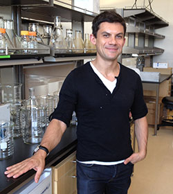 Roberto Zoncu合作论文数Biochemistry, Biophysics and Structural Biology Faculty, Department of Molecular and Cell Biology, University of California, Berkeley3Haridas Dhanya合作论文数3
 Sunil Kumar Sailapu合作论文数Centre for Nanotechnology, Indian Institute of Technology Guwahati3
 M. Sakthivel合作论文数School of Life Sciences, Bharathidasan University3
 Imayavaramban Lakshmanan合作论文数Department of Biochemistry and Molecular Biology, University of Nebraska Medical Center3William M. Lydiatt合作论文数3Zhenhua Miao合作论文数2Pingchen Fan合作论文数2Karen Ebsworth合作论文数2Grace E Kim合作论文数2Peter J. Rohweder合作论文数2R. B. Smith合作论文数2Julian Yano合作论文数2Y Rose Citron合作论文数2Jennifer Shehan合作论文数2
 Vipin Dalal合作论文数Authors contributed equally.2Tyler C Detomasi合作论文数Department of Chemistry, University of California2Justin Zhang合作论文数2
 David W. Dawson合作论文数Department of Pathology and Laboratory Medicine, University of California, Los Angeles2
 Muzafar A. Macha合作论文数Department of Biochemistry and Molecular Biology, College of Medicine, University Nebraska Medical Center2
 Nasser Mohd W合作论文数Department of Pathology, The Ohio State University2
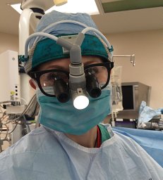 Michele R Aizenberg合作论文数Division of Neurosurgery, The University of Nebraska Medical Center2Dang Ton合作论文数2Yibin Zeng合作论文数2Linda Ertl合作论文数2Penglie Zhang合作论文数2
 Chugh Seema合作论文数Department of Biochemistry and Molecular Biology, University of Nebraska Medical Center2
 Jain Aakriti合作论文数2
 Matthew White合作论文数School of Biosciences, Cardiff University2




