Impact of pCGP and ctDNA dynamics on patient management. A, Swimmer’s plot illustrating the survival of 12 patients with EGFRm NSCLC; these patients were chosen to provide a representative example of situations in which pCGP beyond the first diagnosis was informative for patient management. Examples of how pCGP informed management include the identification of T790M alterations (patients PCGP1, PCGP4, PCGP5, PCGP7, PCGP12); identification of a BRAF alteration (PCGP6); identification of MET amplification (PCGP8 and PCGP9); and identification of a C797S resistance alteration (PCGP2). B and C, The emergence of resistance on pCGP; fish plots illustrate a dynamic, longitudinal representation of molecular alterations identified on pCGP in patients with serial sampling. The Y axis represents the aggregate VAF of all alterations identified at each time point. Each vertical dashed line represents the time point of a pCGP assay, with the number beneath representing the VAF of the most prevalent alteration at that time. B, pCGP serial profiling for a patient with EGFR L858R–driven NSCLC who had five iterations of pCGP. They were on erlotinib up to the second pCGP, which was obtained at the time of radiographic progression. This identified EGFR T790M, and the patient was switched to osimertinib with subsequent suppression of this clone. The third pCGP was at the time of further radiographic progression, no new driver of resistance was identified but EGFR C797S was identified on liver biopsy. They were transitioned from osimertinib to a clinical trial at this point, with corresponding re-expansion of T790M and L858R variants after. Fourth pCGP, after carboplatin/pemetrexed/bevacizumab therapy, identified both C797S and L718Q, an uncommon mutation in EGFR and an additional resistance mechanism. Their final pCGP demonstrated a significant increase in the aggregate VAF of all alterations, and the patient died 2 months later. Separate regimens are separated by semicolons. C, pCGP serial profiling for a patient with ALK-fusion NSCLC that had four iterations of pCGP. Their disease had become resistant to alectinib at the time of the first pCGP, likely attributable to ALK E1210K and I1171T alterations. I1171T is associated with sensitivity to ceritinib, and the patient was switched to ceritinib at this time. After some clinical response with a corresponding contraction of the I1171T variant, expansion of the E1210K variant was seen in addition to an acquired oncogenic PIK3CA resistance variant. The third pCGP is at the time of progression on ceritinib. The patient transitioned to brigatinib initially but progressed after 6 months and transitioned to lorlatinib. Fourth pCGP after 22 months on lorlatinib identified an additional acquired on-target mutation in ALK G1269A. Compound mutations of this kind are common in patients on lorlatinib. Separate regimens are separated by semicolons.
Figure S4. Categorization of EGFR driver variants identified in the EGFRm NSCLC subset.
Figure S3. Actionable alterations identified on first pCGP at first diagnosis (treatment-naive setting).
Supplementary Table S5. Summary of tissue informed clinical management in patients with concurrent pCGP and tissue biopsy.
Abstract Plasma comprehensive genomic profiling (pCGP) is implemented in the clinical care of non–small cell lung cancer (NSCLC). Although less well documented, serial pCGP may also guide the management of oncogene-driven NSCLC after first progression. In this study, we assessed the clinical value of serial pCGP, focusing on EGFR-mutant NSCLC. We conducted a retrospective study of 718 patients with NSCLC who underwent pCGP between 2015 and 2022. Clinical genomic data were programmatically extracted from the data workflows of the Johns Hopkins Lung Cancer Precision Medicine Center of Excellence. After variant annotation and actionability characterization, we examined the prevalence and evolving comutation patterns across serial pCGP, focusing on genomic mechanisms of tyrosine kinase inhibitor (TKI)–acquired resistance in EGFR-mutant NSCLC. A total of 718 patients had 818 instances of pCGP, with 79 patients having longitudinal pCGP (range, 2–5). pCGP uniquely informed management in 13% of patients (n = 92), both at initial diagnosis and on serial genotyping. This occurred predominantly through the identification of actionable mutations when tissue testing was unavailable. Among 214 patients with EGFR-mutant NSCLC, pCGP identified PI3K pathway alterations in 11% after first-line therapy. BRAF V600E (3%) and MET exon 14 skipping mutations (3%) emerged after third-generation TKI therapy. After TKI progression, 31 patients (22%) with EGFR-mutant disease had actionable pCGP findings, of whom 18 (58%) were matched to targeted therapy. Serial pCGP can inform treatment decisions in patients with NSCLC. In those with EGFR-mutant disease, pCGP at progression identifies actionable drivers of therapy resistance, enabling therapeutic intervention. Significance: Our study provides critical insights into the routine implementation of serial pCGP within a thoracic oncology program, supported by a precision oncology informatics framework, in a tertiary healthcare institution. We show that pCGP enables genotyping when tissue testing is not feasible and identifies actionable mutations at resistance. The clinical implementation of pCGP can drive improved clinical outcomes by matching patients with effective interventions in a timely and minimally invasive manner.
Figure S6. Plasma-only variants among patients with simultaneous plasma and tissue NGS.
Figure S5. Mechanisms of resistance to EGFR TKI according to first line of therapy received.
Circulating tumor DNA (ctDNA) analyses are informative as an early indicator of immunotherapy response in advanced non-small cell lung cancer (NSCLC); however, the clinical value of ctDNA molecular response requires further validation. As part of a prospective clinical protocol ( NCT05995821 ), we conducted targeted error-correction sequencing of ctDNA (n=328) and matched WBC DNA (n=109) from 109 patients with metastatic NSCLC who received anti-PD-(L)1 either as monotherapy or in combination. Following cellular origin resolution of 2,818 variants, landmark molecular response (mR) was defined as undetectable ctDNA within 3-9 weeks of treatment initiation. Pre-treatment ctDNA burden, but not blood tumor mutation burden, predicted survival. Implementing a tumor-naïve WBC DNA-informed approach increased the number of evaluable cases without compromising the overall accuracy of landmark ctDNA molecular responses. A direct comparison of single-timepoint on-therapy ctDNA assessment with ctDNA dynamics from baseline to the 3-9-week interval, along with an analysis of heterogeneity in molecular response within the 3-9-week window, showed that undetectable ctDNA at the landmark timepoint can effectively predict survival outcomes. A significant enrichment in landmark ctDNA mR was noted among patients with progression-free survival (PFS) ≥6 months with immunotherapy (p=2.5e-05) and chemo-immunotherapy (p=0.02). Patients in the landmark mR group had longer progression-free (p=1.6e-06) and overall survival (p=2.5e-05) than those with molecular progression. Landmark ctDNA molecular response provides a real-time, accurate approach for monitoring immunotherapy clinical outcomes. Although not currently validated for regulatory use, these findings demonstrate the potential utility of ctDNA as an early endpoint in clinical trials. Employing circulating tumor DNA (ctDNA) dynamics as an early indicator of immunotherapy response requires a roadmap for the next-generation sequencing approach, definition of molecular response and establishment of its clinical sensitivity. In this study, we introduce the concept of a landmark ctDNA molecular response, determined 3-9 weeks after initiation of immunotherapy, that maximizes the number of evaluable patients without sacrificing the specificity of the approach. Notably, when evaluating heterogeneity in ctDNA detection within the landmark 3-9-week window and assessing the impact of landmark interval dynamics on survival, we found that a single ctDNA assessment performed similarly to multiple ctDNA measurements within the landmark window (most notably, regardless of whether the timepoints were concordant or discordant). Our findings demonstrate that a single assessment of early on-therapy landmark ctDNA molecular response, can identify patients at risk of disease progression and enable future intervention and therapy optimization.
Impact of plasma genotyping vs. tissue on patient management. A–D, We considered circumstances in which pCGP was informative and concurrent tissue biopsy uninformative and then the converse, situations in which tissue biopsy was informative and concurrent pCGP uninformative. This latter analysis was limited to second and third assays of genotyping. The total number of patients with tissue biopsy was 31, of whom 22 (71%) had tissue NGS performed as well. Tumor fraction was not calculated as part of the pCGP assays. A, Instances when pCGP was informative and tissue uninformative, clustered according to the iteration of pCGP. For two patients marked as “other,” one had imaging changes that could represent NSCLC recurrence or Mycobacterium avium infection. The patient was hesitant to proceed with tissue biopsy, and the treating physician ordered pCGP, which identified a KRAS G12R mutation. This was considered to increase the likelihood of NSCLC recurrence and to justify the risks of tissue biopsy to obtain confirmation. In the second case, a frail patient had a BRAF V600E alteration on tissue NGS at first diagnosis and progressed through several lines of chemo- and immunotherapy. The treating physician performed repeat pCGP at progression and found no BRAF V600E. Given the toxicities of therapy and concern that the BRAF alteration was no longer present and circulating, the physician was guided not to pursue combined BRAF/MEK inhibition. B, Instances when tissue was informative and contemporaneous pCGP uninformative. C, In the EGFRm population subset, instances when pCGP was informative, whereas tissue NGS was uninformative. D, In the EGFRm population subset, instances when tissue was informative and contemporaneous pCGP uninformative. Although the number of cases was relatively small (n = 15), we found that concurrent tissue biopsy could guide management in more than half of the cases (57%) through information not available on pCGP, including histologic confirmation of radiographically equivocal sites of progression and the diagnosis of small cell histologic transformation. E, Sankey diagram illustrating the mechanisms through which pCGP influenced patient management at the first and second iterations of testing. pCGP was uniquely informative to patient management in the first and second iterations of testing, with benefits accruing to some patients more than once. At the first testing, it outperformed alternative methods for practical reasons, such as rapid turnover and permitting testing in patients who might not otherwise be fit, but it also identified some alterations that were not present on tissue NGS. It continued to be beneficial in the second iteration for patients in which tissue testing was not possible, which may reflect declining fitness for biopsy in this population.
 Kristen A Marrone合作论文数91Christine L. Hann合作论文数80
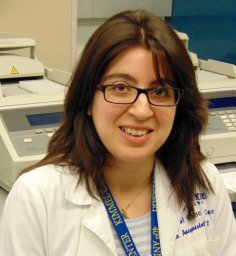 Valsamo Anagnostou合作论文数School of Medicine, Yale University78Patrick M Forde合作论文数69
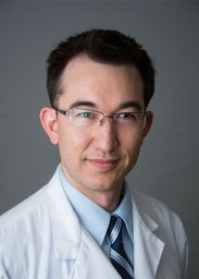 Joseph Murray合作论文数Sch Med, Johns Hopkins Univ68Levy Benjamin合作论文数67Feliciano Josephine合作论文数64
 Archana Balan合作论文数55
 Julie Brahmer合作论文数School of Medicine, Johns Hopkins University;Sidney Kimmel Comprehensive Cancer Center, Johns Hopkins University;Bloomberg~Kimmel Institute for Cancer Immunotherapy, Johns Hopkins University;Johns Hopkins Sidney Kimmel Comprehensive Cancer Center45
 Jarushka Naidoo合作论文数Royal College of Surgeons in Ireland44David S Ettinger合作论文数38Lavanya Sivapalan合作论文数33
 Victor E. Velculescu合作论文数Department of Genetic Medicine, School of Medicine, Johns Hopkins University;Delfi Diagnostics32
 Noushin Niknafs合作论文数Johns Hopkins Univ, Dept Biomed Engn, Baltimore, MD 21218 USA31Gavin Pereira合作论文数31
 James Robert White合作论文数Resphera Biosciences23Jillian Phallen合作论文数22Robert B. Scharpf合作论文数Department of Otolaryngology-Head and Neck Surgery, School of Medicine, Johns Hopkins University22
 Qing Kay Li合作论文数Department of Otolaryngology-Head and Neck Surgery, School of Medicine, Johns Hopkins University22
 Ali H Zaidi合作论文数Institute for the Treatment of Esophageal and Thoracic Disease, Allegheny Health Network22
 Peter B Illei合作论文数Department of Pathology, Memorial Sloan-Kettering Cancer Center22Nisha Rao合作论文数21Akul Goel合作论文数21Karlijn Hummelink合作论文数21Samuel Rosner合作论文数21
 Alessandro Leal合作论文数Johns Hopkins Univ, Sidney Kimmel Comprehens Canc Ctr, Sch Med, Baltimore, MD 21287 USA21Weksler Benny合作论文数21
 David L Bartlett合作论文数Allegheny Health Network Cancer Institute;Surgery Division, Center for Cancer Research, National Cancer Institute21
 Kim Monkhorst合作论文数21
 Nathan Bahary合作论文数Department of Microbiology & Molecular Genetics, School of Medicine, University of Pittsburgh;Division of Oncology, Department of Medicine, School of Medicine, University of Pittsburgh21Catherine A. Shu合作论文数Med Ctr, Columbia Univ19Bestvina Christine M合作论文数19Richard Lee合作论文数18Amy L Cummings合作论文数18Yun Qing合作论文数Dept Biostat, Univ Texas MD Anderson Canc Ctr18
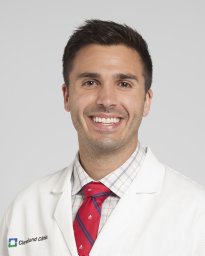 Lukas Delasos合作论文数18Gabriele Minuti合作论文数18
 Giuseppe Lamberti合作论文数Medical Oncology, IRCCS Azienda Ospedaliero-Universitaria di Bologna18
 Do Minh Truong合作论文数18Bironzo Paolo合作论文数Dept Oncol, Univ Turin18Negrao Marcelo V合作论文数Heart Institute (InCor),, University of São Paulo Medical School18
 Ni Ying合作论文数18
 Lorenza Landi合作论文数Medical Oncology Department, Istituto Toscano Tumori;+39 0586223457;f.cappuzzo@gmail.com.;Medical Oncology Department, Istituto Toscano Tumori18
 Mihaela Aldea合作论文数18
 J. Jack Lee合作论文数Division of Discovery Science, Department of Biostatistics, University of Texas MD Anderson Cancer Center18Christopoulos Petros合作论文数18
 Sandip Pravin Patel合作论文数Center for Personalized Cancer Therapy, UC San Diego Moores Cancer Center18
 Biagio Ricciuti合作论文数Lowe Center for Thoracic Oncology, Dana-Farber Cancer Institute;Harvard Medical School18Owen Dwight合作论文数18




