Importance:Recovery after upper extremity peripheral nerve injury (PNI) surgery depends on changes in cortical neural patterns that support sensorimotor control. Task-based functional connectivity (FC) can characterize these changes, yet few studies have explored FC during ecologically fine motor valid tasks after PNI. Objective:To investigate task-based FC with the left primary motor cortex (M1) during right hand drawing in individuals following right hand PNI surgery. Participants:Forty-four right-handed adults, including 12 patients post PNI surgery (n = 8 with nerve repair, n = 4 with nerve transfer) and 32 healthy controls. Methods:All participants underwent fMRI while performing a RH visuomotor precision drawing task. Seed-based connectivity analysis was performed to characterize the pattern of FC between left M1 and all voxels in the brain. We hypothesized that left M1 FC would differ between patients and controls, between Repair and Transfer groups, and covary with time since surgery. Results:Patients (vs. controls) showed greater FC between left M1 and right visual and premotor cortices. Nerve transfer (vs. repair) showed greater FC between left M1 and right inferior parietal areas. Time since surgery was not linearly related to FC, though exploratory analyses suggested a negative association between log-time and FC between left M1 and right inferior parietal lobule. Conclusion:After PNI surgery, visuomotor precision drawing involved distinct and behaviorally relevant neural patterns, which varied by task demand and potentially by surgical group despite clinical heterogeneity. Inferior parietal cortex may be especially engaged in early months after surgery (i.e. log-time). To improve recovery of upper limb function after PNI, clinical recommendations include incorporating early function-specific dexterous training, tailoring rehabilitation across surgical and recovery stages, and using multidimensional assessments of hand function.
Acellular nerve allografts have been used as an alternative to reconstruct nerve gaps. However, regeneration and recovery using long acellular nerve allografts (> 3 cm) is poor in comparison to short acellular nerve allografts (< 3 cm). To understand why long acellular nerve allografts have limited regeneration, we focused on identifying differences in the microenvironment of short (2 cm) and long (4 cm) acellular nerve allografts by comparing the transcriptional profile of these acellular nerve allografts. After repairing the sciatic nerve of Lewis rats using either short or long acellular nerve allografts, we found that the proximal and mid-distal graft regions of long acellular nerve allografts are characterized by an upregulation of metabolic and immune pathways and downregulation of regenerative processes in comparison to the short acellular nerve allografts. Based on these results, we modulated the regenerative and immune microenvironment of long acellular nerve allografts using tacrolimus (FK506). Histomorphometric and muscle force analysis revealed that FK506 increases the number of axons and improves recovery of motor function across long acellular nerve allografts. Transcriptome analysis of the mid-distal graft region of long acellular nerve allografts from animals treated with FK506 revealed upregulation of regenerative pathways and downregulation of immune processes, specifically related to T cell activity. Additionally, FK506 altered the number of macrophages and Schwann cells in the long acellular nerve allografts. From the transcriptome analysis, we identified FK506 upregulates expression of Spp1 (osteopontin) which promotes regeneration of motor neurons after injury. Experiments on cultured Schwann cells revealed that FK506 increases mRNA expression of Spp1. Our data show the development of a degenerative and immune microenvironment within long acellular nerve allografts and demonstrate that FK506 can modulate this microenvironment to improve nerve regeneration across these long acellular nerve allografts.
Background:The COVID-19 pandemic accelerated the adoption of telemedicine for surgical consultations. Although patient satisfaction has been well documented, limited research exists regarding telemedicine's effectiveness in determining the ability to indicate for surgical care compared with in-office visits. We conducted a retrospective review comparing surgical decision-making via telemedicine versus in-person consultations, hypothesizing that in-office visits would be more effective in establishing a surgical plan than telemedicine. Methods:A retrospective review of the medical records of all new patients presenting to the senior author's nerve surgery clinic at a high-volume quaternary referral center between June 2020 and January 2023 was performed. Surgical consultations were categorized as (1) surgery recommended, (2) surgery not recommended, (3) further screening required, or (4) surgery declined. The Fisher exact test compared the distribution of these categories between consultation types and the proportion of patients who underwent surgery after an initial recommendation. Results:Of the 809 patients, 283 (35%) had in-person and 526 (65%) had telemedicine consultations. Surgery was recommended in 49.5% of in-person consultations compared with 38% of telemedicine consultations (P = 0.03). Among those recommended for surgery at the initial visit, 77.9% of in-person and 77.8% of telemedicine patients ultimately underwent surgery (P = 0.10). Of the telemedicine patients subsequently seen in the office and offered surgery, 84.5% ultimately underwent that surgery. Conclusions:Our findings suggest that telemedicine and in-office visits are equally effective in establishing a surgical plan, suggesting that the expansion of telemedicine could be considered for a broader geographic patient base.
BACKGROUND:Deoxycholic acid (ATX-101) is a drug administered by subcutaneous injection for local fat reduction. However, ATX-101 treatment has been reported to cause marginal mandibular nerve injury with noticeable functional deficits when targeting submental fat. As a cytolytic agent with some selectivity for adipocytes, ATX-101 may damage the lipid-rich myelin surrounding peripheral nerves. OBJECTIVES:The aim of this study was to characterize nerve injury caused by ATX-101 injection in an experimental rat sciatic nerve model. METHODS:Injuries to the sciatic nerve caused by intrafascicular and extrafascicular injections of ATX-101, and by lidocaine (positive control) and saline (negative control) injections, were compared. Nerves were harvested at a 2-week endpoint for histomorphometric analysis. RESULTS:The cross-sectional area of nerve injury was significantly increased by ATX-101 injection. The damaged areas amounted to 75% ± 15% with intrafascicular ATX-101 (P < .001), 41% ± 21% with extrafascicular ATX-101 (P < .01), and 38% ± 20% with positive-control lidocaine (P < .01), compared with 7% ± 13% with negative-control saline. Demyelinating injury was a significant mechanism of injury in the affected nerve fibers compared with uninjured nerve fibers (P < .04), but there was no difference in the axon-to-myelin area ratio between the lidocaine and ATX-101 cohorts. After 2 weeks, Wallerian degeneration was evident with only small regenerating nerve fibers present in the ATX-101-injured groups compared with saline (average fiber width, 2.54 ± 0.26 μm vs 5.03 ± 0.44 μm, P < .001). CONCLUSIONS:ATX-101 can cause extensive nerve injury in rats. The mechanism of action for ATX-101 does not preferentially target myelin more than other common neurotoxic agents. Appropriate knowledge of surgical anatomy and injection technique is necessary for any practitioners administering ATX-101 injections.
Introduction:Seddon and Sunderland's original nerve injury classification systems have stood the test of time over the last 70 years and continue to be widely used today. However, since those original descriptions, knowledge of nerve pathophysiology and healing has advanced, electrodiagnostic results have become more refined, and surgical options have increased. Methods: We offer a revisited review of the nerve injury classification to incorporate new knowledge for the modern era of nerve surgery. Results: We offer the addition of grades 0 and VI to the existing classification of nerve injuries based on Sunderland's framework, and we present a simplified classification that is patient and physician oriented, reflecting prognosis, time to recovery, and degree of recovery. Discussion: By following this nerve injury framework, clinicians can better assess, prognosticate, and manage patients.
BACKGROUND:The spinal accessory nerve is occasionally injured during brachial plexus injuries, potentially limiting its use as a donor motor nerve for reconstructive surgery. Assessing spinal accessory nerve function preoperatively is essential to optimize outcomes in brachial plexus reconstruction. The authors examined the demographics of spinal accessory nerve injury, focusing on its prevalence, predisposing factors, and potential for spontaneous recovery in patients with brachial plexus injuries. METHODS:A total of 170 patients were included in this study. At the first visit, the spinal accessory nerve function was assessed by measuring the compound muscle action potential of the upper trapezius muscle. The function of the nerve was classified as follows: no injury (amplitude of the potential ≥50% compared with the unaffected side), partial injury (amplitude <50%), and severe injury (absence of the potential). For the evaluation of spontaneous recovery, patients with partial and severe injuries underwent further assessment at 1 year after the injury and at the final follow-up. RESULTS:The authors found the associated spinal accessory nerve injury in 30 patients (17.6%) (partial, 17; severe, 13). Spinal accessory injury occurred in 13 of 39 patients with phrenic nerve palsy (33.3%). Phrenic nerve palsy may serve as a predisposing factor for this injury. In 18 patients followed up for more than 1 year, 15 (83.3%) showed good spontaneous recovery. CONCLUSIONS:Spinal accessory nerve injury occurred in approximately 18% of patients with brachial plexus injuries. Most cases of spinal accessory nerve injury associated with brachial plexus injury have a potential for good spontaneous recovery.
Surgeons’ ability to interpret electrodiagnostic (EDX) studies has greatly enhanced patient selection and outcome for nerve surgeries as it has informed our understanding of the classification of nerve injury. EDX measures axons present, lost, or demyelinated. It does not measure an area where axons experience chronic ischemia, making a Sunderland Zero nerve injury a clinical diagnosis that should be suspected when patients have normal EDX, decreased recruitment on electromyography (EMG), and no atrophy. Only observation of recovery in the earliest postoperative period would confirm a Sunderland Zero. In this article, we present an expanded classification of nerve injury, highlighting Sunderland Zero or ischemic neurapraxia, and provide a historical and pathophysiological overview of how it came to be recognized. We also offer a user-friendly classification and an equation we call the Nerve Injury Calculator (NIC) that calculates the percent axonal loss, demyelinating loss, and ischemic axons to predict timing and quality of recovery. The clinical utility of the NIC is exemplified with three cases. With the development and introduction of the NIC, there is a paradigm shift amongst surgeons, physiatrists, and neurologists that allows us all to speak the same language of EDX and nerve surgery, enabling improved patient education, prediction of prognosis and timing of recovery, and efficient follow up with respect to patients’ postoperative period.
The use of acellular nerve allografts (ANAs) to reconstruct long nerve gaps (>3 cm) is associated with limited axon regeneration. To understand why ANA length might limit regeneration, we focused on identifying differences in the regenerative and vascular microenvironment that develop within ANAs based on their length. A rat sciatic nerve gap model was repaired with either short (2 cm) or long (4 cm) ANAs, and histomorphometry was used to measure myelinated axon regeneration and blood vessel morphology at various timepoints (2-, 4- and 8-weeks). Both groups demonstrated robust axonal regeneration within the proximal graft region, which continued across the mid-distal graft of short ANAs as time progressed. By 8 weeks, long ANAs had limited regeneration across the ANA and into the distal nerve (98 vs. 7583 axons in short ANAs). Interestingly, blood vessels within the mid-distal graft of long ANAs underwent morphological changes characteristic of an inflammatory pathology by 8 weeks post surgery. Gene expression analysis revealed an increased expression of pro-inflammatory cytokines within the mid-distal graft region of long vs. short ANAs, which coincided with pathological changes in blood vessels. Our data show evidence of limited axonal regeneration and the development of a pro-inflammatory environment within long ANAs.
BACKGROUND:Upper extremity (UE) trauma requiring operative care increases during the summer and fall months, which the authors colloquially refer to as "trauma season."METHODS:CPT databases were queried for codes related to acute UE trauma at a single level-1 trauma center. Monthly CPT code volume was tabulated for 120 consecutive months and average monthly volume was calculated. Raw data were plotted as a time series and transformed as a ratio to the moving average. Autocorrelation was applied to the transformed data set to detect yearly periodicity. Multivariable modeling quantified the proportion of volume variability attributable to yearly periodicity. Subanalysis assessed presence and strength of periodicity in four age groups.RESULTS:A total of 11,084 CPT codes were included. Monthly trauma-related CPT volume was highest in July through October and lowest in December through February. Time-series analysis revealed yearly oscillation in addition to a growth trend. Autocorrelation revealed statistically significant positive and negative peaks at a lag of 12 and 6 months, respectively, confirming yearly periodicity. Multivariable modeling revealed R 2 attributable to periodicity of 0.53 ( P < 0.01). Periodicity was strongest in younger populations and weaker in older populations. R 2 was 0.44 for ages 0 to 17, 0.35 for ages 18 to 44, 0.26 for ages 45 to 64, and 0.11 for ages 65 and older.CONCLUSIONS:Operative UE trauma volumes peak in the summer and early fall and reach a winter nadir. Periodicity accounts for 53% of trauma volume variability. The authors' findings have implications for allocation of operative block time and personnel and expectation management over the course of the year.
Within the past decade, there have been multiple innovations in the field of nerve surgery. In this review, we highlight critical changes and innovations that have helped advance the field and present opportunities for further study. This includes the modification and clarification of the classification schema for nerve injuries which informs prognosis and treatment, and a refined understanding and application of electrodiagnostic studies to guide patient selection. We provide indications for operative intervention based on this nerve injury classification and propose strategies best contoured for varying injury presentations at differing time points. Lastly, we discuss new developments in surgical techniques and approaches based on these advancements.
Background: The arcade of Struthers was first proposed by Kane et al in 1973. Clinical investigations of this structure have been limited to small case series, focusing on the arcade as an isolated cause of compressive ulnar neuropathy. The purpose of our study was to investigate the incidence of this structure in patients undergoing ulnar nerve transposition.Methods: A retrospective chart review of prospectively maintained data in a single surgeon's practice was performed. Records of patients undergoing surgery for compressive ulnar neuropathy at the cubital tunnel were evaluated for documentation of a compressive arcade of Struthers. In addition, a scoping review of the literature was undertaken to better characterize current understanding of this structure and its recognition in clinical practice.Results: A total of 197 patients underwent ulnar nerve transposition. The overall incidence of a compressive arcade of Struthers was noted to be 67 out of 197 (34%). All patients with a compressive arcade were noted to have an internal brachial ligament running below the nerve. Patients undergoing revision surgery were found to have a compressive arcade 51% of the time (20/39), whereas 30% of patients undergoing primary surgery were found to have a compressive arcade (47/158). Only 12 clinical studies examining the arcade of Struthers have been published in the last 20 years, the majority being single case reports.Conclusions: Compression of the ulnar nerve by the arcade of Struthers is a common finding and can contribute to compressive ulnar neuropathy at the elbow both in primary and revision cases.
BACKGROUND:Electrodiagnostic studies are critical for surgical decision-making in nerve injuries. Surgeons typically rely on the electrodiagnostician's reports and lack formal training in electrodiagnostic study interpretation. This knowledge gap highlights a need for accessible and effective educational resources for surgeons to improve their understanding of electrodiagnostic studies and enhance patient care. METHODS:The educational module consisted of a prelecture knowledge assessment, a 42-minute video lecture on interpreting electrodiagnostic study results, and a postlecture knowledge assessment. Knowledge retention was assessed by means of an additional survey distributed 3 months after module completion. RESULTS:This study, involving 119 participants (79% attending surgeons, 8% fellows, 9% surgical residents, and 2% who described their position as "other"), demonstrated that a 42-minute video-based learning module significantly improved knowledge of electrodiagnostic study interpretation. Median scores increased from 7 to 9 ( P < 0.001), with improvement persisting at 3 months (median retention score, 11; P < 0.025). Among surgeons completing the 3-month assessment, 65.5% reported that knowledge gained from the module had changed their clinical practice. CONCLUSION:This study demonstrates that a concise, video-based learning module can effectively enhance surgeon knowledge of electrodiagnostic study interpretation and may serve as a valuable tool for surgical education and improving patient selection in nerve surgery.
Background: Nerve interposition grafting is an important technique in nerve reconstructive surgery that is used when a primary repair is not feasible without significant tension. This study sought to evaluate the long-term morbidity of the medial antebrachial cutaneous (MABC) nerve as an alternative donor nerve in comparison with sural nerve harvest. Methods: A single surgeon and institution retrospective chart review was performed to identify all patients who underwent nerve autografting using the sural and MABC as donor nerves between January 1, 2000 and December 31, 2019. Surveys assessed overall patient satisfaction with surgery, as well as donor and recipient site morbidity, satisfaction, pain, numbness, and cold sensitivity. Results: Of the 73 patients contacted, 54 agreed to participate, and 43 of 73 (58.9%) ultimately completed the survey: 28 MABC (65.1%) and 15 sural (34.9%). There were no significant differences between the sural and MABC groups in overall satisfaction with surgery, donor and recipient site satisfaction, pain, cold sensitivity, and effect on quality of life. Even though 66.7% of sural donor sites and 75% of MABC donor sites had residual numbness, the effect this had on quality of life was very low (2 and 3, respectively). Conclusion: The MABC is a safe alternative to the traditional sural nerve autograft. A small subset of patients undergoing nerve autograft harvest will experience long-term morbidity in the form of pain. Conversely, the more common presence of numbness is not reported as bothersome.
BACKGROUND:Nerve injuries remain a challenging complication after shoulder surgery. While most resolve spontaneously, some require surgical intervention. This study describes the characteristics of patients sustaining nerve injuries following shoulder surgery, evaluates referral patterns to nerve surgeons, and characterizes nerve surgeries performed and their outcomes. Increased awareness of these injuries allows patients and providers to be better informed regarding the appropriate management when these complications occur. METHODS:A retrospective review of referrals with nerve injuries following shoulder surgery between 2007 and 2015 was performed. The final analysis included 65 patients. Data reviewed included demographics, procedure and anesthesia type, and diagnosis of nerve injury. Time to referral to nerve surgeon and proportional changes in the Disabilities of the Arm, Shoulder, and Hand (DASH) scores were determined. Outcomes were categorized as failed, partially successful, and successful based on final follow-up. RESULTS:Patients were referred following arthroscopic shoulder surgeries (35.4%), shoulder arthroplasties (24.6%), open shoulder procedures (21.5%), and combined open and arthroscopic procedures (18.5%). The mean time to referral was 7.6 months. Nerve injuries involved brachial plexus (33) and individual and multiple peripheral nerve branches (23 and 7, respectively). Twenty-five (38%) nerve injuries demonstrated spontaneous recovery. Thirty-five patients underwent surgical intervention, of which 27 were successful, 3 were partially successful, and 3 failed. CONCLUSIONS:This is the largest series of patients with iatrogenic nerve injury following shoulder surgeries to date. Our data demonstrate a lack of timely referral to nerve surgeons, especially after arthroscopy. There continues to be a variable injury pattern even among similar surgeries. Despite this, timely surgical intervention, when indicated, can lead to favorable outcomes.
Background Little is known about how peripheral nerve injury affects human performance, behavior, and life. Hand use choices are important for rehabilitation after unilateral impairment, but rarely measured, and are not changed by the normal course of rehabilitation and daily life. Objective To identify the relationship between hand use (L/R choices), motor performance, and patient-centered outcomes. Methods Participants (n = 48) with unilateral peripheral nerve injury were assessed for hand use via Block Building Task, Motor Activity Log, and Edinburgh Handedness Inventory; dexterity (separately for each hand) via Nine-Hole Peg Test, Jebsen Taylor Hand Function Test, and a precision drawing task; patient-centered outcomes via surveys of disability, activity participation, and health-related quality of life; and injury-related factors including injury cause and affected nerve. Factor Analysis of Mixed Data was used to explore relationships between these variables. The data were analyzed under 2 approaches: comparing dominant hand (DH) versus non-dominant hand (NH), or affected versus unaffected hand. Results The data were best explained by 5 dimensions. Good patient outcomes were associated with NH performance, DH performance (separately and secondarily to NH performance), and preserved function and use of the affected hand; whereas poor patient outcomes were associated with preserved but unused function of the affected hand. Conclusion After unilateral peripheral nerve injury, hand function, hand usage, and patient life arise from a complex interaction of many factors. To optimize rehabilitation after unilateral impairment, new rehabilitation methods are needed to promote performance and use with the NH, as well as the injured hand.
Background: Nerve injuries from gunshot wounds (GSWs) to the upper arm can cause significant morbidity and loss of function. However, indications for surgical exploration and nerve reconstruction remain unclear as both low- and high-grade injuries can present with an abnormal neurological examination. Methods: Adult patients presenting with a history of isolated GSW to the upper arm between 2010 and 2019 at a single urban level 1 trauma center were screened for inclusion in this retrospective study. Patient demographics, neurological examination findings, concurrent injuries, and intraoperative findings were gathered. Bivariate analysis was performed to characterize factors associated with nerve injuries. Results: There were 139 adult patients with isolated brachial GSWs, and 49 patients (35%) presented with an abnormal neurological examination and significantly associated with concurrent humerus fractures (39% vs 21%, P = .026) and brachial artery injuries (31% vs 2%, P < .001). Thirty of these 49 patients were operatively explored. Fifteen patients were found to have observed nerve injuries during operative exploration including 8 patients with nerve transections. The radial nerve was the most commonly transected nerve (6), and among the 16 contused nerves, the median (8) was most common. Conclusion: Nerve injury from upper arm GSWs is common with directly traumatized nerves confirmed in at least 39% and nerve transection in at least 16% of patients with an abnormal neurological examination. Timely referral to a hand and/or peripheral nerve surgeon for close clinical follow-up, appropriate diagnosis, and any necessary surgical reconstruction with nerve grafts, tendon transfers, and nerve transfers is recommended.
 Daniel Hunter合作论文数Division of Plastic Surgery, School of Medicine, Washington University in St Louis112
 Christine Novak合作论文数Division of Plastic & Reconstructive Surgery, The Hospital for Sick Children, University of Toronto;Department of Surgery, Temerty Faculty of Medicine, University of Toronto;Rehabilitation Sciences Institute, Temerty Faculty of Medicine, University of Toronto64
 DA Hunter合作论文数58
 Amy M. Moore合作论文数56Thomas H. Tung合作论文数54Terence M. Myckatyn合作论文数Division of Plastic and Reconstructive Surgery, Washington University School of Medicine47
 Matthew Wood合作论文数Department of Politics and International Relations, Faculty of Social Sciences, The University of Sheffield46
 Philip J Johnson合作论文数Nuvaira, Inc42
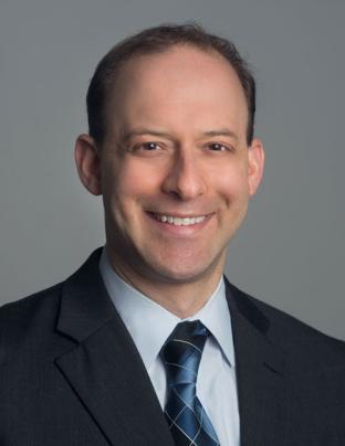 Michael J. Brenner合作论文数Division of Facial Plastic and Reconstructive Surgery/Skull base Surgery, Department of Otolaryngology-Head and Neck Surgery, University of Michigan Medical School;Kresge Hearing Research Institute, University of Michigan Medical School40
 Andrew Yee合作论文数Center for Multiple Myeloma, Massachusetts General Hospital;Harvard Medical School40Snyder-Warwick Alison合作论文数33A. Lee Dellon合作论文数32Ida K. Fox合作论文数Washington University School of Medicine32
 Wilson Z. Ray合作论文数Department of Neurosurgery, Washington University School of Medicine26Lauren Schellhardt合作论文数23Ying Yan合作论文数23Peter J. Evans合作论文数21Santosa Katherine B合作论文数17
 Kristen Davidge合作论文数Department of Surgery, Temerty Faculty of Medicine, University of Toronto;Division of Plastic, Reconstructive & Aesthetic Surgery, Temerty Faculty of Medicine, University of Toronto;Sick Kids Research Institute16
 Shelly Sakiyama Elbert合作论文数Cockrell School of Engineering, The University of Texas16Newton Piyaraj合作论文数16Kahn Lorna C合作论文数16Midha Rajiv合作论文数16J. Megan M. Patterson合作论文数14
 John N Jensen合作论文数14Deng Pan合作论文数Washington University in St. Louis14Gwendolyn M. Hoben合作论文数14Alice Y. Tong合作论文数14Arash Moradzadeh合作论文数14Tj Best合作论文数13
 Gregory H Borschel合作论文数Hosp Sick Children, Div Plast & Reconstruct Surg, Toronto, ON M5G 1X8, Canada13Christine B. Novak合作论文数13VB Doolabh合作论文数13Ee Xueping合作论文数12James B. Lowe合作论文数12
 Louis Poppler合作论文数11
 Kasukurthi Rahul合作论文数School of Medicine, Washington University in St. Louis11
 Ying Yan合作论文数Center for Applied Neural Engineering, Department of Neurosurgery, Washington University School of Medicine in St. Louis11
 Matthew Macewan合作论文数11Stahs Pripotnev合作论文数10Eric M. Genden合作论文数10
 Ayato Hayashi合作论文数Division of Plastic and Reconstructive Surgery, Washington University in St. Louis School of Medicine10M C Hertl合作论文数9Jessica M Hasak合作论文数9
 Hill Elspeth J R合作论文数9
 Elizabeth L Whitlock合作论文数Division of Plastic and Reconstructive Surgery, Washington University School of Medicine9Christina K. Magill合作论文数9Y Nakao合作论文数8M.Wayne Flye合作论文数8




