Phosphatidylinositol transfer proteins (PITPs) are essential lipid-binding proteins that regulate phosphoinositide signaling, membrane trafficking and autophagy through the transport of phosphatidylinositol and other phospholipids between intracellular membranes. Microcolin compounds have been identified as selective inhibitors of class I PITPs, revealing important roles of PITPs in Hippo signaling and autophagy. Here, we report the crystal structure of human PITPα in complex with microcolin H at 2.0 Å resolution. The structure enables a detailed description of the interaction between microcolin H and the lipid-binding cavity. Besides the expected covalent bond to the Cys94 residue, the structure also reveals an extensive network of hydrogen bonds, water bridges and hydrophobic interactions. Importantly, PITPα remains in the open conformation upon binding to microcolin H. Quantitative cavity analysis confirms that the microcolin-bound structure adopts a volume comparable to that of the unliganded PITPα and is markedly larger than that of the lipid-bound state. These findings demonstrate that microcolins selectively trap PITPα in an open conformation and provide a structural basis for their inhibitory mechanism. Furthermore, our results show that ligand binding can profoundly change protein conformation, which underscores the limitation of docking experiments.
Proper functions of cellular organelles require tight control of membrane phospholipid composition, yet the mechanisms by which lipid imbalances are sensed and corrected remain largely unknown. Here, we present evidence of an unexpected metabolic connection between plasma membrane (PM) phosphoinositide metabolism and two key anionic lipids, phosphatidylserine (PS) and phosphatidic acid (PA). Prolonged depletion of PM phosphatidylinositol 4-phosphate (PI4P) by pharmacological inhibition of PI 4-kinase IIIα (PI4KIIIα/PI4KA) increases phospholipase D (PLD) activity and PA levels in the PM. Using lipidomics, RNA-seq, and proximity proteomics, we find that PI4P loss induces a concomitant decrease in PS, activating a reciprocal relationship between PS synthesis and PLD-mediated PA generation. These changes also drive transcriptional and translational upregulation of the small GTPase RhoB, which enhances PLD-mediated PA synthesis and actin cytoskeletal remodeling. Because reduced PI4KA activity underlies numerous hereditary diseases, our studies reveal how perturbation of PM phosphoinositide synthesis triggers an integrated response that maintains the anionic character and structural integrity of the PM.
Although the inner nuclear membrane (INM) is generally considered to be continuous with the outer nuclear membrane (ONM) and connected to the remaining endoplasmic reticulum (ER), it has been well recognized that it is functionally distinct, having a unique protein composition. It has increasingly been recognized, however, that the INM also differs from the ONM and the other ER domains in its lipid composition. It is an intriguing proposition that the unique lipid profile of the INM is intricately linked to its specialized functions related to the nuclear events. Despite rapid progress in recent years in our understanding of the unique lipid profile of the INM and its role in the control of nuclear functions, there is a lot that remains to be understood. This review summarizes recent advances in characterizing the INM lipid composition and lipid synthetic pathways including their possible roles in the control of nuclear functions. Additionally, it discusses current challenges and areas deserving further investigation.
Phosphorylated products of phosphatidylinositol (PI), named Diphosphoinositide (DPI) and triphosphoinositide (TPI) were identified long time ago and found to exhibit high turnover rates based on their rapid 32P-phosphate labeling. The PI kinase activities that were responsible for their production were subsequently identified and found to be associated with different organelle membranes, including the plasma membrane. These activities were then linked with a certain group of cell surface receptors that activated phospholipase C enzymes to hydrolyze PI and used calcium or cGMP as a second messenger. This visionary concept was introduced in the seminal BBA review written by Robert Michell, exactly 50 years ago. The enzymology and functional diversity of PI 4-phosphate (PI4P) (the term that has replaced DPI) has since underwent an expansion that could not have been foreseen. In this review I will attempt to revisit this expansion with some historical reflections celebrating the 50th anniversary of the Michell review.
G protein coupled receptors (GPCRs) are the target of about 35% of FDA-approved drugs, which underlines their importance in physiology and disease. Many GPCRs undergo rapid desensitization upon agonist activation, mediated by β-arrestin binding to the phosphorylated receptors, which prevents their G protein coupling. How such receptors regain their G protein signaling competence is poorly understood. Using the AT1 angiotensin II receptor (AT1R), we show that its rapid re-sensitization requires the sorting of the receptors into an AP2-positive plasma membrane (PM) compartment without receptors having to be internalized. This process requires the scaffolding protein, EFR3 and a dedicated PI(4,5)P 2 pool specifically produced by PIP5KA. While β-arrestin 1 and -2 both can carry the receptors to the AP2 compartment, weaker binding of β-arrestin 1 to the receptor allows some of the receptors to re-sensitize, while strong β-arrestin 2 binding elicits stronger desensitization and directs the receptors for internalization. These results suggest that the rapid phase of re-sensitization of GPCRs occurs without their endocytosis and primarily takes place at the PM at specific steps during clathrin-coated pit maturation. Together with differences observed between the two β-arrestins to interact with CCPs and the receptors, our data suggest that specific PI(4,5)P 2 pools controlled by EFR3A and PIP5K1A determine the balance between β-arrestin1 and -2 receptor interaction and delivery to the AP2 positive compartment, ultimately determining what fraction of the receptors regain their G protein signaling competence.
Cells tightly control the homeostatic levels and subcellular localizations of membrane phospholipids through the regulation of the activities of numerous lipid-metabolizing enzymes and lipid transfer proteins. Yet, the mechanisms by which lipid imbalances are sensed and corrected to establish and maintain homeostasis are, in most cases, unknown. Here we present an expanded view of plasma membrane (PM) phosphoinositide metabolism by revealing an unexpected metabolic connection between two key anionic lipids in this membrane, phosphatidylinositol 4-phosphate (PI4P) and phosphatidic acid (PA). PM pools of PI4P are generated by PI 4-kinase Type IIIα (PI4KIIIα/PI4KA), an essential enzyme whose partial dysfunction leads to numerous hereditary human diseases. We find that depletion of PI4P by pharmacological inhibition of PI4KA increases the activity of phospholipase Ds (PLDs) and the levels of their lipid product, PA, in the PM. Guided by RNA-seq analysis and proximity labeling proteomics, we elucidate how cells connect this PI4P decrease to a compensatory increase in PA levels. Loss of PM PI4P induces a concomitant decrease of phosphatidylserine (PS) levels, and this metabolic rewiring activates a reciprocal relationship between PS synthesis and PLD-mediated PA generation. These metabolic changes also lead to transcriptional and translational upregulation of the small GTPase RhoB, which enhances PLD-mediated PA synthesis and subsequent actin cytoskeletal remodeling. Our study reveals how disease-relevant perturbation of phosphoinositide synthesis induces an integrated response that ultimately boosts levels of PA, a key anionic lipid and metabolic intermediate in phosphoinositide resynthesis.
BACKGROUND:Biallelic loss-of-function variants in PI4K2A have been associated with a neurodevelopmental disorder characterized by seizures and movement disorders, including orofacial dyskinesia. However, only 4 cases have been reported. Orolingual dyskinesia-defined as involuntary movements of the mouth and tongue-is observed in various pediatric neurodevelopmental disorders (NDD) but remains under-recognized. OBJECTIVES:The aims were to highlight orolingual dyskinesia as a core feature of PI4K2A-related disorder (PI4K2A-RD) and explore its presence across other NDDs. METHODS:We described two new families with PI4K2A-RD and reviewed the clinical features of four previously reported cases. A focused literature search was also conducted to identify other neurogenetic conditions associated with orolingual dyskinesia. RESULTS:All individuals with PI4K2A deficiency exhibited orolingual dyskinesia, along with developmental delay, movement abnormalities, and variable seizures. The literature review confirmed frequent underreporting of this feature in NDDs. CONCLUSIONS:Orolingual dyskinesia is a relevant but under-recognized clinical sign in PI4K2A-RD and other neurogenetic conditions, with potential diagnostic value. © 2025 The Author(s). Movement Disorders published by Wiley Periodicals LLC on behalf of International Parkinson and Movement Disorder Society.
Mitochondrial dynamics are orchestrated by protein assemblies that directly remodel membrane structure, however the influence of specific lipids on these processes remains poorly understood. Here, using an inducible heterodimerization system to selectively modulate the lipid composition of the outer mitochondrial membrane (OMM), we show that local production of diacylglycerol (DAG) directly leads to transient tubulation and rapid fragmentation of the mitochondrial network, which are mediated by isoforms of endophilin B (EndoB) and dynamin-related protein 1 (Drp1), respectively. Reconstitution experiments on cardiolipin-containing membrane templates mimicking the planar and constricted OMM topologies reveal that DAG facilitates the membrane binding and remodeling activities of both EndoB and Drp1, thereby independently potentiating membrane tubulation and fission events. EndoB and Drp1 do not directly interact with each other, suggesting that DAG production activates multiple pathways for membrane remodeling in parallel. Together, our data emphasizes the importance of OMM lipid composition in regulating mitochondrial dynamics.
Phosphatidylinositol 4 kinase IIIα (PI4KIIIα/PI4KA) is an essential lipid kinase that plays a critical role in regulating plasma membrane identity. PI4KA is primarily recruited to the plasma membrane through the targeted recruitment by the proteins, EFR3A and EFR3B, which bind to the PI4KA accessory proteins TTC7 (TTC7A/B) and FAM126 (FAM126A/B). Here we characterised how both EFR3 isoforms interact with all possible TTC7-FAM126 combinations and developed a nanobody that specifically blocked EFR3-mediated PI4KA recruitment in TTC7B containing complexes. Most EFR3-TTC7-FAM126 combinations show similar binding affinities, with the exception of EFR3A-TTC7B-FAM126A, which binds with a ∼10-fold higher affinity. Moreover, we showed that EFR3B phosphorylation markedly decreased binding to TTC7-FAM126. Using a yeast display approach, we isolated a TTC7B selective nanobody that blocked EFR3 binding. Cryo-electron microscopy and hydrogen deuterium exchange mass spectrometry showed an extended interface with both PI4KA and TTC7B that sterically blocks EFR3 binding. The nanobody caused decreased membrane recruitment both on lipid bilayers and in cells, with decreased PM production of PI4P. Collectively, these findings provide new insights into PI4KA regulation and provide a tool for manipulating PI4KA complexes, that may be valuable for therapeutic targeting.
Phosphatidylinositol (PI) 3,5-bisphosphate (PI(3,5)P2) is a minor inositol-containing phospholipid that serves as an important regulator of endolysosomal functions. However, the precise sites of subcellular enrichment and molecular targets of this regulatory lipid are poorly understood. Here, we describe the generation and detailed characterization of a short engineered catalytic fragment of the human PIKfyve enzyme, which potently converts PI 3-phosphate to PI(3,5)P2. This novel tool allowed for the evaluation of reported PI(3,5)P2-sensitive biosensors and showed that the recently identified phox homology (PX) domain of the Dictyostelium discoideum (Dd) protein, SNXA, can be used to monitor the production of PI(3,5)P2 in live cells. Modification and adaptation of the DdSNXAPX-based probes into compartment-specific bioluminescence resonance energy transfer-based biosensors allows for the real-time monitoring of PI(3,5)P2 generation within the endocytic compartments of entire cell populations. Collectively, these molecular tools should allow for exciting new studies to better understand the cellular processes controlled by localized PI(3,5)P2 metabolism.
 Péter Várnai合作论文数Technopolis Group63
 László Hunyady合作论文数Department of Physiology, Faculty of Medicine, Semmelweis University47
 Yeun Ju Kim合作论文数45
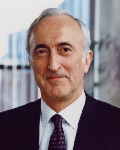 Kevin John Catt合作论文数National Institute of Child Health and Human Development, National Institutes of Health41
 Pemberton Joshua G合作论文数37A Spät合作论文数31Andras Balla合作论文数Institut für Physiologische Chemie, Ruhr-Universität Bochum30A J Baukal合作论文数28
 Marko Jovic合作论文数Nicoya Lifesci Inc24
 Daniel J Toth合作论文数22P Enyedi合作论文数18
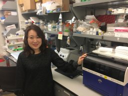 Mira Sohn合作论文数Eunice Kennedy Shriver Natl Inst Child Hlth & Hum, Sect Mol Signal Transduct, Program Dev Neurosci, NIH, Bethesda, MD 20892 USA17
 Gergo Gulyas合作论文数16
 Evzen Boura合作论文数15Guzmán-Hernández Maria Luisa合作论文数National Institutes of Health, University of Geneva14Gerald R. V. Hammond合作论文数12Zsofia Szentpetery合作论文数12Kevin J. Catt合作论文数12
 Gyorgy Hajnoczky合作论文数Department of Pathology & Genomic Medicine, Sidney Kimmel Medical College, Thomas Jefferson University11Nivedita Sengupta合作论文数10T Bondeva合作论文数9
 Marek Korzeniowski合作论文数NICHD, NIH9
 Tibor Rohacs合作论文数Department of Pharmacology, Physiology & Neuroscience, New Jersey Medical School, Rutgers, The State University of New Jersey9
 Zachary Knight合作论文数Howard Hughes Medical Institute;Department of Physiology, University of California, San Francisco8
 Eva Wisniewski合作论文数8
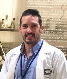 Alvarez-Prats Alejandro合作论文数National Institute of Diabetes and Digestive and Kidney Diseases, National Institutes of Health8
 Sergio Grinstein合作论文数Department of Biochemistry, Temerty Faculty of Medicine, University of Toronto;Research Institute, The Hospital for Sick Children;Keenan Research Centre, Li Ka Shing Knowledge Institute, St. Michael's Hospital8
 Stanko S. Stojilkovic合作论文数Stanko Stojilkovic Lab, National Institutes of Health, US Department of Health and Human Services8
 Amrita Mandal合作论文数NICHD, National Institutes of Health7Takashi Baba合作论文数Section on Molecular Signal Transduction, Program for Developmental Neuroscience, Eunice Kennedy Shriver National Institute of Child Health and Human Development, NIH, Bethesda, MD 20892, USA7Bojjireddy Naveen合作论文数7
 Gyorgy Csordas合作论文数Department of Pathology, Anatomy and Cell Biology, Thomas Jefferson University7
 Anne-Claude Gingras合作论文数Lunenfeld Tanenbaum Research Institute, Mount Sinai Hospital;Department of Molecular Genetics, University of Toronto7G Tuymetova合作论文数6
 J Tóth合作论文数Institute for Nuclear Research (ATOMKI), P.O. Box 51, H-4001, Debrecen, Hungary6
 Julie Brill合作论文数The Hospital for Sick Children Research Institute;Department of Molecular Genetics, University of Toronto6
 Balazs Istvan Toth合作论文数Dept Physiol, Univ Debrecen6
 Kevan M. Shokat合作论文数Department of Chemistry, College of Chemistry, University of California Berkeley;Helen Diller Family Comprehensive Cancer Center, University of California, San Francisco;Department of Cellular and Molecular Pharmacology, University of California, San Francisco Medical Center6
 John E Burke合作论文数5M Bor合作论文数5Kean Michelle J合作论文数5
 John York合作论文数Department of Biochemistry, School of Medicine, Vanderbilt University5
 John E. Heuser合作论文数Department of Cell Biology & Physiology, School of Medicine, Washington University in St. Louis;Roy and Diana Vagelos Division of Biology & Biomedical Sciences, Washington University in St. Louis5S G Rhee合作论文数4Morri Feldman合作论文数4N. Sengupta合作论文数4
 Meredith Jenkins合作论文数Univ Victoria, Dept Biochem & Microbiol, Victoria, BC V8W 2Y2, Canada4
 Roger Williams合作论文数Medical Research Council, Laboratory of Molecular Biology4Erdélyi László Sándor合作论文数4




