Purpose The Clinical Genome Resource (ClinGen) CDH1 Variant Curation Expert Panel (VCEP) developed specifications for CDH1 variant curation with a goal to resolve variants of uncertain significance (VUS) and with ClinVar conflicting interpretations for effective medical care. In addition, the CDH1 VCEP continues to update these specifications in keeping with evolving clinical practice and variant interpretation guidelines. Methods CDH1 variant classification specifications were modified based on updated genetic testing clinical criteria, new recommendations from ClinGen, and expert knowledge from ongoing CDH1 variant curations. Trained biocurators curated 273 variants using updated CDH1 interpretation guidelines and incorporated published and unpublished data provided by diagnostic laboratories. All variants were reviewed by the ClinGen VCEP and classifications submitted to ClinVar. Results Updated CDH1 -specific variant interpretation guidelines include eleven major modifications since the initial specifications from 2018. Using the refined guidelines, 97% (36/37) of variants with ClinVar conflicting interpretations were resolved into benign, likely benign, likely pathogenic, or pathogenic, and 35% (15/43) of VUS were resolved into benign or likely benign. Overall, 88% (239/273) of curated variants had non-VUS classifications. Conclusion The development and evolution of CDH1- specific criteria by the expert panel results in decreased uncertain and conflicting interpretations of variants in this clinically actionable gene.
We report a case of a Hispanic male presenting with acute onset of bilateral lower extremity weakness, without any antecedent viral or bacterial illness, dietary changes, infiltrative orbitopathy, diffuse goiter, infiltrative dermopathy, and family history of periodic paralysis, who was later found to have Graves' disease. This demonstrates a rare case of periodic paralysis as the initial presentation of hyperthyroidism. Thyrotoxic hypokalemic periodic paralysis is common in Asian and Hispanic individuals and uncommon in whites and African Americans.
We perfused iron as FeCl(3) directly into the striatum of normal rats and used the in vivo microdialysis technique to monitor striatal levels of dopamine (DA). KCl was perfused to assess the functional integrity of the DA receptors at the end of each dialysis experiment. Cu(+2) (as CuSO(4)) and Cl(-) (as NaCl) were perfused to compare the effects of Fe(+3) to that of other heavy metal and donors of Cl(-) anion. Perfusion of FeCl(3) (1 mM for 15 min) produced a 250% increase in striatal levels of DA. Perfusion of CuSO(4) (1 mM for 15 min) or NaCl (10 mM for 15 min) did not affect striatal DA levels. There was a significant increase in DA levels with KCl stimulation (56 mM for 15 min) after perfusion with FeCl(3). We conclude that iron releases DA from striatal nerve endings without the immediate destruction of the DA terminals. The implications of chronic release of dopamine as a cause of dopaminergic cell death are discussed.
A paucity of studies are available concerning the comparative therapeutic effectiveness of presently available dopamine agonist agents in the control of Parkinson symptoms. To provide a basis for resolving this issue, we measured the circling response in unilaterally nigrotomized (6-OHDA) rats following the administration of ropinirole, pramipexole, pergolide, bromocriptine, and cabergoline. Cabergoline, and to a lesser extent pergolide, produced the most vigorous and longest lasting circling response. This response was sustained with administration of these agents over a nine day period. Bromocriptine, pramipexole and ropinirole were all less effective. These results suggest that dopamine agonists whose effect is primarily on D1 and D2 receptors are more effective than those whose actions do not include D1 activation.
Movement DisordersVolume 14, Issue 2 p. 368-370 Clinical/Scientific Notes Focal tremor following striatal infarct—A case report† Timothy Brannan MD, Corresponding Author Timothy Brannan MD Department of Neurology, Jersey City Medical Center, Jersey City, New Jersey, U.S.A. Department of Neurology, JFK Neuroscience Institute, Edison, New Jersey, U.S.A.Jersey City Medical Center, Division of Neurology, 50 Baldwin Ave., Jersey City, NJ 07304, U.S.A.Search for more papers by this authorMelvin D. Yahr MD, Melvin D. Yahr MD Department of Neurology, Mount Sinai School of Medicine, New York, NY, U.S.A.Search for more papers by this author Timothy Brannan MD, Corresponding Author Timothy Brannan MD Department of Neurology, Jersey City Medical Center, Jersey City, New Jersey, U.S.A. Department of Neurology, JFK Neuroscience Institute, Edison, New Jersey, U.S.A.Jersey City Medical Center, Division of Neurology, 50 Baldwin Ave., Jersey City, NJ 07304, U.S.A.Search for more papers by this authorMelvin D. Yahr MD, Melvin D. Yahr MD Department of Neurology, Mount Sinai School of Medicine, New York, NY, U.S.A.Search for more papers by this author First published: 17 January 2001 https://doi.org/10.1002/1531-8257(199903)14:2<368::AID-MDS1030>3.0.CO;2-0Citations: 4 † A videotape accompanies this article. AboutPDF ToolsRequest permissionExport citationAdd to favoritesTrack citation ShareShare Give accessShare full text accessShare full-text accessPlease review our Terms and Conditions of Use and check box below to share full-text version of article.I have read and accept the Wiley Online Library Terms and Conditions of UseShareable LinkUse the link below to share a full-text version of this article with your friends and colleagues. Learn more.Copy URL Citing Literature Volume14, Issue2March 1999Pages 368-370 RelatedInformation
We report a patient with a parkinsonian syndrome induced by sertraline (Zoloft®), an SSRI antidepressant, whose symptoms resolved after the drug was discontinued. This case prompted us to investigate the effect of sertraline on dopamine metabolism in animals. Sertraline (30 mg/kg, IP) or placebo (vehicle) was administered to two groups of six normal, anesthetized rats and using cerebral microdyalisis extracellular striatal levels of dopamine, the dopamine metabolites (HVA and DOPAC), as well as the serotonin metabolite 5-HIIA were monitored. In animals pre-treated with sertraline, DOPAC, HVA, and 5-HIAA levels were significantly decreased compared to control animals (p < 0.01). These data indicate that sertraline has an effect on dopamine metabolism, which may alter function in the striatum and induce a parkinsonian syndrome.
We tested the circling response to l-DOPA and apomorphine administration in rats with unilateral 6-hydroxydopamine (6-OHDA) lesions of the substantia nigra. Rats demonstrated a progressively diminished circling response when l-DOPA-carbidopa was repeatedly administered at 120 min intervals. This decreasing response was not present when apomorphine was administered under the same conditions. We also perfused l-DOPA directly into the striatum in vivo of rats with an ipsilateral 6-OHDA nigrotomy at 60 min intervals and monitored striatal dopamine levels with the technique of brain microdialysis. Dopamine formation increased from the first to the fifth trial. This may be secondary to the decrease in uptake sites which accompanies the loss of striatal dopamine nerve terminals. We postulate that the continued presence of dopamine at striatal receptor sites conditions a short-term loss of dopamine receptor sensitivity and a consequent decreased circling response. The observation that desensitization (as measured by decreasing circling) was not present following repeated apomorphine administration may be attributable to its shorter duration of action. We also perfused l-DOPA into the striatum of normal rats and noted a progressive decrease in striatal dopamine levels from the first to the fifth trial. Since this occurred following direct administration of l-DOPA into the striatum, the decrease could not be accounted for by peripheral pharmacodynamics or bioavailability of l-DOPA in the striatum. Since this decrease in dopamine formation was seen only in the normal striatum, its relevance to the diminished behavioral response is unclear.
Inhibitors of the enzyme catechol-O-methyl transferase (COMT) may be useful adjuncts to L-DOPA in the treatment of Parkinson's disease as they offer the possibility of increasing the availability of the amino acid. It is unknown whether a COMT inhibitor which penetrates the blood-brain barrier is preferable to one restricted to extra-cerebral inhibition. We measured liver and brain COMT activity two hours following administration of two COMT inhibitors: entacapone (ENT), mainly peripherally acting, and dinitrocatechol (DNC), peripheral and central acting. As expected, the full spectrum inhibitor DNC (30 mg/kg) induced a near total inhibition of liver and brain COMT activity. Unexpectedly, however, ENT, at 30 mg/kg, produced the same degree of liver and brain COMT inhibition as DNC; using 10mg/kg, ENT still inhibited both liver and brain COMT activity by 80%. Only at 2.5 and 5 mg/kg did ENT achieve a differential inhibition of liver (80% inhibition) versus brain (10-30% inhibition) COMT activity. In a second series of experiments, we administered ENT (2.5, 10, and 30 mg/kg) and DNC (30 mg/kg) to rats and monitored extracellular striatal dopamine and dopamine metabolite levels with cerebral microdialysis both under basal conditions and following L-DOPA/carbidopa administration. No compound modified basal striatal levels of dopamine. ENT at 30 mg/kg (but not 2.5 or 10 mg), as well as DNC, decreased striatal levels of the methylated dopamine metabolite homovanillic acid (HVA). When L-DOPA/carbidopa was administered, dopamine formation was greatest and HVA formation least in animals pretreated with DNC and 30 mg/kg ENT (but not 2.5 or 10 mg/kg ENT). The finding that ENT at doses relatively specific for peripheral enzyme inhibition did not promote dopamine or inhibit HVA formation is most likely due to the 20% residual liver COMT activity present when the inhibitor was used at less than full doses. Our data indicate that DNC and ENT both inhibit striatal HVA formation and increase dopamine formation from exogenously administered L-DOPA. The dopamine promoting effect of ENT is only present, however, at doses which inhibit central as well as peripheral COMT activity.
We used the technique of cerebral microdialysis to monitor the metabolism of exogenously administered L-dopa and compared dopamine and dopamine metabolite formation in the striatum (a site containing abundant dopamine nerve terminals and dopa-decarboxylase (DDC) activity) versus the cerebellum and occipital cortex (sites with limited dopaminergic innervation and DDC activity). The concentrations of dopamine and the major dopamine metabolites dihydroxyphenylacetic acid (DOPAC) and homovanillic acid (HVA) increased in each brain region following L-dopa perfusion; however, dopamine formation was 90% less in the occipital cortex as compared to the striatum and 95% less in the cerebellum. DOPAC formation was 57% less in the occipital cortex and 74% less in the cerebellum. HVA formation was 42% less in the occipital cortex and 70% less in the cerebellum. The levels of the L-dopa metabolite 3-O-methyldopa and the serotonin metabolite 5-hydroxyindoleacetic acid (5HIAA) were identical in the striatum, occipital cortex, and cerebellum both before and after L-dopa administration. We conclude that brain areas with marked reductions in dopamine nerve terminals and DDC activity have a diminished capacity to synthesize dopamine and also lack storage mechanisms to protect the newly synthesized dopamine from degradative metabolism. The relevance of these findings to the use of L-dopa in treating Parkinson's disease is discussed.
The D stereoisomer of dihydroxyphenylalanine (D-DOPA) and its alpha-keto acid metabolite 3,4-dihydroxyphenylpyruvic acid (DHPPA) when infused into the striatum, significantly increased in vivo extracellular dopamine levels. Following D-DOPA administration, the cumulative increase in dopamine levels was 30% of the increase following L-DOPA: following DHPPA it was 11% that of L-DOPA. Rats with unilateral 6-hydroxydopamine-induced lesions of the substantia nigra demonstrated brisk contralateral turning following each compound. The turning, however, was delayed by 10-20 min and total turning was 40% less following D-DOPA and 57% less following DHPPA than it was following L-DOPA. These data indicate that exogenously administered D-DOPA can be metabolized in vivo within the brain to dopamine and suggest this may occur via a transamination pathway in which DHPPA is an intermediary metabolite. The possible relevance of these findings to the treatment of Parkinson's disease is discussed.
edited by L. Battistin, G. Scarlato, T. Caraceni, and S. Ruggieri, 640 pp., ill., Philadelphia, Lippincott-Raven Publishers, 1996, $165.00. This book is a multiauthored account of the 11th Annual International Symposium on Parkinson's Disease held in Rome in March 1994 under the auspices of the World Federation of Neurology. The proceedings of this symposium, which has been held at three year intervals since 1959, have been published in the Advances in Neurology …
The long-term effect of selegiline (L-deprenyl) in the treatment of Parkinson's disease has not been clearly delineated. We report on a group of patients whose treatment was initiated with selegiline (n = 43) and then subsequently included L-dopa-carbidopa (Sinemet) and in whom an extended period of observation was carried out; they are compared to a group of patients whose treatment consisted of L-dopa-carbidopa alone (n = 39). In each, serial observations of the parkinsonian state and the response to treatment on a yearly basis for a period of 5 years were performed. No significant difference in the Hoehn-Yahr stage or in the motor subscores of tremor, rigidity, bradykinesia, and gait-posture was found between the two groups, nor was there a significant difference in the incidence of fluctuating responses or dyskinesias. The group that received combination therapy required less L-dopa than did the group that received L-dopa-carbidopa alone during the first 3 years of treatment and a similar trend was evident in years 4 to 5. We conclude that minimal benefits accrued to the parkinsonian patients from long-term use of selegiline. No clinical evidence to support the claim of "neuroprotective" properties was found. Selegiline's major usefulness is to modify the fluctuating therapeutic response seen with L-dopa-carbidopa.
Utilizing the cerebral microdialysis technique, we have compared in vivo the effects of selective MAO-A, MAO-B, and nonselective MAO inhibitors on striatal extracellular levels of dopamine (DA) and DA metabolites (DOPAC and HVA). The measurements were made in rats both under basal conditions and following L-DOPA administration. Extracellular levels of dopamine were enhanced and DA metabolite levels strongly inhibited both under basal conditions and following L-DOPA administration by pretreatment with the nonselective MAO inhibitor pargyline and the MAO-A selective inhibitors clorgyline and Ro 41-1049. The MAO-B inhibitor deprenyl had no effect on basal DA, HVA, or DOPAC levels. Nevertheless, deprenyl significantly increased DA and decreased DOPAC levels following exogenous L-DOPA administration, a finding compatible with a significant glial metabolism of DA formed from exogenous L-DOPA. We conclude that DA metabolism under basal conditions is primarily mediated by MAO-A. In contrast, both MAO-A and MAO-B mediate DA formation when L-DOPA is administered exogenously. The efficacy of newer, reversible agents which lack the ''cheese effect'' such as Ro 41-1049 are comparable to the irreversible MAO-A inhibitor clorgyline. The possible relevance of these findings for the treatment of Parkinson's disease is discussed.
Dopamine (DA) is released in large quantities from the striatum during cerebral ischemia. Along with excitatory neurotransmitters, DA plays a role in cellular neuronal ischemic injury. In this study we examined the role of nitric oxide (NO) in the ischemia-induced release of DA. A microdialysis probe was stereotactically placed into the corpus striatum of 16 Sprague-Dawley rats for DA, 3,4-dihydroxyphenylacetic acid (DOPAC), and homovanillic acid (HVA) level determinations. After probe stabilization, the animals received either NG-nitro-L-arginine-methyl ester (L-NAME), a NO synthase inhibitor, or vehicle through the microdialysis probe. Temporary global forebrain ischemia was induced using bilateral carotid artery ligature tightening and controlled hemorrhagic hypotension for 15 min. L-NAME administration caused a reduction in ischemic estimated extraneuronal DA concentration by 60% (P < 0.005) compared with control. There was an increase in both DOPAC and HVA concentrations during the recovery period compared to baseline values in the control group (P < 0.05). L-NAME also caused a reduction in HVA concentration compared to vehicle administration during the latter part of recovery (P < 0.05). These data support the concept that ischemic dopamine release may be mediated by NO. This NO-modulated DA release may contribute to the previously reported deleterious neurotoxic effects of NO during ischemia.
We administered Ro 41-1049, an inhibitor of the enzyme monoamine oxidase type A (MAO-A) to rats and monitored extracellular catecholamine levels in the corpus striatum before and after the intraperitoneal (IP) administration of a bolus of L-dopa. Acute administration of Ro 41-1049 (1–50 mg/kg IP) produced a dose-dependent decrease in basal levels of the dopamine metabolites 3,4-dihydroxyphenylacetic acid (DOPAC) and homovanillic acid (HVA) and an increase in basal levels of dopamine. In rats treated with Ro 41-1049 (20 mg/kg IP), L-dopa administration (100 mg/kg IP) produced a greater increase in striatal levels of dopamine than it did in controls, while DOPAC and HVA formation was attenuated. We conclude that inhibition of central MAO-A activity promotes synaptic accumulation of dopamine following administration of pharmacological doses of L-dopa.
Dopamine (DA) is released in large quantities into the corpus striatum during cerebral ischemia and may exacerbate tissue damage. Using cerebral microdialysis, we studied the effect of etomidate on in vivo ischemia-induced DA release in rat corpus striatum. Reversible cerebral ischemia was induced by using carotid ligatures and hypovolemic hypotension, and monitored with laser Doppler flowmetry. After baseline measurements, 20 normothermic, anesthetized rats were subjected to three separate periods of cerebral ischemia, interrupted by 45- to 75-min periods of reperfusion. The rats were randomized into two groups. All rats received 400 mg/kg of intraperitoneal chloral hydrate for induction of anesthesia. In Group I (n = 10) anesthesia was maintained using additional intraperitoneal chloral hydrate 100 mg/kg every 2 h. Group II received etomidate 0.6 mg/kg 10 min before the first episode of cerebral ischemia, followed by an infusion of 60 micrograms.kg-1 x min-1. Before each subsequent period of induced ischemia, an additional dose of etomidate (0.6 mg/kg) was administered. DA levels were approximately 350 times above baseline in Group I during the three ischemic episodes (IS1, IS2, and IS3). In Group II, ischemia-induced DA release was significantly attenuated (by 79%) during IS1, IS2, and IS3 compared to Group I (P < 0.01). DA levels did not significantly change in magnitude during the three ischemic episodes in either group.(ABSTRACT TRUNCATED AT 250 WORDS)
On rare occasions, torsion dystonia can rapidly worsen and produce life-threatening symptoms. We present reports on two children who had generalized dystonia and who demonstrate the management difficulties of ''dystonic storms.''
Log in or Register Subscribe to journalSubscribe Get new issue alertsGet alerts Enter your Email address: Wolters Kluwer Health may email you for journal alerts and information, but is committed to maintaining your privacy and will not share your personal information without your express consent. For more information, please refer to our Privacy Policy. Subscribe to eTOC Secondary Logo Journal Logo All Articles Images Videos Podcasts Blogs Advanced Search Toggle navigation Subscribe Register Login Articles & Issues Current IssuePrevious IssuesPublished Online First For Authors Submit a ManuscriptInformation for AuthorsLanguage Editing ServicesAuthor Permissions Journal Info About the JournalEditorial BoardAdvertisingOpen AccessSubscription ServicesReprintsRights and Permissions All Articles Images Videos Podcasts Blogs Advanced Search
We present two families with adult‐onset focal dystonias with marked variability in disease expression, discuss the clinical findings in relation to the literature, and comment on the difficulties in determining the pattern of inheritance.
 J. Martínez-Tica合作论文数18
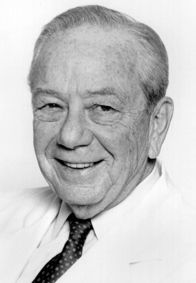 Melvin Yahr合作论文数The Mount Sinai Medical Center15Jesse M Weinberger合作论文数14Alla Prikhojan合作论文数13Hs Maker合作论文数7P. Knott合作论文数5Robert Koorn合作论文数5David L Reich合作论文数Department of Anesthesiology, Mount Sinai School of Medicine, New York, New York5Ronald A. Kahn合作论文数4M Yahr合作论文数4Md Yahr合作论文数3C Weiss合作论文数3
 Horacio Kaufmann合作论文数Department of Neurology, Grossman School of Medicine, New York University;Dysautonomia Center, NYU Langone Health;Division of Autonomic Disorders, NYU Langone Health3
 Juan Narbona合作论文数3Melvin D. Yahr合作论文数3R. Weiser合作论文数2RA KAHN合作论文数2
 José A. Obeso合作论文数Centro Integral de Neurociencias AC;Universidad CEU-San Pablo2A. Bhardwaj合作论文数2Alessandro Di Rocco合作论文数2D. Togasaki合作论文数2H. Maker合作论文数2A SLIVKA合作论文数1J. A. Obeso合作论文数1I TAFF合作论文数1S. Sousa合作论文数1EP LAPIN合作论文数1JT CAROSCIO合作论文数1J Vaamonde合作论文数1M. A. García合作论文数1John Mandeli合作论文数1Grace-Ann Fasaye合作论文数1Carolina Pardo合作论文数1JG YOUNG合作论文数1
 Carla C Oliveira合作论文数1Michael J. Anderson合作论文数1Sainan Wei合作论文数1H. K. Lee合作论文数1P Quesada合作论文数1J. Vaamonde合作论文数1M. Gudin合作论文数1R Ibáñez合作论文数1GM LEHRER合作论文数1Katherine Dixon合作论文数1
 J Artieda合作论文数IdiSNA, Instituto de Investigación Sanitaria de Navarra1
 Arjen R Mensenkamp合作论文数1
 Kasmintan (Intan) Schrader合作论文数Department of Medical Genetics, Faculty of Medicine, The University of British Columbia1
 Brian H. Shirts合作论文数University of Washington Medical Center;Laboratory Medicine, University of Washington1




