Traumatic spinal cord injury (SCI) results in complex neurological dysfunction. Astrocytes react in response to SCI by undergoing proliferation and migrating to the injury site, resulting in astrogliosis. Neural stem cells (NSCs) have been proposed as a promising therapeutic strategy to promote neurogenesis following transplantation post SCI. In this study the antigenic and morphological phenotype of astrocytes, and the pattern of galectin expression was examined following SCI. Female Sprague Dawley rats were contused at thoracic (T) level T9 and treated with Cyclosporin-A (CsA) and/or NSCs 1 week post-injury. CsA and NSC treatment led to increased glial fibrillary acidic protein (GFAP), brain lipid binding protein (BLBP) and vimentin expression. Quiescent and reactive astrocytes were the most abundant cell morphology observed. Astrocytes co-localised with galectin-1 (Gal-1) and galectin-3 (Gal-3). Gal-1 expression was increased in both CsA and CSA + NSC treated animals 2 weeks post-treatment. Treatment with CsA and/or NSCs offers many prospective beneficial effects. A detailed revelation of how astrocytes are heterogeneously responsive to SCI, in addition to their affinity to galectins and response to CsA and NSCs is shown in this study. The current study’s findings support the potential application of NSCs as a novel therapeutic tool in the treatment of SCI.
Human pluripotent stem cells (hPSC) represent a unique opportunity to study fundamental biological processes in a human- and cell-specific setting. Its translational potential and the impact on human health makes this technology revolutionary. The possibility to generate stem cells from almost any somatic cell, and their capacity to be differentiated in virtually all cells of the body has been demonstrated extensively during the last decade of research. Target-centric as well as phenotypic screenings using differentiated cells have become a reality, while the use of these cells for "disease modelling" is still challenging due to the paucity of relevant and reproducible phenotypes. The combination of hPSCs with gene editing technologies aiming to e.g. reduce immunogenic response has enabled promising clinical trials that will eventually demonstrate their therapeutic potential in tissue regeneration and cancer treatment. Maximizing the therapeutic applications of hPSCs requires systematic data comparison, consensus between scientists and health care professionals, as well as a close collaboration between research labs, clinics, and regulators. The goal of this review is to provide a comprehensive outlook of the current use of hPSCs in drug development and regenerative medicine for the treatment of central nervous system (CNS) disorders. In the first part, we analyse how hPSCs are currently used in drug development and discuss their use in challenging paradigms such as neurodegeneration. In the second part we review the status of hPSCs in regenerative medicine. Finally, key challenges and pitfalls of the technology will be discussed, and strategies proposed to improve hPSC research and to benefit patients across different therapeutic areas.
A fast track "Hot Start" process was implemented to launch the European Bank for Induced Pluripotent Stem Cells (EBiSC) to provide early release of a range of established control and disease linked human induced pluripotent stem cell (hiPSC) lines. Established practice amongst consortium members was surveyed to arrive at harmonised and publically accessible Standard Operations Procedures (SOPs) for tissue procurement, bio-sample tracking, iPSC expansion, cryopreservation, qualification and distribution to the research community. These were implemented to create a quality managed foundational collection of lines and associated data made available for distribution. Here we report on the successful outcome of this experience and work flow for banking and facilitating access to an otherwise disparate European resource, with lessons to benefit the international research community. ETOC: The report focuses on the EBiSC experience of rapidly establishing an operational capacity to procure, bank and distribute a foundational collection of established hiPSC lines. It validates the feasibility and defines the challenges of harnessing and integrating the capability and productivity of centres across Europe using commonly available resources currently in the field.
Achieving consistency in standards of access to and quality of human induced pluripotent stem cells has lagged behind their use. In Europe, a network of academic and industrial partners has been established to overcome this challenge. The experience reveals the devil in the detail of worthy ambitions informing future efforts.
The promise of regenerative medicine is that of truly regenerating function in damaged tissue and organs, even curing disease – transformational aspects in treating disease that traditional biopharmaceuticals and medical devices can only dream about. However, the reality is that this sector has fallen short of expectations. Initial investment rounds did not translate into significant value-creating products, the lack of 'blockbuster' cell-based products made many multinationals reluctant to consider strategic portfolio programmes in cell-based regenerative medicines. To this day, significant game-changing efficacy remains to be demonstrated in large-scale Phase III randomised, placebo-controlled clinical trials. In recent years, however, there has been a resurgence of interest in the use of stem cells as a therapy, mainly catalysed by the maelstrom of commercial activity in the engineered T-cell immunotherapy sector. To explain the deal status of the sector, how and why high profile transactions took place are analysed, in order to comprehend the drivers that led to this. Benchmarking is also undertaken with successful trends in other areas, such as anti-hepatitis C virus medicines, to identify comparables. Finally, perspectives and lessons based on the compiled material are summarised into a coherent set of reflections on what the future holds from the transactional aspect for this exciting category of new medicines in development.
For a long time it has been assumed that the only role of sperm at fertilization is to introduce the male genome into the egg. Recently, ideas have emerged that the epigenetic state of the sperm nucleus could influence transcription in the embryo1,2. However conflicting reports have challenged the existence of epigenetic marking of sperm genes3,4, and there are no functional test supporting the role of sperm epigenetic marking on embryonic gene expression. Here, we show that sperm is epigenetically programmed to regulate embryonic gene expression. By comparing the development of spermand spermatid-derived frog embryos we show that the programming of sperm for successful development relates to its ability to regulate transcription of a set of developmentally important genes. During spermatid maturation into sperm, these genes lose H3K4me2/3 and retain H3K27me3 marking. Experimental removal of these epigenetic marks, at fertilization, deregulate gene expression in the resulting embryos in a paternal chromatin dependent manner. This demonstrates that epigenetic instructions delivered by the sperm at fertilization are required for correct regulation of gene expression in the future embryos. The epigenetic mechanism of developmental programming revealed here are likely to relate to the mechanisms involved in transgenerational transmission of acquired traits. Understanding how parental experience can influence development of the progeny has broad potential for improving human health.
This paper describes a decisional tool that is designed to identify cost-effective process designs for drug screening products derived from human induced pluripotent stem cells (hiPSC). The decisional tool comprises a bioprocess economics model linked to a search algorithm to assess the financial impact of manual and automated bioprocessing strategies that use 2D-planar tissue culture technologies. The tool was applied to a case study that examines the production of patient-specific iPSC-derived neurons for drug screening. The production strategies were compared across three analytical drug screening methods, each requiring cell production at a distinct scale (manual patch-clamp analysis, high throughput screening and plate-based pharmacology), as well as different annual cell line utilization requirements ('throughputs') (between 10 and 100 lines) so as to represent different industry scenarios. The tool determined the critical cell line throughput where the most cost-effective production strategy switched from the manual to automated workflow. The key process economics driver was the number of iPSC expansion stages required. Stochastic modelling of the bioprocess illustrated that the automated was more robust than the manual workflow in the scenarios investigated. The tool predicted the level of performance improvements required in iPSC expansion and differentiation as well as reductions in indirect costs and media costs so as to achieve an acceptable cost of goods (COG). (C) 2015 The Authors. Published by Elsevier B.V.
A major goal of immunotherapy remains the control of pathogenic T cell responses that drive autoimmunity and allograft rejection. Adherent progenitor cells, including mesenchymal stromal cells (MSCs) and multipotent adult progenitor cells (MAPCs), represent attractive immunomodulatory cell therapy candidates currently active in clinical trials. MAPCs can be distinguished from MSCs on the basis of cellular phenotype, size, transcriptional profile, and expansion capacity. However, despite their ongoing evaluation in autoimmune and allogeneic solid organ transplantation settings, data supporting the immune regulatory potential of clinical-grade MAPCs are limited. In this study, we used allogeneic islet transplantation as a model indication to assess the ability of clinical-grade MAPCs to control T cell responses that drive immunopathology in human autoimmune disease and allograft rejection. MAPCs suppressed T cell proliferation and Th1 and Th17 cytokine production while increasing secretion of IL-10 and were able to suppress effector functions of bona fide autoreactive T cells from individuals with type 1 diabetes mellitus, including killing of human islets. Furthermore, MAPCs favored the proliferation of regulatory T cells during homeostatic expansion driven by γ-chain cytokines and exerted a durable, yet reversible, control of T cell function. MAPC suppression required licensing and proceeded via IDO-mediated tryptophan catabolism. Therefore, the common immune modulatory characteristics of clinical-grade MAPCs shown in this study suggest that they can be regarded as an alternative source of adult progenitor cells with similar clinical usefulness to MSCs. Taken collectively, these findings may guide the successful deployment of both MSCs and MAPCs for the amelioration of human autoimmunity and allograft rejection.
The utilization of neural stem cells and their progeny in applications such as disease modelling, drug screening or safety assessment will require the development of robust methods for consistent, high quality uniform cell production. Previously, we described the generation of adherent, homogeneous, non-immortalized mouse and human neural stem cells derived from both brain tissue and pluripotent embryonic stem cells (Conti et al., 2005; Sun et al., 2008). In this study, we report the isolation or derivation of stable neurogenic human NS (hNS) lines from different regions of the 8-9 gestational week fetal human central nervous system (CNS) using new serum-free media formulations including animal component-free conditions. We generated more than 20 adherent hNS lines from whole brain, cortex, lobe, midbrain, hindbrain and spinal cord. We also compared the adherent hNS to some aspects of the human CNS-stem cells grown as neurospheres (hCNS-SCns), which were derived from prospectively isolated CD133(+)CD24(-/lo) cells from 16 to 20 gestational week fetal brain. We found, by RT-PCR and Taqman low-density array, that some of the regionally isolated lines maintained their regional identity along the anteroposterior axis. These NS cells exhibit the signature marker profile of neurogenic radial glia and maintain neurogenic and multipotential differentiation ability after extensive long-term expansion. Similarly, hCNS-SC can be expanded either as neurospheres or in extended adherent monolayer with a morphology and marker expression profile consistent with radial glia NS cells. We demonstrate that these lines can be efficiently genetically modified with standard nucleofection protocols for both protein overexpression and siRNA knockdown of exogenously expressed and endogenous genes exemplified with GFP and Nestin. To investigate the functional maturation of neuronal progeny derived from hNS we (a) performed Agilent whole genome microarray gene expression analysis from cultures undergoing neuronal differentiation for up to 32 days and found increased expression over time for a number of drugable target genes including neurotransmitter receptors and ion channels and (b) conducted a neuropharmacology study utilizing Fura-2 Ca2+ imaging which revealed a clear shift from an initial glial reaction to carbachol to mature neuron-specific responses to glutamate and potassium after prolonged neuronal differentiation. Fully automated culture and scale-up of select hNS was achieved; cells supplied by the robot maintained the molecular profile of multipotent NS cells and performed faithfully in neuronal differentiation experiments. Here, we present validation and utility of a human neural lineage-restricted stem cell-based assay platform, including scale-up and automation, genetic engineering and functional characterization of differentiated progeny. (C) 2011 Elsevier Ltd. All rights reserved.
BACKGROUND AIMS:Spinal cord injury is a devastating injury that impacts drastically on the victim's quality of life. Stem cells have been proposed as a therapeutic strategy. Neural stem (NS) cells have been harvested from embryonic mouse forebrain and cultured as adherent cells. These NS cells express markers of neurogenic radial glia.METHODS:Mouse NS cells expressing green fluorescent protein (GFP) were transplanted into immunosupressed rat spinal cords following moderate contusion injury at T9. Animals were left for 2 and 6 weeks then spinal cords were fixed, cryosectioned and analyzed. Stereologic methods were used to estimate the volume and cellular environment of the lesions. Engraftment, migration and differentiation of NS cells were also examined.RESULTS:NS cells integrated well into host tissue and appeared to migrate toward the lesion site. They expressed markers of neurons, astrocytes and oligodendrocytes at 2 weeks post-transplantation and markers of neurons and astrocytes at the 6-week time-point. NS cells appeared to have a similar morphologic phenotype to radial glia, in particular at the pial surface.CONCLUSIONS:Although no functional recovery was observed using the Basso Beattie Bresnahan (BBB) locomotor rating scale, NS cells are a potential cellular therapy for treatment of injured spinal cord. They may be used as delivery vehicles for therapeutic proteins because they show an ability to migrate toward the site of a lesion. They may also be used to replace lost or damaged neurons and oligodendrocytes.
The last decade has seen a dramatic rise in the development of new cellular therapeutics in a wide range of indications. There have been acceptable safety profiles reported in early studies using blood-derived and adherent stem cell products, but also an inconsistent efficacy record. Further expansion has been hindered in part by a lack of capital (both private and public) and delayed entry into the cell therapy space by large healthcare and pharmaceutical companies, those members of the industry most reliably able to initiate and maintain advanced-phase clinical trials. With recognition that the International Society for Cellular Therapy (ISCT) is uniquely positioned to serve the global translational regenerative medicine research community as a network hub for scientific standards and policy, the ISCT commissioned the establishment of an Industry Task Force (ITF) to address current and future roles for industry. The objectives of the ITF were to gather information and prioritize efforts for a new Commercialization Committee (CC) and to construct innovative platforms that would foster constructive and synergistic collaborations between industry and ISCT. Recommendations and conclusions of the ITF included that the new CC: (1) foster new relationships with therapeutic and stem cell societies, (2) foster educational workshops and forums to cross-educate and standardize practices, (3) create industry subcommittees to address priority initiatives, with clear benchmarks and global implementation, and (4) establish a framework for a greater industry community within ISCT, opening doors for industry to share the new vision for commercialization of cell therapy, emphasizing the regenerative medicine space.
The last decade has seen a dramatic rise in the development of new cellular therapeutics in a wide range of indications. There have been acceptable safety profiles reported in early studies using blood-derived and adherent stem cell products, but also an inconsistent efficacy record. Further expansion has been hindered in part by a lack of capital (both private and public) and delayed entry into the cell therapy space by large healthcare and pharmaceutical companies, those members of the industry most reliably able to initiate and maintain advanced-phase clinical trials. With recognition that the International Society for Cellular Therapy (ISCT) is uniquely positioned to serve the global translational regenerative medicine research community as a network hub for scientific standards and policy, the ISCT commissioned the establishment of an Industry Task Force (ITF) to address current and future roles for industry. The objectives of the ITF were to gather information and prioritize efforts for a new Commercialization Committee (CC) and to construct innovative platforms that would foster constructive and synergistic collaborations between industry and ISCT. Recommendations and conclusions of the ITF included that the new CC: (1) foster new relationships with therapeutic and stem cell societies, (2) foster educational workshops and forums to cross-educate and standardize practices, (3) create industry subcommittees to address priority initiatives, with clear benchmarks and global implementation, and (4) establish a framework for a greater industry community within ISCT, opening doors for industry to share the new vision for commercialization of cell therapy, emphasizing the regenerative medicine space.
Regenerative medicine research is focussed on the discovery of novel therapies (small molecules, biologics or cells) that restore function in damaged or aging tissues and organs. The field is underpinned by developments in stem cell science and the modulation of stem cells using small molecules is now providing unique insights into stem cell regulation and developmental biology. It has been shown that small molecules can be used to help drive somatic cell reprogramming, maintain induced pluripotent states and also directly control lineage specification and proliferation events. Small molecules have the potential to help enable the development of viable cell therapies and small molecule oral drugs can be envisaged that can control endogenous cell populations to support regeneration. This review seeks to illustrate the key role small molecules have to play in modulation of stem cells for regenerative medicine and describes some future potential directions in the field.
Neural stem cells, cells which have the ability to self-renew and to differentiate into three neural lineages, hold great potential for many applications in biomedical research, cellular therapy and drug discovery. However, the use of neural stem cells for these applications has been hampered by the lack of availability of stable, pure cell lines. In this review we describe the recent generation of homogenous, pluripotent, stable mouse and human neural stem (NS) cell lines. These cell lines, which can be generated from both foetal and adult tissue and ES cells, possess many attributes which make them amenable to use in the biopharmaceutical industry, such as ease of scale-up, purity and a stable phenotype over extended passage. These characteristics are discussed, along with the technical, safety and ethical issues associated with the use of NS cells in therapeutic and drug discovery applications.
Sarah Adler, Timothy Allsopp, Susanne Bremer, Leonora Buzanska, Rosemary Drake, Ulrik Frandsen, Laura Gribaldo, Karin Gmeiner, Linda Harkness, Thomas Hartung, Lyn Healy, Jürgen Hescheler, Holger Knaut, CarlFredrik Mandenius, Patricia Pazos, Anna Price, Cristian Pellizzer, Martin Pera, Benjamin Reubinoff, Tomas Stojanov, Raimund Strehl, Tina Stummann, James Trosko, Marina Hasiwa(Rapporteur), Glyn Stacey(Chair).
The process of bringing a new pharmacologically active drug to market is laborious, time consuming, and costly. From drug discovery to safety assessment, new methods are constantly sought to develop faster and more efficient procedures to eliminate drugs from further investigation because of their limited effectiveness or high toxicity. Because in vitro cell assays are an important arm of this discovery process, it is therefore somewhat unsurprising that there is an emerging contribution of embryonic stem (ES) cell technology to this area. This technology utilizes the in vitro differentiation of ES cells into somatic cell target populations that, when coupled to the use of "lineage selection" protocols, allows for the production of infinite numbers of pure populations of the desired cells for both bioactivity and toxicological screens. Unlike the use of transformed cell lines, ES-derived cells remain karyotypically normal and therefore better reflect the potential responses of cells in vivo, and when selected are more homogeneous than those obtained using primary cultures. In this chapter we discuss the use of ES cell-derived somatic cells in pharmacological screens, with particular emphasis on neural cells, and describe the methods and protocols associated with the development of ES cell-derived neural cell assays.
ES cells are extraordinary cells, capable of proliferating in a pluripotent state indefinitely and of differentiating spontaneously into all cell types in vivo and many in vitro. However, the manipulation and modification of ES cells by processes such as directed differentiation and genetic modification have placed ES cells at the forefront of many biological studies and could lead to their application in biopharmaceutical areas such as cellular therapy and drug screening. Here we describe some of the ES cell based technologies that have lead to this realisation of ES cell potential.
Caspase 3 activation has been implicated in cell death following a number of neurodegenerative insults. To determine whether caspase genes can affect the susceptibility of cells to neurodegeneration, a transgenic mouse line was created, expressing human caspase 3 under control of its own promoter. The human gene was regulated by the murine homeostatic machinery and human procaspase 3 was expressed in the same tissues as mouse caspase 3. These novel transgenic mice appeared phenotypically and developmentally normal and survived in excess of 2 years. Behavioural assessment using the 5-choice serial reaction time task found no differences from wild-type littermates. Caspase activity was found to be tightly regulated under physiological conditions, however, significantly larger lesions were obtained when transgenic mice were subjected to focal cerebral ischaemia/reperfusion injury compared to wild-type littermates. These data demonstrate that mice overexpressing human caspase 3 are essentially normal, however, they have increased susceptibility to degenerative insults.
 John Fazakerley合作论文数The Peter Doherty Institute for Infection and Immunity, Faculty of Medicine, Dentistry and Health Sciences, The University of Melbourne5
 John Sharkey合作论文数4
 Lyn E Healy合作论文数Francis Crick Inst, London NW1 1AT, England3Lilian Hook合作论文数3
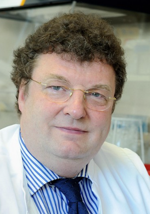 Jürgen Hescheler合作论文数Institute of Neurophysiology, University of Cologne3
 Sean Wyatt合作论文数3
 Ailsa Mcgregor合作论文数Nervous System Research, Novartis Pharma AG3
 Robert J. Deans合作论文数Department of Microbiology, University of Michigan3Martina F Scallan合作论文数Department of Microbiology, University College Cork3
 Paul A. De Sousa合作论文数Department of Biology, University of Pennsylvania3
 Alun Davies合作论文数School of Biosciences, Cardiff University3J S Kelly合作论文数2Lorraine E Kerr合作论文数2Scott R Burger合作论文数2Tina C Stummann合作论文数2Dawn Driscoll合作论文数2Norma Fulton合作论文数2Beate Kreisel合作论文数2Andreas Ebneth合作论文数2Patricia Pazos合作论文数2Ralf Huss合作论文数2Kurt C. Gunter合作论文数2Rachel Steeg合作论文数2
 John E. J. Rasko合作论文数Faculty of Medicine, University of Sydney2
 Chris Mason合作论文数Department of Biochemical Engineering, University College London2
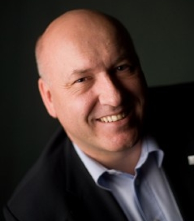 Thomas Hartung合作论文数Department of Environmental Health and Engineering, Johns Hopkins University Bloomberg School of Public Health2Moss D J合作论文数2Neubiser Richard合作论文数2Lardenoije René合作论文数2Field Ed合作论文数2Clark Tara合作论文数2
 Charles S Cox合作论文数2
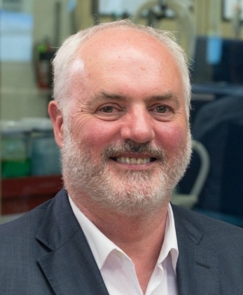 Frank Barry合作论文数Regenerative Medicine Institute, University of Galway2Ulrik Frandsen合作论文数2
 Paul Jones合作论文数Division of Neuroscience and Biomedical Systems, University of Glasgow2
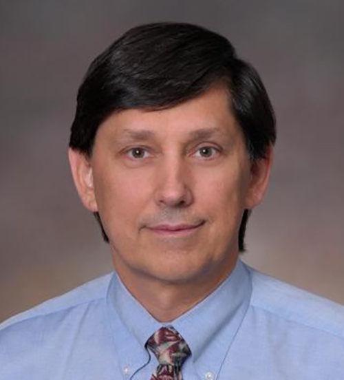 Richard T. Maziarz合作论文数Oregon Health and Science University2
 Holger Knaut合作论文数Department of Molecular and Cellular Biology, Harvard University2Benjamin E. Reubinoff合作论文数2
 Leonora Buzanska合作论文数2
 Marina Hasiwa合作论文数2
 Glyn Stacey合作论文数School of Allied Health Sciences, University of Suffolk;International Stem Cell Banking Initiative;SSCBio Ltd;University of Bedfordshire;Institute of Zoology, Chinese Academy of Sciences2
 Laura Gribaldo合作论文数2
 Carl-Fredrik Mandenius合作论文数Department of Physics, Chemistry and Biology, Linköping University2
 Martin Pera合作论文数The Jackson Laboratory2Tomas Stojanov合作论文数2
 Mark Bonyhadi合作论文数2Friedrich Bonhoeffer合作论文数2
 Jon A. Rowley合作论文数Aastrom Biosciences, Inc.2Linda Harkness合作论文数2




