Background/Objectives: Disrupted intracellular calcium (Ca2+i) regulation and renin–angiotensin system (RAS) activation are pathogenetic factors in diabetic cardiomyopathy, a major complication of type 1 (T1D) and type 2 (T2D) diabetes. This study explored their potential link in diabetic rat hearts. Methods: Experiments were conducted on T1D and T2D Sprague-Dawley rats induced by streptozotocin and fructose-rich diet, respectively. In T1D, rats were treated with Enalapril (Ena) or Losartan (Los) for six weeks, whereas T2D animals received high-dose (HD) or low-dose (LD) Ena for 8 weeks. Heart function was assessed via echocardiography, Ca2+i transients by Indo-1 fluorometry in Langendorff-perfused hearts, and key Ca2+i cycling proteins by Western blot. Data: mean ± SD. Results: Diabetic hearts exhibited reduced contractile performance that was improved by RAS inhibition both in vivo (ejection fraction (%): T1D model: Control: 79 ± 7, T1D: 54 ± 11, T1D + Ena: 65 ± 10, T1D + Los: 69 ± 10, n = 18, 18, 15, 10; T2D model: Control: 73 ± 8, T2D: 52 ± 6, T2D + LDEna: 62 ± 8, T2D + HDEna: 76 ± 8, n = 9, 8, 6, 7) and ex vivo (+dPressure/dtmax (mmHg/s): T1D model: Control: 2532 ± 341, T1D: 2192 ± 208, T1D + Ena: 2523 ± 485, T1D + Los: 2643 ± 455; T2D model: Control: 2514 ± 197, T2D: 1930 ± 291, T2D + LDEna: 2311 ± 289, T2D + HDEna: 2614 ± 268). Analysis of Ca2+i transients showed impaired Ca2+i release and removal dynamics and increased diastolic Ca2+i levels in both models that were restored by Ena and Los treatments. We observed a decrease in sarcoendoplasmic reticulum Ca2+-ATPase2a (SERCA2a) expression, accompanied by a compensatory increase in 16Ser-phosphorylated phospholamban (P-PLB) in T2D that was prevented by both LD and HD Ena (expression level (% of Control): SERCA2a: T2D: 36 ± 32, T2D + LDEna: 112 ± 32, T2D + HDEna: 106 ± 30; P-PLB: T2D: 557 ± 156, T2D + LDEna: 129 ± 38, T2D + HDEna: 108 ± 42; n = 4, 4, 4). Conclusions: The study highlights the critical role of RAS activation, most likely occurring at the tissue level, in disrupting Ca2+i homeostasis in diabetic cardiomyopathy. RAS inhibition with Ena or Los mitigates these disturbances independent of blood pressure effects, underlining their importance in managing diabetic heart failure.
Angiotensin II (Ang II) has various cardiac effects and causes vasoconstriction. Ang II activates the type-1 angiotensin receptor—Gq/11 signaling pathway resulting in the release of 2-arachidonoylglycerol (2-AG). We aimed to investigate whether cardiac Ang II effects are modulated by 2-AG-release and to identify the role of type-1 cannabinoid receptors (CB1R) in these effects. Expression of CB1R in rat cardiac tissue was confirmed by immunohistochemistry. To characterize short-term Ang II effects, increasing concentrations of Ang II (10−9–10−7 M); whereas to assess tachyphylaxis, repeated infusions of Ang II (10−7 M) were administered to isolated Langendorff-perfused rat hearts. Ang II infusions caused a decrease in coronary flow and ventricular inotropy, which was more pronounced during the first administration. CB agonist 2-AG and WIN55,212-2 administration to the perfusate enhanced coronary flow. The flow-reducing effect of Ang II was moderated in the presence of CB1R blocker O2050 and diacylglycerol-lipase inhibitor Orlistat. Our findings indicate that Ang II-induced cardiac effects are modulated by simultaneous CB1R-activation, most likely due to 2-AG-release during Ang II signalling. In this combined effect, the response to 2-AG via cardiac CB1R may counteract the positive inotropic effect of Ang II, which may decrease metabolic demand and augment Ang II-induced coronary vasoconstriction.
ObjectiveEstrogens enhance ischemia tolerance (IT) in the myocardium, the mechanism of which remains unclear. We investigated the effects of long-term estrogen deprivation on the intracellular calcium (Ca-i(2+)) transient of the heart and its possible influence on IT.MethodsHearts of ovariectomized (OVX) and sham-operated (control) adult female rats (some receiving estrogen therapy) were studied 10 weeks after surgical operation: control (n = 8), OVX (n = 10), sham-operated estrogen-substituted (n = 7), and ovariectomized estrogen-substituted (n = 9). In vivo heart function was assessed by echocardiography, whereas Ca-i(2+) transients were recorded, concomitantly with left ventricular pressure and coronary flow, by Indo-1 surface fluorometry in isolated Langendorff-perfused hearts. Isolated hearts were subjected to a 30-minute global ischemia-30-minute reperfusion protocol. Left ventricular expression of myocardial sarcoendoplasmic reticulum Ca2+-ATPase (SERCA2a), phospholamban (PLB), and Ser16-phosphorylated PLB was measured.ResultsOvariectomy did not influence resting cardiac function in vivo or ex vivo. However, Ca2+ removal was slower. During ischemia, Ca-i(2+) elevation and ischemic contracture were more pronounced after ovariectomy. Postischemic restitution of inotropic function (developed pressure; +dP/dt(max)) and lusitropic function (-dP/dt(max)) and Ca-i(2+) transient recovery (amplitude; +/- dCa(i)(2+)/dt(max)) were decreased in OVX hearts. Sarcoendoplasmic reticulum Ca2+-ATPase expression was unaltered, whereas PLB and Ser16-phosphorylated PLB levels were higher after ovariectomy. All effects of ovariectomy were restored by estrogen therapy.ConclusionsOvariectomy impairs myocardial Ca2+ removal by increasing the expression of the SERCA2a inhibitor PLB. Defective Ca2+ transport causes ischemic Ca-i(2+) overload and insufficient postischemic recovery of Ca-i(2+) transients, which entail depressed hemodynamic restitution. Protection of intact Ca2+ cycling in the myocardium by estrogens plays a major role in enhancing IT.
AIM:Various components of metabolic syndrome associate with cardiac intracellular calcium (Cai 2+) mishandling, a precipitating factor in the development of heart failure. We aimed to provide a thorough description of early stage Cai 2+-cycling alterations in the fructose-fed rat, an experimental model of the disorder, where insulin resistance, hypertension and dyslipidaemia act cooperatively on the heart.METHOD:Rats were fed with fructose-rich chow. After 6 weeks, echocardiography was performed, which was followed by measurements of myocardial Cai 2+ transients recorded by Indo-1 surface fluorometry in isolated perfused hearts. Sarcoplasmic reticulum (SR) Ca(2+) -ATPase (SERCA2a) activity was assessed by administration of its inhibitor cyclopiazonic acid (CPA). Mathematical model analysis of Cai 2+ transients was used to estimate kinetic properties of SR Ca(2+) transporters. Protein levels of key Ca(2+) handling proteins were also measured.RESULTS:Echocardiography showed signs of cardiac hypertrophy, but in vivo and ex vivo haemodynamic performance of fructose-fed rat hearts were unaltered. However, a decline in Ca(2+) sequestration capacity (-dCai 2+/dt and decay time of Cai 2+ transients) was observed. Model estimation showed decreased affinity for Ca(2+) (higher K(m) ) and elevated V(max) for SERCA2a. Diseased hearts were more vulnerable to CPA application. Fructose feeding caused elevation in SERCA2a and phosphorylated phospholamban (PLB) expression, while total PLB level remained unchanged.CONCLUSION:In early stage, metabolic syndrome primarily disturbs SERCA2a function in the heart, but consequential haemodynamic dysfunction is prevented by upregulation of SERCA2a protein level and phosphorylation pathways regulating PLB. However, this compensated state is very vulnerable to a further decline in SERCA2a function.
The muscle Lim protein knock-out (MLP-KO) mouse model is extensively used for studying the pathophysiology of dilated cardiomyopathy. However, explanation is lacking for the observed long survival of the diseased mice which develop until adulthood despite the gene defect, which theoretically predestines them to early death due to heart failure. We hypothesized that adaptive changes of cardiac intracellular calcium (Ca i 2+ ) handling might explain the phenomenon. In order to study the progression of changes in cardiac function and Ca i 2+ cycling, myocardial Ca i 2+ -transients recorded by Indo-1 surface fluorometry were assessed with concomitant measurement of hemodynamic performance in isolated Langendorff-perfused hearts of 3- and 9-month old MLP-KO animals. Hearts were challenged with β-agonist isoproterenol and the sarcoplasmic reticular Ca2+-ATPase (SERCA2a) inhibitor cyclopiazonic acid (CPA). Cardiac mRNA content and levels of key Ca2+ handling proteins were also measured. A decline in lusitropic function was observed in 3-month old, but not in 9-month old MLP-KO mice under unchallenged conditions. β-adrenergic responses to isoproterenol were similar in all the studied groups. The CPA induced an increase in end-diastolic Ca i 2+ -level and a decrease in Ca2+-sequestration capacity in 3-month old MLP-KO mice compared to age-matched controls. This unfavorable condition was absent at 9 months of age. SERCA2a expression was lower in 3-month old MLP-KO than in the corresponding controls and in 9-month old MLP-KO hearts. Our results show time-related recovery of hemodynamic function and an age-dependent compensatory upregulation of Ca i 2+ handling in hearts of MLP-KO mice, which most likely involve the normalization of the expression of SERCA2a in the affected hearts.
The endothelium‐mediated vasomotor function of skeletal muscle arterioles has been shown to be impaired in HHcy. Thus we hypothesized that HHcy modulates the reactive hyperemic response of coronary circulation following occlusion. Rats received 1g/kg of body weight methionine daily, for 5 weeks in the drinking water to induce mild HHcy (~ 30 μmol vs. ~ 7 μmol). In Langendorff‐perfused hearts reactive hyperemia (RH) after 5 min of global ischemia was studied prior to and after the simultaneous administration of the nitric oxide synthase inhibitor L‐NAME (10−4M) and the cyclooxygenase inhibitor indomethacin (INDO 10−5M) into the perfusate. We have found that the early peak values of reactive hyperemia were not significantly different in HHcy and control (C) groups (at 2 min of RH: HHcy: 139±14 vs. C: 147±19% of basal flow). The later phase of reactive hyperemia however, was significantly diminished in HHcy hearts (at 8 min of RH: HHcy: 116±7 vs. C: 130±14% of basal flow). Presence of L‐NAME+INDO eliminated the difference between the reactive hyperemia of control and HHcy hearts. On the basis of these findings we propose that cardiac reactive hyperemia is impaired in HHcy, primarily due to reduced release of endothelium‐derived dilator factors. (Grants: Hungarian Sci. Res. Funds/OTKA T48376, 61694, 68502, T67984; and Am. Heart Assoc. NE Aff. 0555897T, USA).
Acta PhysiologicaVolume 194, Issue 2 p. 171-173 Teaching workshop on ‘Implications of the Bologna Declaration for Teaching Physiology in Medical Education’ at the joint meeting of the German Physiological Society and the Federation of European Physiological Societies, Cologne, 2–5 March 2008 L. H. E. H. Snoeckx, L. H. E. H. Snoeckx Department of Physiology, Maastricht University, Maastricht, the NetherlandsSearch for more papers by this authorT. Ivanics, T. Ivanics Institute of Human Physiology and Clinical Experimental Research, Semmelweiss University, Budapest, HungarySearch for more papers by this authorL. Peltonen, L. Peltonen Department of Biomedicine/Physiology, University of Helsinki, Helsinki, FinlandSearch for more papers by this authorU. Decking, U. Decking Department of Cardiovascular Physiology, University of Düsseldorf, Düsseldorf, GermanySearch for more papers by this authorJ. H. Ravesloot, J. H. Ravesloot Department of Physiology, Academic Medical Center of the University of Amsterdam, Amsterdam, the NetherlandsE-mail: l.snoeckx@fys.unimaas.nlSearch for more papers by this author L. H. E. H. Snoeckx, L. H. E. H. Snoeckx Department of Physiology, Maastricht University, Maastricht, the NetherlandsSearch for more papers by this authorT. Ivanics, T. Ivanics Institute of Human Physiology and Clinical Experimental Research, Semmelweiss University, Budapest, HungarySearch for more papers by this authorL. Peltonen, L. Peltonen Department of Biomedicine/Physiology, University of Helsinki, Helsinki, FinlandSearch for more papers by this authorU. Decking, U. Decking Department of Cardiovascular Physiology, University of Düsseldorf, Düsseldorf, GermanySearch for more papers by this authorJ. H. Ravesloot, J. H. Ravesloot Department of Physiology, Academic Medical Center of the University of Amsterdam, Amsterdam, the NetherlandsE-mail: l.snoeckx@fys.unimaas.nlSearch for more papers by this author First published: 05 September 2008 https://doi.org/10.1111/j.1748-1716.2008.01886.xRead the full textAboutPDF ToolsRequest permissionExport citationAdd to favoritesTrack citation ShareShare Give accessShare full text accessShare full-text accessPlease review our Terms and Conditions of Use and check box below to share full-text version of article.I have read and accept the Wiley Online Library Terms and Conditions of UseShareable LinkUse the link below to share a full-text version of this article with your friends and colleagues. Learn more.Copy URL Share a linkShare onFacebookTwitterLinked InRedditWechat Volume194, Issue2October 2008Pages 171-173 RelatedInformation
A jelen munkaban a PARP feherje szerepet vizsgaltuk a cukobetegseg szovődmenyeinek kialakulasaban. Kimutattuk, hogy az erek es a vese karosodasaban fontos szerepe van ennek a feherjenek, es a feherje aktivitasanak gatlasa allatokban javitja a betegseg lefolyasat. Osszefuggest talaltunk a PARP aktivacioja, valamint a rosszul beallitott cukorbetegseg kozott, es kimutattuk kulonfele sejten beluli szignal transzdukcios ut szerepet ezekben a folyamatokban. A munka szamos nemzetkozi szintű kozlemenyt eredmenyezett. | In the present project, we have investigated the role of the PARP enzyme in the pathogenesis of diabetic complications. We have demonstrated that the vascular and renal damage associated with diabetes is importantly mediated by the activation of the PARP enzyme. We have demonstrated a relationship between poorly controlled diabetes and the activation of PARP and we have demonstrated the role of several intracellular signal transduction pathways in the process. The work has resulted in a number of internationally significant publications.
AIM:The objective was to characterize cross-bridge kinetics from the cytoplasmic calcium ion concentration ([Ca2+](i)) and the left ventricular pressure (LVP) in the early-stage diabetic rat heart under baseline conditions and upon beta-adrenergic stimulation.METHODS:Four weeks after the induction of diabetes in rats by the injection of streptozotocin, the hearts were perfused according to Langendorff, and [Ca2+](i) was obtained by epifluorescence measurements using Indo-1 AM. [Ca2+](i) and LVP were measured simultaneously at a temporal resolution of 200 Hz. The input/output relationship between the Ca2+ and the pressure transients was described by a mathematical model representing the chemical binding of Ca2+ to troponin C on the actin myofilament (TnCA), and the subsequent cooperative force-producing cross-bridge formation of the Ca2+-TnCA complex with myosin. The kinetic parameters of this model were evaluated using a numerical optimization algorithm to fit the model equations to the experimental data. beta-adrenergic stimulation of the hearts with increasing doses of isoproterenol allowed quantification of the model parameters over an extended dynamic range, because isoproterenol administration increased developed pressure, heart rate, as well as [Ca2+](i) amplitude in a dose-dependent manner.RESULTS:Model analysis of the experimental data indicates that beta-adrenergic stimulation of healthy hearts resulted in a decreased sensitivity of TnCA for Ca2+, increased rates of cross-bridge cycling and decreased cooperativity. By contrast, the responses in cross-bridge kinetic parameters to isoproterenol stimulation were blunted in the 4-week diabetic heart.CONCLUSION:We conclude from our modelling results that myocardial cross-bridge cycling is impaired at the early stage of diabetes.
A palyazat fő celkitűzese a kulonboző etiologiaju szivizom elegtelenseg hattereben allo koros intracellularis kalcium homeosztazis felderitese volt. A kiserleteink soran vizsgalt kardiomiopatiak magukba foglaltak a diabetesz talajan kialakulo szivizom elegtelenseget, a toxikus eredetű es a dilatativ kardiomiopatiat. A kardialis inszufficiancia es Ca2+i homeosztazis kapcsolatat iszkemia/reperfuzio soran is vizsgaltuk. A kardiomiopatias elvaltozasokat patkany es genetikailag modositott eger modellekben vizsgaltuk. Alap modszerkent a Langendorff szerint perfundalt szivet alkalmaztuk, vizsgalt parametereink a sziv hemodinamikai teljesitmenye, a felszini fluorimetriaval nyert Ca2+i szint, valamint a Ca2+i homeosztazisban reszt vevő nehany kulcsfeherje szoveti tartalma volt. Kiserleteink eredmenyekent feltartuk a kulonboző szivelegtelenseg modellekben bekovetkező karakterisztikus Ca2+i homeosztazis valtozasokat. Ez lehetőve tette eredmenyeink molekularis szintű interpretalasat. | Funds of this grant allowed us to study the pathological changes of myocardial intracellular calcium (Ca2+i) cycling in cardiomyopthy of various origin. Experimental models used in this series of experiments included diabetic, toxic and dilatative cardiomyopthy. We also studied the relationship between Ca2+i homeostasis and cardiac insufficiency in an ischemia/reperfusion model. The experiments were performed in rat and genetically modified mouse models using Langendorff perfused hearts. The main methodology included measurements of hemodynamic performance of the hearts, Ca2+i measured by surface flouorometry and the determination of tissue content of some key proteins involved in Ca2+i handling. Our experiments allowed the determination of characteristic changes of Ca2+i homeostasis in cardiomyopathy of varoius etiology. These results enabled us a molecular interpretation of pathological Ca2+i handling in heart failure.
 Zsuzsanna Miklos合作论文数22
 L LIGETI合作论文数19László Ligeti合作论文数13
 Andras J. Toth合作论文数Department of Manufacturing Engineering, Budapest University of Technology and Economics11Ger J van der Vusse合作论文数Cardiovascular Research Institute Maastricht, Department of Physiology, Maastricht University10
 Robert S Reneman合作论文数Cardiovascular Research Institute Maastricht, Maastricht University9
 László Dézsi合作论文数Department of General Pharmacology, Gedeon Richter Ltd.9Mark Kollai合作论文数9
 Attila Tóth合作论文数Centre for Neuroscience, University of Pécs9Zs Miklos合作论文数6Orsolya Szenczi合作论文数6
 Csaba Szabó合作论文数Department of Medicine, Université de Fribourg;Department of Oncology, Microbiology and Immunology, University of Fribourg6
 Jorn op den Buijs合作论文数5
 Natal A W van Riel合作论文数Eindhoven Univ Technol, Dept Biomed Engn, NL-5600 MB Eindhoven, Netherlands5Batkai Sandor合作论文数Hannover Med Sch, Inst Mol & Translat Therapeut Strategies5
 Dick W. Slaaf合作论文数Department of Biophysics, Maastricht University5
 Ger J. Van Der Vusse合作论文数Department of Physiology, Maastricht University5Z Ruttner合作论文数4N Doliba合作论文数4Naw Van Riel合作论文数4Péter Kemecsei合作论文数4Dw Slaaf合作论文数4Kemecsei P合作论文数4
 Maria Szekeres合作论文数4Zoltan Ruttner合作论文数4D ZHANG合作论文数4K Ikrényi合作论文数3Drozgyik A合作论文数3Benis É合作论文数3
 Zsolt Lohinai合作论文数Semmelweis University Institute of Human Physiology and Experimental Research3G Van Der Vusse合作论文数2Christina M Prestia合作论文数2Akos Koller合作论文数2A MAYEVSKY合作论文数2Md Osbakken合作论文数2
 Balazs Istvan Toth合作论文数Dept Physiol, Univ Debrecen2
 Andras Attila Kiss合作论文数2nd Department of Pathology, Semmelweis University2Theo H. M. Roemen合作论文数2L. H. E. H. Snoeckx合作论文数2
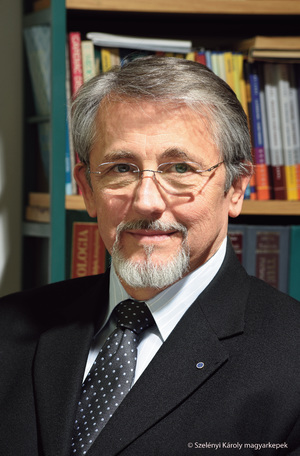 Laszlo Rosivall合作论文数Institute of Pathophysiology, Faculty of Medicine, Semmelweis University2Mary Osbakken合作论文数2
 Péter Kempler合作论文数1st Department of Internal Medicine, Semmelweis University2
 Nandor Acs合作论文数Obstetrics and Gynecology Department, Faculty of Medicine, Semmelweis University2Péter Vörös合作论文数2Jorn Op Den Buijs合作论文数2Rita Marincsák合作论文数2P P Nánási合作论文数1R. Benko合作论文数1




