Sodium glucose cotransporter 2 (SGLT2) inhibitors are expected to have, renoprotective effects based on the possible mechanism that SGLT2 inhibitors reduce hyperfiltration by blocking proximal tubule sodium reabsorption and activating tubuloglomerular feedback (TGF) and may decrease inflammatory, fibrotic, and hyperplastic response of proximal tubular cells by blocking glucose reabsorption into proximal tubular cells. Although SGLT2 inhibition is associated with an acute reduction in estimated glomerular filtration rate (eGFR) by about 4-6 mL/min per 1.73m2, eGFR typically has a tendency to return to baseline and remains stable thereafter.
Human endometrial mucosa is a dynamically remodeling tissue, undergoing cyclical morphologic and functional changes in response to fluctuating sex steroid hormones each menstrual cycle during a woman's reproductive life. Postmenopausal endometrium responds similarly to exogenous estrogen. Cyclical endometrial regeneration also occurs in nonmenstruating rodents, although to a lesser extent. The recent identification of rare populations of endogenous epithelial progenitor cells, mesenchymal stem/stromal cells (MSCs), the side population (SP) cells, and label-retaining cells (LRCs) suggests these stem/progenitor cell populations may play a key role in endometrial regeneration during menstrual and estrus cycles. This review summarizes the identification of epithelial progenitors, MSC, SP, and LRC, and discusses their contribution to endometrial tissue regeneration, maintaining tissue homeostasis, decidualization, and placentation. Markers for human endometrial MSC have been identified, revealing their perivascular location in both the functionalis and basalis layers. These markers also allow their purification from biopsy tissue and menstrual blood. These findings have advanced our understanding of normal endometrial physiology and will provide new insight into endometrial proliferative disorders (endometriosis, endometrial cancer). The ability to prospectively isolate endometrial MSC will enable their utilization in cell-based therapies for reproductive tract pathologies.
Study question: To compare the pregnancy rate in patients submitted to FET (Frozen Embryos Transfer) of cleavage stage embryos (Group 1), or at the blastocyst stage (Group 2).Summary answer: Our data showed that the transfer of frozen blastocysts has a better pregnancy rate than cleavage embryos.What is known already: The first birth of a baby arising from a cryopreserved embryo occurred in 1983.Since then, improvements in embryo cryopreservation have occurred.However since early 90 ′ s a vitrification technique has also been developed to cryopreserve embryos in any stage of development.Study design, size, duration: Retrospective cohort study during 2012, with a total of 132 cycles.Participants/materials, setting, methods: In the group 1 the embryos had been frozen on the 2 nd , 3 rd or 4 th day after fertilization, while in the group 2 the embryos were frozen on 5 th or 6 th day at blastocyst stage.All embryos were cryopreserved by vitrification followed Kuwayama et al, 1998 protocol.The pregnancy rates were compared through the chi-square test (p , 0.05).Main results and the role of chance: The Group 1 consisted of 74 patients, which pregnancy and clinical pregnancy rate were 27.0% (20/74) and 21.6% (16/74), respectively.The Group 2 had 58 patients, and pregnancy and clinical pregnancy rate were 50.0%(29/58) and 43.1% (25/58) respectively.There was statistical difference between the groups (P¼ 0.0107).The mean age were 34.8 and years old for groups 1 and 2 respectively.Limitations, reason for caution: The study was conducted during a short period of time.Wider implications of the findings: This result suggests that the transfer of embryos at the blastocyst stage would be the best choice for IVF clinics.Study funding/competing interest(s): There were no competing interests in this study.
The human uterus is composed of the endometrial lining and the myometrium. The endometrium, in particular the functionalis layer, regenerates and regresses with each menstrual cycle under hormonal control. A mouse xenograft model has been developed in which the functional changes of the endometrium are reproduced. The myometrium possesses similar plasticity, critical to permit the changes connected with uterine expansion and involution associated with pregnancy. Regeneration and remodeling in the uterus are likely achieved through endometrial and myometrial stem cell systems. Putative stem/progenitor cells in humans and rodents recently have been identified, isolated and characterized. Their roles in endometrial physiology and pathophysiology are presently under study. These stem/progenitor cells ultimately may provide a novel means by which to produce tissues and organs in vitro and in vivo.
Uterine tissue engineering can be considered a possible option for fertility preservation in patients with uterine factor infertility. Several bioengineered organs including heart have recently made through recellularization of decellularized scaffolds (DS) prepared from the equivalent organs. The purpose of this study was to develop a technique for decellularization of rat uterus and to elucidate potential of the resulting DS for uterine regeneration. DS were prepared from uteri collected from Fischer adult rats and recellularized in vitro by uterine cells isolated from Fischer adult and neonatal rats. DS were also transplanted to the defective area of the uterine horns of Fischer adult rats. Rat uteri were excised along with large vessels and perfused with detergent to prepare DS. DS was injected and incubated with rat uterine cells in vitro for recellularization. Recipient rats underwent segmental uterine excision and were subjected to placement of DS onto the defective area. Histological and electron microscopic analysis revealed that DS contained no intact cells but retained extracellular matrix and microvasculature. Recellularization of DS resulted in regeneration of endometrium comprised in stroma and epithelium, as determined by immunohistochemistry. Uterine tissue consisting of endometrium and myometrium was also regenerated at the transplantation site of DS in vivo 28 days after surgery. Recipient rats were mated with male rats 28 days after surgery, and 1.3 ± 0.5 fetuses (mean ± SD) per uterine horn were found in the late pregnancy stage. We have developed a decellularization method to prepare rat uterine DS and found that the DS have potentials as a 3D scaffold to regenerate uterine tissues in vivo and in vitro through recellularization. These results implicate DS as a possible ideal material for uterine regeneration for patients with uterine factor infertility.
BACKGROUND Little is known about the effects of recurrent pregnancy loss (RPL) on the psychological adjustment of couples. The aim of this study was to elucidate psychological adjustment and RPL-associated psychosocial stress affecting Japanese couples with a history of RPL, focusing on gender differences and quality of the marital relationship. METHODS The study included 76 RPL couples who visited the outpatient clinic of a tertiary hospital. They completed self-administered questionnaires that assessed RPL-associated stress, quality of their marital relationship (Quality Marriage Index, QMI), depression (Beck Depression Index) and anxiety (State-Trait Anxiety Inventory). RESULTS Women showed significantly higher levels of depression, anxiety and RPL-associated personal and social stress compared with men. Although there were no differences in QMI scores and RPL-associated marital stress between men and women, women with a low perception of marital relationship quality (low QMI) had significantly higher levels of depression and anxiety compared with women with a moderate or high QMI. In contrast, depression and anxiety scores did not differ according to the quality of the marital relationship among men. Of 76 couples, 26 men (34%) and 45 women (59%) who had considered professional mental health consultations regarding their RPL status but had not yet initiated the process were more depressed and anxious than 48 men and 24 women, respectively, who had never considered such consultation. CONCLUSIONS Women were significantly more distressed than men. Poor quality of the marital relationship was significantly associated with impaired psychological adjustment among women, but not among men. These gender discrepancies may foster a mutual worsening of psychological adjustment and marital relationships in RPL couples. The need to seek help not only in women but also in a substantial portion of men suggests the importance of couple-based psychological care in the management of RPL.
The Wnt/β-catenin signaling is essential for various organogenesis and is often implicated during tumorigenesis. Dysregulated β-catenin signaling is associated with the formation of endometrial adenocarcinomas (EACs), which is considered as the common form of endometrial cancer in women. In the current study, we investigate the downstream target of Wnt/β-catenin signaling in the uterine epithelia and the mechanism leading to the formation of endometrial hyperplasia. We report that conditional ablation and activation of β-catenin in the uterine epithelia lead to aberrant epithelial structures and endometrial hyperplasia formation, respectively. We demonstrate that β-catenin regulates Foxa2 with its candidate upstream region for the uterine epithelia. Furthermore, knockdown of Foxa2 leads to defects in cell cycle regulation, suggesting a possible function of Foxa2 in the control of cell proliferation. We also observe that β-catenin and Foxa2 expression levels are augmented in the human specimens of complex atypical endometrial hyperplasia, which is considered to have a greater risk of progression to EACs. Thus, our study indicates that β-catenin regulates Foxa2 expression, and this interaction is possibly essential to control cell cycle progression during endometrial hyperplasia formation. Altogether, the augmented expression levels of β-catenin and Foxa2 are essential features during the formation of endometrial hyperplasia.
Primary ovarian insufficiency (POI) is a heterogeneous condition defined by the triad of oligo/amenorrhea, elevated gonadotropins and estrogen deficiency in women under the age of 40 years. Although autoimmune abnormalities appear to be involved in the development of POI, there are only a few studies with respect to human leukocyte antigen (HLA). The objective of this study was to identify an HLA allele(s) and/or haplotype(s) constituting a susceptibility factor(s) for POI. We examined 83 Japanese women with apparently idiopathic isolated POI. For controls, Japanese HLA reference data registered in the HLA Laboratory were utilized. No significant association was found for a total of 94 alleles for HLA-A, B, C, DRB1, and DQB1 loci, after both stringent Bonferroni correction and less stringent Benjamini- Hochberg (B-H) correction for multiple comparisons. By contrast, of 86 haplotypes identified for MHC class I (HLA-A, B, and C) and 31 haplotypes detected for MHC class II (HLA-DRB1 and DQB1), a single haplotype (A*24:02-C*03:03-B*35:01) remained significant after Bonferroni and B-H corrections (frequency: 4.82% in women with POI and 1.06% in the control data; p = 0.00049). The results imply that a specific HLA haplotype (A*24:02-C*03:03-B*35:01) constitutes a susceptibility factor for apparently isolated POI in Japanese women.
BACKGROUND The transcription factor, octamer-binding transcription factor 4 (OCT4)/POU5F1, is expressed in embryonic stem cells, germ cells and some types of adult stem cells. Human OCT4 encodes two isoforms, OCT4A and OCT4B. While OCT4A plays a crucial role in the maintenance of stem cell properties, including pluripotency, whereas OCT4B does not. We previously reported that human myometrium contains side population cells (myoSP) with a Hoechst 33 342 low-fluorescent profile. These cells exhibit phenotypic and functional characteristics of myometrial stem cells. The objective of this study was to investigate the comparative expression of OCT4 in the stem/progenitor cell population of the human myometrium. METHODS Human myometrial tissue samples were collected from 18 consenting patients who underwent hysterectomy because of benign gynecological diseases. The resultant isolated or cultured myometrial cells and isolated myoSP were subjected to semi-quantitative and real-time RT-PCR analyses, immunoblot analyses and immunohistochemistry. RESULTS RT-PCR and immunoblot analyses revealed that OCT4 mRNA and OCT4 protein were detectable in some (but not all) myometrial samples. Immunohistochemistry showed that OCT4 protein was confined to the nuclei of relatively few cells in myometrial tissues expressing OCT4 mRNA. OCT4 and OCT4A transcripts, but not those of OCT4B, were more abundant in myoSP than in non-myoSP, as determined by real-time and semi-quantitative RT-PCR analyses. CONCLUSIONS Relatively few myometrial cells express OCT4 protein. OCT4 mRNA, in particular OCT4A mRNA, is up-regulated in myoSP that have been reported to exhibit stem cell-like properties. Taken together, the present results indicate that the myoSP population is enriched in OCT4 mRNA.
 Takao Saruta合作论文数45A Shimada合作论文数39A Kasuga合作论文数34Yasunori Yoshimura合作论文数Yoshimura Bioteic Literacy Institute27Izumi Takei合作论文数22
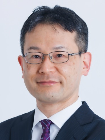 Hiroyuki Uchida (内田裕之)合作论文数Department of Neuropsychiatry, School of Medicine, Keio University21T. Arase合作论文数14T Kasatani合作论文数12
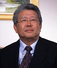 Yuji Matsuzawa (松澤佑次)合作论文数Division of Metabolic Medicine, Osaka University;Sumitomo Hospital12Yuri Ozawa合作论文数11T Yanagawa合作论文数10T. Nagashima合作论文数10H. Masuda合作论文数10Tsunehisa Makino合作论文数10
 Hitoshi Masuda合作论文数Department of Urology, National Cancer Center Hospital East9N. Sakai合作论文数8C Maruyama合作论文数8
 Masanori Ono合作论文数Keio University School of Medicine Department of Obstetrics and Gynecology 35 Shinanomachi 160-8582 Shinjuku-ku, Tokyo Japan8Kaoru Miyazaki合作论文数8
 Sergio Nozawa合作论文数8
 Mitsuru Murata (村田満)合作论文数School of Medicine, Keio University8Yutaka Motohashi合作论文数8Shunichi Yamashita合作论文数8Koichi Matsubara合作论文数8Hiroshi Hirose合作论文数8T Sugi合作论文数7H. Matsubayashi合作论文数7M Tsushima合作论文数7N Ozawa合作论文数7Ryuji Suzuki合作论文数7K Kataoka合作论文数6T Kajitani合作论文数6A Kanatsuka合作论文数6M. Ishigami合作论文数6K Iwasaki合作论文数6Yamada S合作论文数6Eiji Kawasaki合作论文数Department of Metabolism/Diabetes and Clinical Nutrition, Nagasaki University Hospital of Medicine and Dentistry6Hiromi Oda合作论文数6J Morimoto合作论文数5Y Asaba合作论文数5A Imagawa合作论文数5T. Hanafusa合作论文数5Kagami M合作论文数5Takeshi Arai合作论文数5Yoichi Oikawa合作论文数5T Nakamori合作论文数4H Makino合作论文数4Ki Hirano合作论文数4Nomaguchi H合作论文数4




