Growth of Caco-2 and many cancer cells is inhibited by 1,25(OH)(2)D(3). Whereas TGF-beta 1 inhibits normal colonic epithelial cell growth, most human colon cancer-derived cells, including Caco-2 and SW480 cells, are resistant to it. The mechanisms underlying these antiproliferative actions and resistance to TGF-beta growth inhibition are largely unknown. We observed that 1,25-dihydroxyvitamin D(3) [1,25(OH)(2)D(3)] sensitized Caco-2 and SW480 cells to TGF-beta 1 growth inhibitory effects. Versus 1,25(OH)(2)D(3) alone, the combination of 1,25(OH)(2)D(3) and TGF-beta 1 significantly reduced cell numbers. Also, the amount of active TGF-beta 1 was increased (~4-fold) by this secosteroid in conditioned media from Caco-2 cells. The 1,25(OH)(2)D(3) increased the expression of IGF-II receptors (IGF-IIR), which facilitated activation of latent TGF-beta 1, and was found to activate TGF-beta signaling in Caco-2 cells. By using neutralizing antibodies to human TGF-beta 1, we showed that this cytokine contributes to secosteroid-induced inhibition of Caco-2 cell growth. Also, 1,25(OH)(2)D(3) was found to enhance the type I TGF-beta receptor mRNA and protein abundance in Caco-2 cells. Whereas the 1,25(OH)(2)D(3)-induced sensitization of Caco-2 cells to TGF-beta 1 was IGF-IIR independent, the type I TGF-beta 1 receptor was required for this sensitization. Thus 1,25(OH)(2)D(3) treatment of Caco-2 cells results in activation of latent TGF-beta 1, facilitated by the enhanced expression of IGF-IIR by this secosteroid. Also, 1,25(OH)(2)D(3) sensitized Caco-2 cells to growth inhibitory effects of TGF-beta 1, contributing to the inhibition of Caco-2 cell growth by this secosteroid.
We have previously demonstrated a modulation of Na+/H+ exchange (NHE) activity by vitamin D3 in the rat ileum and Caco-2 cells. However, the molecular mechanism(s) of action of vitamin D3 on NHE are still not understood. The current studies were undertaken to understand the regulation of individual NHE isoforms on mRNA levels in two distinct models of vitamin D3 deficiency. Acute D3 deficiency was induced secondary to streptozotocin-induced diabetes mellitus, while chronic D3 deficiency was induced by feeding a D3-deficient diet in an environment devoid of fluorescent light. Vitamin D3 deficiency in both models increased the initial rates of rat ileal brush-border membrane (BBM) Na+/H+ exchange by 2.5-fold compared to D-repleted controls. In parallel to the increased exchanger activity, NHE3 mRNA abundance was increased about twofold in both acute and chronic D deficiency compared to control. There was no change in NHE1 or NHE2 abundance in vitamin D3-deficient rat ileum. These findings indicate that vitamin D3 regulates Na+/H+ exchange activity in rat ileum by influencing the mRNA levels of NHE3, the predominant luminal membrane isoform involved in vectorial Na+ transport.
Apobec-1 catalyzes C to U editing of apolipoprotein B (apoB) mRNA in the mammalian intestine. Rat apobec-1 is transcribed from three distinct promoters, which contain distinct 5' untranslated regions (5'UTRs) accompanied by variable numbers of in-frame upstream AUGs (uAUGs). We have observed a shift in apobec-1 promoter usage in an experimental model of colon carcinogenesis, resulting in transcripts loaded with 5'AUGs. In colon cancer, apobec-1 protein levels decreased by 90% in the cancer tissue as compared to normal tissue, suggesting an inhibitory effect of the 5'UTR on apobec-1 translation. We investigated the effects of these different 5'UTRs by site-directed mutagenesis coupled with in vitro translation studies. These studies established that the uAUGs within the 5'UTR of the alternative transcripts inhibit apobec-1 translation. This effect was independent of the length of the 5'UTR. Further analysis demonstrated that these uAUGs altered the polysome distribution, shifting the mRNA towards a denser, post-polyribosomal fraction. These findings were confirmed in transient transfection studies in vivo using HepG2 cells, where functional expression of apobec-1 was restored by mutagenesis of the uAUGs. Taken together, these data imply that rat apobec-1 gene expression is downregulated through alternative promoter usage. This dominant translational control of apobec-1 gene expression is most plausibly exerted through uAUGs.
transfection with its complementary DNA(cDNA) on SC-236-induced apoptosis and apoptosisrelated genes was further investigated.Results: SC-236 induced apoptosis in AGS cells dosedependently.Treatment with SC-236 decreased the protein expression of PKO-/~l,increased expression of PKC~ and PKC~I, but did not alter the expression of the other PKC isoforms in AGS cells.Overexpression of PKC-/~I attenuated the apoptotic response of AGS cells to SC-236, associated with overexpreseion of both p21 "af~c' p~ mRNA and protein.Inhibition of PKO-,81-mediated overexpression of p21 ,~f~c~p~ partially reduced the antiapoptotic effect of PKC-/~I.Conclusions: SC-236-induced apoptosis in gastric cancer cells is partly mediated by differential regulation of PKC isoform expression.Enhanced expression of exogenous PKC-,81 protects against SC-236-induced apoptosis through upregulation of p21 ~=,~.
BACKGROUND: Studies in both sporadic human colon cancer, and in experimental models of this malignancy, indicate that bile acids, such as cholic acid, may promote colonic carcinogenesis.In contrast, we have previously shown that the bile acid, ursodeoxycholic acid (UDCA), when administered continuously during both tumor initiation and progression/promotion, inhibited the development of azoxymethane (AOM)-induced colon cancer in rats.More recently, we have found that UDCA also inhibited AOM-induced hyperprolifaration, as well as aberrant crypt formation and growth.In an extension of these studies we asked whether this chemopreventive effect of UDCA would occur if administered only during the initiation phase, or during the post-initiation (promotion/progression} phase.METHODS: Male Fisher 344 rats (180) were divided into six treatment groups: Groups1&2 received standard rat chow (AIN 76A) throughout the study.Groups 3&4 received UDCA supplementation (0.4% w/w) only during the first 5 weeks (initiation phase).Groups 5&6 received UDCA supplementation (0.4% w/ w) only after 5 weeks (promotion/progression phase).Two wks after starting the diets, groups 2, 4 & 6 (40 rats each) received azoxymethane 20 mg/kg body weight per wk x 2 wks, while groups 1, 3 and 5 (20 rats each) received saline (AOM-vehicle) as controls.Thirty-two wks later, rats were sacrificed and colons resected.VisibE tumors were harvested and fixed in 10% buffered formalin for histologic characterization.RESULTS: There were no significant differences in weight gain among the groups.In the carcinogen-treated dietary control group, tumor incidence was 72.3%; and tumor multiplicity was 1.9 tumors per tumor bearing rat (TBR).Compared with the AOM-treated unsupplemented group, UDCA, administered only during the initiation, or in the post initiation phase, each significantly decreased tumor incidence to 46.2% and 38.4% (p
Vitamin D(3) metabolites and analogues have recently been shown to play an important role in the regulation of a number of important cellular processes, including proliferation, differentiation, and apoptosis, in addition to their established roles in mineral homeostasis. The actions of these secosteroids involve both rapid, nongenomic effects and genomic effects; the latter mediated via the vitamin D receptor and other transcription factors. Their effects have been described in a variety of cell types, including normal and malignant colonocytes. This article summarizes the rapid and genomic actions of vitamin D(3) metabolites and analogues on normal and pathologic processes in the colon, with particular emphasis on the potential of these secosteroids to prevent colon cancer.
BACKGROUND & AIMS:Previous studies showed decreased protein kinase C (PKC)-delta expression in azoxymethane-induced rat and sporadic human colonic tumors. To elucidate the role of PKC-delta on the neoplastic phenotype of human colon cancer cells, we established stable transfectants of this isoenzyme in CaCo-2 cells.METHODS:Human PKC-delta complementary DNA was subcloned into 2 distinct metallothionein-regulated expression vectors. Polyclonal populations of PKC-delta transfectants were characterized by Western blotting. PKC-delta activity was measured in situ using a PKC-delta-specific substrate. Proliferation was determined by Coulter counter, and cell cycle distribution was analyzed by flow cytometry. In vitro transformation was assessed by growth in soft agar and differentiation by changes in alkaline phosphatase and sucrase isomaltase. Apoptosis was evaluated by 4',6-diamidino-2-phenylindole dihydrochloride and terminal deoxynucleotidyl transferase-mediated deoxyuridine triphosphate nick-end labeling staining.RESULTS:In the presence of Zn(2+), PKC-delta transfectants expressed a 4-fold increase in the protein and a 2-fold increase in activity of PKC-delta. PKC-delta transfectants exhibited a 30% decrease (P < 0.05) in cell growth and an enhanced differentiation phenotype. Increased PKC-delta expression induced a significant G0/G1 arrest, inhibited anchorage-independent growth (50%, P < 0.05), and caused a 2-fold increase in apoptosis (P < 0.05).CONCLUSIONS:Our studies show that increased expression of PKC-delta inhibits anchorage-dependent and -independent growth, while inducing cellular differentiation and limiting survival of this human colon cancer cell line.
Na(+)/H(+) exchange (NHE) activity has been shown to be regulated by various external signals and protein kinases in many tissues and cell types. A family of six NHE isoforms has been identified. Three isoforms, NHE1, NHE2, and NHE3, have been shown to be expressed in the human intestine. The present studies were designed to study regulation of these human NHE isoforms by the alpha-isoform of protein kinase C (PKC) in the Caco-2 cell line. The mRNA levels of the NHE isoforms in Caco-2 cells were initially measured by a semiquantitative RT-PCR technique in response to PKC downregulation by long-term exposure to 1 microM 12-O-tetradecanoylphorbol-13-acetate (TPA) for 24 h. PKC downregulation resulted in an approximately 60% increase in the mRNA level for NHE3, but not for NHE1 or NHE2. Utilizing dichlorobenzimidazole riboside, an agent to block the synthesis of new mRNA, we demonstrated that the increase in the NHE3 mRNA in response to downregulation of PKC was predominantly due to an increase in the rate of transcription, rather than a decrease in the NHE3 mRNA stability. Consistent with the mRNA results, our data showed that amiloride-sensitive (22)Na(+) uptake was increased after incubation of Caco-2 cells with 1 microM TPA for 24 h. To elucidate the role of PKC-alpha, an isoform downregulated by TPA, the relative abundance of NHE isoform mRNA levels and the apical NHE activity were assessed in Caco-2 cells over- and underexpressing PKC-alpha. Our results demonstrated that NHE3, but not NHE1 or NHE2, mRNA was downregulated by PKC-alpha and that apical NHE activity was higher in cells underexpressing PKC-alpha and lower in cells overexpressing PKC-alpha than in control cells. In conclusion, these data demonstrate a differential regulation of NHE3, but not NHE2 or NHE1, expression by PKC in Caco-2 cells, and this regulation appears to be predominantly due to PKC-alpha.
BACKGROUND: We previously demonstrated that the bile acid, ursodeoxycholic acid (UDCA), and an analogue of 1,25(0H)~ vitamin D3 (FED3), inhibit cydooxygensse-2 (COX-2) mRNA and protein expression in azoxymethane (AOM)-induced colonic tumors.We have now extended these studies by investigating the effects of these chemopreventive agents on several transcription factors implicated in the regulation of COX-2 gene expression.We have also examined AOi-inducad tumors for the effects of UDCA and FeD3 on VEGF, an angioganic factor and downstream target of COX-2, important in tumor progression.To further dissect the molecular mechanisms by which UOCA and FeD3 inhibit COX-2 expression, we have examined the effects of these agents on basal and COX-2 stimulated expression by the tumor promoting deoxycholic acid (DC) in HCA-7 ceils, derived from a human colon carcinoma~ METHODS: Tumors were induced by AOM in rats fad standard chow (AIN-76A); or chow supplemented with 0.4% UDCA, or with Fe-D3 (2.5 nmol/kg chow).After 32 weeks, rats were sacrificed and tumors harvested.A section of each tumor was used to characterize tumor histology.A separate portion was homogenized and proteins extracted in Laemmli buffer.HCA-7 cells were treated with DC alone, or with UDCA, or FED3, and cells ware then lysod in RIPA buffer.Proteins from cellular and tumor lysatss were separated by SOS-PAGE Expression levels of C/EBPs, VEGF and COX-2 were measured by qua,i;;,,~;ve Western bluffing.RESULTS: In AOM-induced tumors, compared to normal colon, OEBP-,8 and VEGF expression levels increased 8_+2-fold, and 20_+4-fold respectively (p <0.05).In contrast, OEBP-a and -8 were not changed in these tumors.In tumors from rats supplemented with UOCA or Fs-D3, we observed a 60 -10% decrease in the expression of C/EBP-p;, in these tumors, moreover, F6-D3, caused a 50-+20% decrease in VEGF, compared to carcinogen-induced tumors from unsupplemented animals (p
1,25(OH)zD 3 ] , the major active biological metabolite of vitamin D 3 , is a potential chemopreventive agent for human colon cancer.Alterations in TGF-f3, and its receptors, TGF-I3RI, & RII, have been implicated in the development of colon cancer.While the growth of normal colonic epithelial cells is inhibited by TGF-f3I, many colon cancer-derived cell lines, including Caco-2 cells, are resistant to the antiproliferative effects of TGF-I31.The mechanisms by which this occurs, however, remain largely unknown.The aim of this study was to evaluate the potential ability of 1,25(OHhD 3 _ to sensitize Caco-2 cells to the growth inhibitory effects of TGF-I31.METHODS: Cell proliferation was measured by an MTS assay.Promoter-reporter constructs of targeted genes were used to study the promoter activation in transient transfections.RNase protection assays (RPA) were used to measure rnRNA abundance.Protein expression and activity of the MAP kinases were determined by quantitative Western blots.RESULTS: While TGF-131 alone failed to alter Caco-2 cell growth, 1,25(OH)zD 3 (100 nM) restored the ability of TGF-f31 (2.5 ng/ml) to inhibit the growth of these cells (-25%) as determined by the MTS assay.In transient transfections, 1,25(OHhD 3 induced TGF-I3RI, but not TGF-I3RII, promoter activation.Increases in TGF-f3RI mRNA and protein abundance in 1,25(OH)zD 3-treated cells were also determined by RPA and Western blotting, respectively.Additional studies indicated that activation of both ERK and JNK were required for the 1,25(OH)zD r induced increase in TGF-I3RI gene expression in these cells.CONCLU-SION: These studies suggest that the chemopreventive actions of 1,25(OHhD 3 may involve sensitization of resistant colon cancer cells to the growth inhibiting effects of TGF-I3I.
Previous studies by our laboratory have shown that a noncalcemic fluorinated analog of 1alpha,25-dihydroxyvitamin D3, 1alpha,25-dihydroxy-16-ene-23-yne-26,27-hexafluorocholcal ciferol (F6-D3), significantly reduced the frequency of colonic adenomas and completely abolished the development of colonic adenocarcinomas in rats treated with azoxymethane. The mechanisms involved in this analog's chemopreventive actions, however, remain unclear. In the present study, we now show that although both 1alpha,25-dihydroxyvitamin D3 and F6-D3 inhibited the proliferation of CaCo-2 cells, a human colonic adenocarcinoma cell line, by increasing their doubling times, only F6-D3 caused an arrest of these cells in the G1 phase of their cell cycle. This arrest was accompanied by an increase in the expression of the cyclin-dependent kinase (cdk) inhibitor proteins, p2Waf1 and p27Kip1, which served to decrease the activity of cyclin-dependent kinase 2 and cyclin-dependent kinase 6, whereas the expression and phosphorylation of pRB were unchanged. In contrast to the increased expression of these cdk inhibitors, the expression of cyclin E was decreased, which further inhibited the activity of cyclin-dependent kinase 2. Collectively, the inhibition of these cyclin-dependent kinases served to arrest the CaCo-2 cells, independent of changes in pRB. Furthermore, antibody neutralization studies suggest that transforming growth factor-beta may mediate the coassociations between cdk2 and p27Kip1 and cyclin E induced by F6-D3. These data indicate that cell cycle arrest may, at least in part, underlie the chemopreventive actions of F6-D3 observed in the azoxymethane model of colon cancer. Furthermore, if the antiproliferative action observed in CaCo-2 cells also occurs in human colonic epithelium, F6-D3 may have chemopreventive potential against human colon cancer, as well.
PTEN gene expression is suggested to be due to hypermethylation of precoding region, but direct evidence has not been reported.Using colon cancer cell line, we purposed to certify this possibility that expression of PTEN gene is regulated by methylation of DNA.MaterialslMethods: We have analyzed 6 colon cancer cell lines, T-84, HT-29, SW480, SW620, CaC02, and Col0320DM provided from ATCC.Expression of PTEN gene transcript and protein expression were examined by RT-PCR-Southern analysis and Western blot with polyclonal goat anti-PTEN antibody (Santa Cruz Biotechnology), respectively.Analysis of DNA sequence was performed by direct sequencing technique.Results: RT-PCR-Southern analysis showed that PTEN gene transcript was expressed in CaC02, HT-29, SW480, and SW620 cells, but significantly reduced in T-84 and Col0320DM cells.Protein expressions of PTEN examined by Western blot were also reduced in these two cell lines.However, mRNA and protein expression of PTEN were augmented by demethylating agent 5-Azadeoxycitidine, added to the culture medium in T-84 and Col0320DM cells.Direct sequencing of 800 bps upstream to start codon in exon I of PTEN gene, which was treated with bisulfite-converting reagents revealed that over 90% of CpG sequence of PTEN gene in T-84 cells were methylated.In contrast, CpGs in this region of CaC02 cells were not methylated, and those of HT-29 cells were partially (54%) methylated.Conclusions: These results indicate that expression of PTEN gene is regulated by hypermethylation of precoding region in some colon cancer cell lines.However, PTEN gene product is weakly expressed even in T-84 cells.This result suggests that suppression of PTEN gene seems to be not essential in carcinogenesis, but it may be important to tumor cell survival in colon cancer.
Background: Treatment with a COX-2 inhibitor causes regression of intestinal polyps in size in APClJ.716knockout mice.In this tumor model, COX-2 expression is mainly up-regulated in stromal fibroblasts.How does COX-2 expressed in stromal fibroblasts influence neoplasia?Vascular endothelial growth factor (VEGF) is well known as a major factor involved in tumor angiogenesis and its expression is significantly up-regulated in tumor tissue compared with adjacent normal tissue.Hepatocyte growth factor (HGF) is a known mitogen in digestive tissues and its production is reported to increase in gastrointestinal tumors.It has been reported that prostaglandin E z stimulates HGF production in fibroblasts.Aim: To investigate the effect of COX-I and COX-2 in fibroblasts on tumor growth, fibroblasts were prepared from wild type, COX-lor COX-2 knockout mice.VEGF and HGF production were measured and the mitogenic effect of these fibroblasts on co-cultured endothelial cells (HUVEC) and colon cancer cells (colon-38; mouse colon cancer derived cell line) was determined.Materials and Methods: Fibroblasts were prepared from wildtype, COX-I and COX-2 knockout mice and cultured in serum free DMEM for 12 hours.Conditioned media was collected for measurement of PGE z, VEGF, and HGF levels.Using a Boyden chamber with fibroblasts in the outer chamber, proliferation of HUVEC or colon-38 cells co-cultured in the inner chamber was evaluated using the MIT assay.Results: Skin fibroblasts prepared from COX-2 knockout mice produced little VEGF compared with those derived from wild type or COX-I knockout mice (1.8 vs. 300 or 270 pgllOO,OOO cells, respectively).HGF production in COX-2 knockout fibroblasts was significantly less than those in wild type or COX-I knockout fibroblasts (0.36 vs. I.2 or 0.74 nmolellOO,OOO cells, respectively).PGE z productions in COX-I and COX-2 knockout fibroblasts were similar, but significantly lower than that in wild type.Wild type and COX-I knockout fibroblasts stimulated proliferation of co-cultured HUVEC and colon-38 cells, whereas COX-2 knockout fibroblast showed little effect on co-cultured HUVEC or colon-38 cell proliferation.Conclusion: These results suggest that COX-2 expression in fibroblasts plays an important role in the regulation of VEGF and HGF production, whereas COX-I does not.VEGF and HGF production might be independent of PGE z.This indicates the possibility that COX-2 inhibition can suppress tumor growth via inhibition of angiogenesis and HGF production.
would go into crises after approximately 25 passages.These SV40 T-Ag transfected gastric epithelial cells (HAE, HFE) at passage 18 were transfected with human telomerase (hTRT-145).The subsequent cell lines (HAE 145, HFE 145) have gone through more than 40 passages and are growing well, doubling approximately every 24 hours.These cells grow well in DMEM culture medium with high glucose with 2-5% fetal bovine serum.Characterization has been performed on the HFE 145 cell line.These cells express the SV40 Large T-antigen and have telomerase activity significantly higher than the parental cells.These cells are strongly positive for cytokeratin 10,11,18 and weakly positive for cytokeratin 13, 16 and 20, which is almost identical to cytokeratin staining of the parental cell line (HFE).Periodic Acid Schiff (PAS) staining with digestion shows that the cells secrete neutral mucins, and alcian blue staining( which stains acidic mucin) is negative, consistent with normal gastric epithelial cells.Cell growth was inhibited when cells were placed in soft agar, suggesting that these cells are not tumorigenic.The cells constitutively express mRNA from muc 5ac, muc 5b, muc 6 genes which is consistent with normal gastric epithelial cells.Electron microscopy shows that these cells form tight junctions when grown as monolayers on plastic tissue culture dishes and on glass slides.These cell lines will be useful models for studying mechanisms of gastric diseases, including gastric cell injury caused by infectious agents such as H. pylori.These cells will also be useful for chemical cytotoxicity, and carcinogenic investigations.
Protein kinase C (PKC)consists of a gene family of serine/threonine kinases which regulate numerous events including cellular proliferation and differentiation. Studies in human and experimental models of colon cancer suggest that alterations in the expression of specific isoforms of PKC are involved in colonic malignant transformation. Previously, our laboratory has shown that PKC/3II protein expression was altered in azoxymethane-induced rat colonic tumors. The specific aim of this study was to characterize the functional consequences of alterations in PKC/311 in CaCo-2 cells, a colonic adenocarcinoma cell line. Methods: Full length human PKC/3II cDNA, and a kinase-dead isoform mutated in the catalytic domain, were cloned into MRE, a Zn2+ inducible metallothionine expression vector. Mutant and wild type sequences were confirmed by direct sequencing. CaCo-2 cells were transfected, selected with G418 and polyclonal populations were expanded . Cell Iysates were analyzed by western blotting for isoform specific PKC expression. Following immunoprecipitation with PKC/3II specific antibodies, kinase activity was measured using acetylated myelin basic protein substrate. Proliferation was analyzed by cell counting. Differentiation was assessed by alkaline phosphatase . Results: In the presence of 175 p,M Zn2+, CaCo-2 cells transfected with both wild type PKC/3II and kinase-dead PKC/3II exhibited 3-4 -fold increases in the expression of the isoform compared to empty vector (EV) transfectants as assessed by Western blotting. Transfection with kinase-dead PKC/3II significantly inhibited (p :s 0.05) kinase activity by nearly 70%. Importantly, the mutant and wild type clones exhibited no significant changes in the expression of non-targeted PKC isoforms, including PKCa,/3I,/l or ~. While the wild type PKC/3II transfected CaCo-2 cells did not differ in proliferation compared to the EV cells, kinase-dead transfectants showed more than a 30% increase (p :s 0.05) in cell growth compared to EV control s. In addition, by day I I post-plating, these kinase-dead PKC/3II transfected cells demonstrated a 25% decrease in alkaline phosphatase activity compared to EV controls. In conclusion, we have shown that Caco-2 cells, transfected with a kinase-dead PKC/3II cDNA and possessing decreased kinase activity, demonstrate enhanced proliferation and inhibited differentiation. These studies suggest that PKC/3II may be involved in the regulation of these important cellular processes in human colon cancer cells.
 Pk Dudeja合作论文数72
 Marc Bissonnette合作论文数The University of Chicago Medical Center62Md Sitrin合作论文数54R K Wali合作论文数46
 Sharad Khare合作论文数Department of Medicine, University of Chicago32Xy Tien合作论文数23B. Scaglione-Sewell合作论文数Department of Medicine, University of Chicago22R Wali合作论文数16R Dahiya合作论文数15Mjg Bolt合作论文数14
 Nicholas O. Davidson合作论文数Division of Gastroenterology, John T. Milliken Department of Medicine, Washington University School of Medicine in St. Louis;Digestive Disease Research Core Center, Washington University13M Sitrin合作论文数11John Hart合作论文数Department of Medicine, The University of Chicago11Allan G. Halline合作论文数11S Skarosi合作论文数11Es Foster合作论文数10
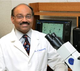 Ramaswamy Kalyanasundaram合作论文数Department of Biomedical Sciences, University of Illinois College of Medicine at Rockford10
 Bruce Marc Bissonnette合作论文数9JM HARIG合作论文数9Sf Skarosi合作论文数8
 Russell F. Jacoby合作论文数William S. Middleton Memorial Veterans Hospital8Hk Roy合作论文数7Ba Scaglionesewell合作论文数7S Niedziela合作论文数7Roy H合作论文数7
 Pradeep K Dudeja合作论文数Jesse Brown VA Medical Center, Chicago, IL 60612, USA7C Abraham合作论文数6B. Frawley合作论文数6Sharon M. Niedziela合作论文数6Dennis P. Wilson合作论文数Department of Economics;University of Texas - Arlington;Department of Economics, University of Texas - Arlington6S C Hartmann合作论文数5Bh Davis合作论文数5
 Xavier Llor合作论文数Digestive Diseases, Internal Medicine, Yale School of Medicine, Yale University5Baum C L合作论文数5Sandra Cerda合作论文数5Ar Safa合作论文数4S Hartmann合作论文数4Brendan P. Frawley合作论文数4F C von Lintig合作论文数3Matthew R. Lyons合作论文数3AW NORMAN合作论文数3Gerard R. Boss合作论文数3H Holubec合作论文数3Ap Chen合作论文数3Deborah Nelson合作论文数3
 David M. Wilson合作论文数Laboratory of Molecular Gerontology, GRC, National Institute on Aging, IRP, NIH3B Aquino合作论文数3Yingchun Zhang合作论文数3




