Achieving bone union remains a significant clinical dilemma. The use of osteoinductive agents, specifically bone morphogenetic proteins (BMPs), has gained wide attention. However, multiple side effects, including increased incidence of cancer, have renewed interest in investigating alternatives that provide safer, yet effective bone regeneration. Here we demonstrate the robust bone healing capabilities of the main megakaryocyte (MK) growth factor, thrombopoietin (TPO), and second-generation TPO agents using multiple animal models, including mice, rats, and pigs. This bone healing activity is shown in two fracture models (critical-sized defect [CSD] and closed fracture) and with local or systemic administration. Our transcriptomic analyses, cellular studies, and protein arrays demonstrate that TPO enhances multiple cellular processes important to fracture healing, particularly angiogenesis, which is required for bone union. Finally, the therapeutic potential of thrombopoietic agents is high since they are used in the clinic for other indications (eg, thrombocytopenia) with established safety profiles and act upon a narrowly defined population of cells.
Blunt thoracic trauma (BTT) and the resultant isolated mitral papillary muscle avulsion, pericardial rupture, and cardiac herniation injuries are each rarely diagnosed clinical entities. We describe the first case of combined pericardial tear with cardiac herniation and ruptured mitral papillary muscles following BTT. Preoperative transesophageal echocardiography (TEE) diagnosed the delayed mitral papillary muscle rupture while all previous diagnostic modalities failed to delineate the pericardial rupture and cardiac herniation. Particular emphasis is placed on the clinical and radiologic aspects of the case that would heighten clinical suspicion in the emergency setting where blunt cardiac injury sequelae are suspected and frequently missed.
Preparation of mineralized tissue specimens for bone-specific staining encompasses a critical sequence of histological techniques that provides visualization of tissue and cellular morphology. Bone specimens are fixed in 10% neutral buffered formalin (NBF), dehydrated in graded ethanol (EtOH) solutions (and optionally cleared in xylene), infiltrated and embedded in polymethyl methacrylate (methyl methacrylate or MMA), classically sliced into 4-10 micrometer (mu m) sections, and stained with bone-specific histological stains such as von Kossa (with either nuclear fast red solution counterstain or MacNeal's tetrachrome counterstain), modified Goldner's trichrome, Alizarin Red S, Safranin O, and tartrate-resistant acid phosphatase (TRAP) stain. Here, we describe the tissue processing of mineralized mouse bones from dissection to staining for histological analysis by light microscopy.
Preparation of mineralized tissue specimens for bone-specific staining encompasses a critical sequence of histological techniques that provides visualization of tissue and cellular morphology. Bone specimens are fixed in 10 % neutral-buffered formalin, dehydrated in graded ethanol (EtOH) solutions (and optionally cleared in xylene), infiltrated and embedded in polymethyl methacrylate (methyl methacrylate), classically sliced into 4-10 micrometer (μm) sections, and stained with bone-specific histological stains such as von Kossa (with either nuclear fast red solution counterstain or MacNeal's tetrachrome counterstain), modified Goldner's trichrome, and alizarin red S stain. Here, we describe the tissue processing of mineralized mouse bones from dissection to staining for histological analysis by light microscopy.
Current and future advances in orthopedic treatment are aimed at altering biological interactions to enhance bone healing. Currently, several clinical scenarios exist for which there is no definitive treatment, specifically segmental bone loss from high-energy trauma or surgical resection — and it is here that many are aiming to find effective solutions. To test experimental interventions and better understand bone healing, researchers employ critical size defect (CSD) models in animal studies. Here, an overview of CSDs is given that includes the specifications of varying models, a discussion of current scaffold and bone graft designs, and current outcome measures used to determine the extent of bone healing. Many promising graft designs have been discovered along with promising adjunctive treatments, yet a graft that offers biomechanical support while allowing for neovascularization with eventual complete resorption and remodeling remains to be developed. An overview of this important topic is needed to highlight current advances and provide a clear understanding of the ultimate goal in CSD research — develop a graft for clinical use that effectively treats the orthopedic conundrum of segmental bone loss.
 Melissa A. Kacena合作论文数School of Medicine, Yale University5Harris Jonathan S合作论文数4Ying-Hua Cheng合作论文数2
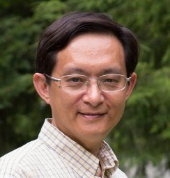 Tien-Min Gabriel Chu合作论文数Sch Dent, Indiana Univ2Millikan Patrick D合作论文数2
 Monique Bethel合作论文数2Wessel Alexander R合作论文数2Keith W. Condon合作论文数2Krista L Jackson合作论文数1David C Scofield合作论文数1Kaitlyn S Griffin合作论文数1Karl D. Shively合作论文数1Aamir Tucker合作论文数1Kelli Manikowski合作论文数1
 Jeff Rytlewski合作论文数1
 Roman M Natoli合作论文数1W Christopher Shelley合作论文数1Ross McCauley合作论文数Indiana Univ Sch Med1Faisal Shariff合作论文数1Patrick L. Mulcrone合作论文数1Edward Evans合作论文数1
 Joydeep Ghosh合作论文数Dept Med, Indiana Univ Sch Med1
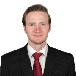 Jeffery J Nielsen合作论文数Dept Med Chem & Mol Pharmacol, Purdue Univ1Ushashi C. Dadwal合作论文数1Anglen Jeffrey O合作论文数1McKinley Todd O合作论文数1
 Low Stewart A合作论文数Department of Bioengineering, University of Utah1
 Aarti Gautam合作论文数Integrative Systems Biology, US Army Center for Environmental Health Research1
 Thompson Mark E合作论文数Children's Hospital of Colorado, University of Colorado1
 Mark D Walsh合作论文数1
 Jonathan W Lowery合作论文数Harvard School of Dental Medicine, Marian University College of Osteopathic Medicine1
 Patrick K Davis合作论文数1
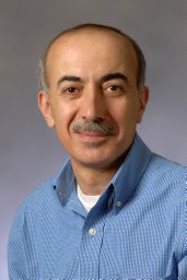 Edward F. Srour合作论文数School of Medicine, Indiana University1
 Mervin Yoder合作论文数Department of Surgery, School of Medicine, University of Pittsburgh1
 Jiliang Li合作论文数1
 Philip S. Low合作论文数Department of Chemistry Purdue University1
 Rasha Hammamieh合作论文数Division of Pathology, Walter Reed Army Institute of Research1Marta Alvarez合作论文数School of Medicine, Indiana University1
 Nabarun Chakraborty合作论文数Division of Pathology, Walter Reed Army Institute of Research1
 Stacy-Ann Miller合作论文数1
 Paul Childress合作论文数1
 Alexander Brinker合作论文数1




