PURPOSE:To assess general population perspectives on pre-biopsy disclosure of serious risks and factors associated with a preference for disclosure. METHODS:2,407 adults from the Dutch general population completed 3,565 case vignettes describing hypothetical patients requiring biopsies, each reporting the risk of serious complications (requiring additional treatment or prolonged hospitalization, or resulting in death). Vignettes varied by patient age (9, 26, 46, or 71 years), probability of serious complications (<0.1%, <1%, <5%, or unspecified), and biopsy purpose (primary diagnosis, staging, or therapy response). Respondents indicated agreement with "If I were in this situation, I would want to be informed" on a four-point Likert scale from strongly disagree to strongly agree. RESULTS:Most responses (93.1%) favored disclosure (agreed/strongly agree), while 4.0% opposed (disagree/strongly disagree). For <0.1% risk vignettes, 91.8% of responses favored disclosure. Women were less likely than men to favor disclosure (odds ratio (OR) 0.760, P < 0.001). Agreement was higher for respondents with secondary (OR 1.274, P = 0.028) and tertiary education (OR 1.689, P < 0.001) compared with those with primary education. Agreement was lower for vignettes with a 46-year-old patient versus a 9-year-old (OR 0.778, P = 0.032) and for staging biopsies versus primary diagnosis (OR 0.780, P = 0.006), and higher when risk was < 5% or unspecified versus < 0.1% risk vignettes (OR 1.304, P = 0.023; OR 1.618, P < 0.001). CONCLUSION:Most Dutch people want disclosure of serious biopsy risks, including very low-risk events, though a small minority preferred not to be informed, with preferences influenced by respondent characteristics and clinical context.
Artificial intelligence (AI) is rapidly integrating into clinical radiology. As primary diagnosticians, radiologists increasingly interpret AI-generated analyses and are expected to oversee the monitoring and governance of deployed AI systems. Although AI literacy among radiologists is improving, several technical aspects of AI remain insufficiently accessible. One such concept is uncertainty quantification (UQ), which estimates the reliability of AI predictions and can signal when outputs should be interpreted with caution. This review introduces key UQ concepts relevant to radiology, distinguishing between aleatoric uncertainty and epistemic uncertainty arising from data variability and knowledge gaps. We summarize commonly used UQ approaches in current research and practice. Furthermore, through a narrative review of selected recent AI imaging studies, we illustrate how UQ methods are applied in practice and highlight methodological trends, findings, and limitations. Although UQ has the potential to improve the safety and interpretability of AI-assisted screening, challenges remain, including calibration, threshold selection, computational cost, and the need for prospective clinical validation.
Purpose: To evaluate how often history taking and physical examination are omitted before MRI referral and whether their omission is associated with clinical reasoning quality and MRI diagnostic yield. Materials and Methods: In this prospective study, adults undergoing MRI at a tertiary academic hospital were surveyed before imaging to determine whether the referring clinician had taken their history and performed a physical examination. Multivariable regression was used to assess determinants of omission and associations with clinical reasoning quality (defined as agreement between the suspected diagnosis and MRI findings) and MRI positivity (defined as findings relevant to the indication). Results: Among 275 patients (median age 61 years; 50.0% male), history taking was omitted in 18.2% of cases and physical examination was omitted in 70.9%. History taking was less likely during surveillance than during new/first visits (odds ratio (OR) 0.140, p < 0.001) and more likely when MRI was requested by residents rather than medical specialists (OR 4.645, p = 0.018). Physical examination was more likely when MRI was requested by residents (OR 3.174, p = 0.007) or nurse specialists/physician assistants (OR 3.145, p = 0.033), and less likely during follow-up visits (OR 0.183, p < 0.001) and surveillance visits (OR 0.061, p < 0.001). Omission of physical examination was not associated with clinical reasoning quality (p = 0.370). Neither omission of history taking nor omission of physical examination was associated with MRI positivity (p = 0.430 and p = 0.286, respectively). Conclusions: History taking and physical examination were often omitted before MRI referral. Although no statistically significant association was observed between omission of bedside assessment and clinical reasoning quality or MRI positivity, reduced bedside assessment may limit the clinical context informing referral and interpretation.
Artificial Intelligence (AI) for detecting clinically significant prostate cancer (csPCa) on MRI has achieved diagnostic performance comparable to that of radiologists. By autonomously interpreting examinations, AI could improve workflow efficiency and help address increasing imaging demands and radiologist shortages. Despite this promise, autonomous AI has not been implemented in clinical practice. This narrative review explores remaining technical and societal barriers to deploying autonomous csPCa detection. We focus on three key domains: limitations in the current evidence base, safety issues and mitigation strategies, and the perspectives of patients and radiologists. Our findings highlight the need for evidence from large, multicenter, prospective trials and evaluation frameworks that reflect the consequences of clinical decision-making, as well as further exploration of safeguards to monitor and address mismatches between training data and incoming scans during deployment. Moreover, patients and radiologists show limited acceptance of autonomous AI, although this may improve with greater transparency, targeted education, and clearer guidelines on medico-legal responsibilities. Addressing these challenges is essential to the responsible deployment of autonomous AI and to realizing its efficiency gains in clinical practice.
To evaluate how policies under the current Trump administration (2025-present) have affected the work of nuclear medicine researchers. An online survey was distributed to corresponding authors who published in the three highest-ranked general nuclear medicine journals between 2021 and 2024. The survey included demographics and items on perceived effects of the current Trump administration, tailored to respondents’ location. Data were summarized descriptively, and a multivariable ordinal logistic regression evaluated factors (age, sex, geographic location, academic degree and rank, and years of research involvement) associated with expected research progress. Of 2,570 reachable authors, 117 completed the survey. Among U.S.-based respondents, 68.8
PURPOSE:To assess radiology researchers' perceptions of whether, and to what extent, the political context during the current Trump administration (2025-present) affected their research activities, collaborations, work environment, motivation, mobility, and overall research progress. METHODS:Corresponding authors who published in the 12 top-ranked general radiology journals in 2024 were invited to complete an anonymized online questionnaire. The survey collected demographic characteristics and insights related to the broader research environment under the current Trump administration (2025-present). Data were summarized using descriptive statistics, and ordinal regression analysis was performed to examine associations between participant characteristics and expected research progress. Free-text responses were analyzed qualitatively to identify recurring themes. RESULTS:A total of 176 authors participated. Most respondents were aged 35-44 years (33.0%), male (68.8%), from Europe (55.1%), held a medical doctor degree (71.6%), were full professors (31.3%), and had >10 years of research experience (79.0%). Most U.S.-based participants reported that funding, collaboration, and research motivation were negatively affected by the current Trump administration. Most non-U.S. respondents similarly perceived negative impacts on willingness to work, study, attend conferences, and collaborate with U.S. researchers. Overall, 47.7% anticipated slower research progress and 26.1% much slower progress. Male gender was the only factor associated with more optimistic expectations, with an odds ratio of 2.130 (P = 0.038). Free-text comments emphasized disrupted funding, administrative barriers, safety concerns, and shifts of research opportunities to non-U.S. regions. CONCLUSION:The current Trump administration is perceived to negatively affect radiology research advancement, funding, collaboration, and researcher motivation.
To assess public attitudes toward prospective disclosure of diagnostic error risk in radiologic imaging (i.e., informing patients in advance about the possibility a radiologic examination may yield an incorrect finding, rather than disclosure of an error after it has occurred) and to identify factors influencing these preferences. A population-based survey was conducted in the Netherlands. Participants aged ≥ 18 years (n = 1524) responded to CT-scan vignettes describing pre-procedural potential diagnostic errors, varying by patient age (9, 26, 46, or 71 years) and probability of incorrect findings (< 0.1
BackgroundMyxofibrosarcoma (MFS) and undifferentiated soft tissue sarcoma (USTS) are common sarcoma subtypes with overlapping molecular features. Both are treated with neoadjuvant radiotherapy followed by surgery, yet radiotherapy response is variable and unpredictable. This study investigated DNA methylation and copy number variation (CNV) profiles obtained from pre-radiotherapy biopsies as predictive biomarkers of radiotherapy response.Patients and methodsPre-radiotherapy biopsies and post-radiotherapy resections were obtained from 49 patients (27 MFS, 22 USTS). Radiotherapy response was assessed on the resection specimens using the EORTC-STBSG 5-tier system; grades A-C (<10% viable tumor) were classified as responders, D-E (≥10% viable tumor) as non-responders. Genome-wide DNA methylation and CNV data were generated from the pre-radiotherapy biopsies using Illumina MethylationEPIC BeadChips and were correlated with response grades.ResultsDNA methylation profiling yielded evaluable results in 23/49 tumors (15 MFS, 8 USTS), with 9 responders and 14 non-responders. Unsupervised methylation clustering, incorporating public datasets, showed that MFS, USTS, and pleomorphic liposarcomas formed a single, heterogeneous cluster. Similarly, CNV profiles did not distinguish MFS from USTS. Methylation patterns did not significantly differ between responders and non-responders. CNV profiles were largely comparable between responders and non-responders, except of a significantly higher frequency of chromosome 11q24.1 loss in responders compared to non-responders (100% vs 33%; P = 0.0039).ConclusionsOur findings support the concept that MFS and USTS represent a spectrum of the same disease. We could not demonstrate the value of DNA methylation profiling in radiotherapy response prediction. However, 11q24.1 loss may represent a potential predictive biomarker and merits further validation.
OBJECTIVE:To investigate how often referring physicians perform history taking and physical examination before PET/computed tomography (PET/CT) referral, and whether their absence influences clinical reasoning quality or diagnostic yield. METHODS:Patients undergoing PET/CT at a tertiary academic hospital were asked whether their referring physician had conducted history taking and physical examination before referral. Associations between omission of clinical assessment and clinical reasoning quality or PET/CT positivity (defined as PET/CT findings relevant to the clinical indication) were analyzed using multivariable regression. RESULTS:Among 288 patients (median age: 66 years; 54% male), history taking was omitted in 18.2% and physical examination in 47.7%. For history taking, only referrals from pulmonology were significantly more likely to include history taking [odds ratio (OR): 4.708, P = 0.033]. For physical examination, referrals from residents were more likely to include physical examination (OR: 2.355, P = 0.022), as were surgical departments (OR: 8.527, P = 0.030), requests for new clinical complaints (OR: 2.303, P = 0.008), and total-body PET/CT scans (OR: 1.870, P = 0.037), whereas urology referrals were less likely (OR: 0.019, P = 0.008). Neither omission of history taking nor physical examination was associated with clinical reasoning quality or PET/CT positivity (all P > 0.15). CONCLUSION:A relevant proportion of patients referred for PET/CT did not receive history taking or physical examination. While omission of these assessments did not appear to affect the clinical reasoning quality of the referring physicians or PET/CT yield, it may limit the contextual information available for interpreting imaging results.
Although undersampling combined with deep learning (DL)-based reconstruction shortens MRI acquisition, it increases the chance of inaccuracies, highlighting the need for quantifiable uncertainty measures. Two inference-time perturbation strategies, echo-train dropout (ET-Drop) and Gaussian noise Monte Carlo sampling (GN-MC), were compared in terms of the correlation between their variance-based uncertainty maps and absolute reconstruction error in DL-accelerated T2w prostate MRI. This retrospective multi-center study used a publicly available dataset with 312 k-spaces from NYU for training and a dataset with 120 k-spaces from University Medical Center Groningen for external validation. Fully sampled 3 T data were retrospectively undersampled to acceleration factors R = 3 and R = 6 and reconstructed by a vSHARP model. Per slice, five GN-MC perturbations were reconstructed by adding complex noise at 2.5σ, and five ET-Drop perturbations, created by omitting non-central echo trains. Voxel-wise aleatoric uncertainty was defined as the variance (σ2) across these reconstructions and correlated with absolute reconstruction error over whole slices and within the prostate. Both uncertainties yielded moderate slice-level correlations with absolute error. At R = 3, ET-Drop slightly outperformed GN-MC (median ρ = 0.39 vs 0.35; p < 0.001). At R = 6, the ranking reversed (0.44 vs 0.40; p < 0.001). Correlations within the prostate fell to 0.10–0.15. ET-Drop variance maps were dominated by coil sensitivities. Both perturbation strategies yield variance-based uncertainty maps that correlate moderately with voxel-wise error. More importantly, they consistently highlighted acquisition-related fragility, supporting the role of uncertainty mapping as a useful quality-control tool in prostate MRI.
To determine past 15-year trends in workload, negative findings, and incidental findings in acute neuroradiology during on-call hours at a European tertiary care center. This study analyzed a sample of 2494 CT and 264 MRI scans of the head and/or neck performed during on-call hours at a tertiary care center on random dates between 2009 and 2023. The workload significantly increased by 130
To evaluate the repeatability of AI-based automatic measurement of vertebral and cardiovascular markers on low-dose chest CT. We included participants of the population-based Imaging in Lifelines (ImaLife) study with low-dose chest CT at baseline and 3–4 month follow-up. An AI system (AI-Rad Companion chest CT prototype) performed automatic segmentation and quantification of vertebral height and density, aortic diameters, heart volume (cardiac chambers plus pericardial fat), and coronary artery calcium volume (CACV). A trained researcher visually checked segmentation accuracy. We evaluated the repeatability of adequate AI-based measurements at baseline and repeat scan using Intraclass Correlation Coefficient (ICC), relative differences, and change in CACV risk categorization, assuming no physiological change. Overall, 632 participants (63 ± 11 years; 56.6
Journal Article Accepted manuscript Addressing Spinal Implant Infections: Emerging Options and Unresolved Challenges Get access Don Bambino Geno Tai, Don Bambino Geno Tai Division of Infectious Diseases and International Medicine, University of Minnesota, Minneapolis, MN, USA Corresponding author: Don Bambino Geno Tai, MD, 420 Delaware Street SE, Minneapolis, Minnesota, USA, 55401 Email: [email protected] https://orcid.org/0000-0003-0581-8015 Search for other works by this author on: Oxford Academic PubMed Google Scholar Robin Patel, Robin Patel Division of Clinical Microbiology, Department of Pathology, Mayo Clinic, Rochester, MN, USADivision of Public Health, Infectious Diseases, and Occupational Medicine, Mayo Clinic, Rochester, MN USA https://orcid.org/0000-0001-6344-4141 Search for other works by this author on: Oxford Academic PubMed Google Scholar Francis Lovecchio, Francis Lovecchio Department of Orthopedic Surgery, Hospital for Special Surgery, New York City, NY, USA https://orcid.org/0000-0001-5236-1420 Search for other works by this author on: Oxford Academic PubMed Google Scholar Thomas Kwee, Thomas Kwee Department of Radiology, University Medical Center Groningen, University of Groningen, Groningen, The Netherlands Search for other works by this author on: Oxford Academic PubMed Google Scholar Marjan Wouthuyzen-Bakker Marjan Wouthuyzen-Bakker Department of Medical Microbiology and Infection Prevention, University Medical Center Groningen, University of Groningen, Groningen, The Netherlands Alternate contact authors: Marjan Wouthuyzen-Bakker, MD, PhD University Medical Center Groningen Hanzeplein 1, 9713 GZ, Groningen, the Netherlands Email: [email protected] https://orcid.org/0000-0001-7866-2467 Search for other works by this author on: Oxford Academic PubMed Google Scholar Clinical Infectious Diseases, ciaf065, https://doi.org/10.1093/cid/ciaf065 Published: 12 February 2025 Article history Received: 14 January 2025 Revision received: 22 January 2025 Editorial decision: 03 February 2025 Accepted: 11 February 2025 Published: 12 February 2025
Abstract Bladder cancer is the 10th most common and 13th most deadly cancer worldwide, with urothelial carcinomas being the most common type. Distinguishing between non-muscle-invasive bladder cancer (NMIBC) and muscle-invasive bladder cancer (MIBC) is essential due to significant differences in management and prognosis. MRI may play an important diagnostic role in this setting. The Vesical Imaging Reporting and Data System (VI-RADS), a multiparametric MRI (mpMRI)-based consensus reporting platform, allows for standardized preoperative muscle invasion assessment in BCa with proven diagnostic accuracy. However, post-treatment assessment using VI-RADS is challenging because of anatomical changes, especially in the interpretation of the muscle layer. MRI techniques that provide tumor tissue physiological information, including diffusion-weighted (DW)- and dynamic contrast-enhanced (DCE)-MRI, combined with derived quantitative imaging biomarkers (QIBs), may potentially overcome the limitations of BCa evaluation when predominantly focusing on anatomic changes at MRI, particularly in the therapy response setting. Delta-radiomics, which encompasses the assessment of changes (Δ) in image features extracted from mpMRI data, has the potential to monitor treatment response. In comparison to the current Response Evaluation Criteria in Solid Tumors (RECIST), QIBs and mpMRI-based radiomics, in combination with artificial intelligence (AI)-based image analysis, may potentially allow for earlier identification of therapy-induced tumor changes. This review provides an update on the potential of QIBs and mpMRI-based radiomics and discusses the future applications of AI in BCa management, particularly in assessing treatment response. Critical relevance statement Incorporating mpMRI-based quantitative imaging biomarkers, radiomics, and artificial intelligence into bladder cancer management has the potential to enhance treatment response assessment and prognosis prediction. Key Points Quantitative imaging biomarkers (QIBs) from mpMRI and radiomics can outperform RECIST for bladder cancer treatments. AI improves mpMRI segmentation and enhances radiomics feature extraction effectively. Predictive models integrate imaging biomarkers and clinical data using AI tools. Multicenter studies with strict criteria validate radiomics and QIBs clinically. Consistent mpMRI and AI applications need reliable validation in clinical practice. Graphical Abstract
To assess the knowledge of internal medicine, surgery, and radiology residents of medical imaging costs at a university hospital in the Netherlands. A survey was conducted among internal medicine, surgery, and radiology residents at a tertiary care university hospital to determine their knowledge and view on medical imaging costs. Participants were asked to estimate the costs of a two-view chest X-ray, unenhanced CT of the brain, unenhanced MRI of the brain, contrast-enhanced CT of the chest and abdomen, ultrasound of the complete abdomen, and FDG-PET and PSMA-PET torso. Estimates within ± 25
PurposeTo assess nuclear medicine researchers' experiences and attitudes toward image fraud, as well as their perspectives on preventive measures.MethodsThis survey targeted corresponding authors who published in three nuclear medicine journals between 2021 and 2024. Participants were asked about their experiences related to medical image fraud, as well as their views on its prevalence, causes, and potential preventive measures.ResultsOf the 2,837 corresponding authors invited, 284 (10.0%) completed the survey. Most of the 284 respondents were mid-career European male MDs with over 10 years of research experience. While 91% reported never feeling pressured to falsify medical images, 13.7% admitted doing so in the past five years, and 38.7% had witnessed colleagues engaging in such practices. Common forms included cherry-picking, unauthorized image reuse, and misleading enhancements. In the past five years, 1.1% admitted using AI to falsify medical images, while 2.8% reported witnessing colleagues do so. No demographic factors were significantly associated with misconduct. Key drivers cited were publication pressure, competition, and aesthetic expectations. Respondents emphasized the need for greater transparency, oversight, and cultural change. Current safeguards were generally considered ineffective. Stricter policies, increased awareness, and AI tools were suggested as potential solutions.ConclusionsImage fraud in nuclear medicine research appears to be relatively prevalent. It is more frequently witnessed among other colleagues than self-reported by individual researchers. The findings highlight the need to fostering a culture of research integrity and for stronger preventive measures, including greater awareness, stricter journal policies, and improved control.
Purpose To validate a deep learning (DL) model for predicting the risk of prostate cancer (PCa) progression based on MRI and clinical parameters and compare it with established models. Materials and Methods This retrospective study included 1607 MRI scans of 1143 male patients (median age, 64 years; IQR, 59-68 years) undergoing MRI for suspicion of clinically significant PCa (csPCa) (International Society of Urological Pathology grade > 1) between January 2012 and May 2022 who were negative for csPCa at baseline MRI. A DL model was developed using baseline MRI and clinical parameters (age, prostate-specific antigen [PSA] level, PSA density, and prostate volume) to predict the time to PCa progression (defined as csPCa diagnosis at follow-up). Internal and external testing was performed. The model's ability to predict progression to csPCa was assessed by Cox regression analyses. Predictive performance of the DL model up to 5 years after baseline MRI in comparison with the European Randomized Study of Screening for Prostate Cancer (ERSPC) future-risk calculator, Prostate Cancer Prevention Trial (PCPT) risk calculator, and Prostate Imaging Reporting and Data System (PI-RADS) was assessed using the Harrell C-index. Optimized follow-up intervals were derived from Kaplan-Meier curves. Results DL scores predicted csPCa progression (internal cohort: hazard ratio [HR], 1.97 [95% CI: 1.61, 2.41; P < .001]; external cohort: HR, 1.32 [95% CI: 1.14, 1.55; P < .001]). The model identified a subgroup of patients (approximately 20%) with risks for csPCa of 3% or less, 8% or less, and 18% or less after 1-, 2-, and 4-year follow-up, respectively. DL scores had a C-index of 0.68 (95% CI: 0.63, 0.74) at internal testing and 0.56 (95% CI: 0.51, 0.61) at external testing, outperforming ERSPC and PCPT (both P < .001) at internal testing. Conclusion The DL model accurately predicted PCa progression and provided improved risk estimations, demonstrating its ability to aid in personalized follow-up for low-risk PCa. Keywords: MRI, Prostate Cancer, Deep Learning Supplemental material is available for this article. ©RSNA, 2025.
Background: Computed tomography urography (CTU) is routinely used to evaluate the upper urinary tract in patients with hematuria. CTU may detect incidental findings outside the urinary tract, but it remains unclear if this adds value. This study aimed to develop a deep learning algorithm that automatically segments and selectively visualizes the urinary tract on CTU. Methods: The urinary tract (kidneys, ureters, and urinary bladder) was manually segmented on 2 mm dual-phase CTU slices of 111 subjects. With this dataset, a deep learning-based AI was trained to automatically segment and selectively visualize the urinary tract on CTU scans (including accompanying unenhanced CT scans), which we dub “focused view CTU”. Focused view CTU was technically optimized and tested in 39 subjects with hematuria. Results: The technically optimized focused view CTU algorithm provided complete visualization of 97.4% of kidneys, 80.8% of ureters, and 94.9% of urinary bladders. All urinary tract organs were completely visualized in 66.6% of cases. In these cases (excluding 33.3% of cases with incomplete visualization), focused view CTU intrinsically achieved a sensitivity, specificity, positive predictive value, and negative predictive value of 100.0%, 92.3%, 92.9%, and 100.0% for lesions in the urinary tract compared to unmodified CT, although interrater agreement was moderate (κ = 0.528). All incidental findings were successfully hidden by focused view CTU. Conclusions: Focused view CTU provides adequate urinary tract segmentation in most cases, but further research is needed to optimize the technique (segmentation does not succeed in about one-third of cases). It offers selective urinary tract visualization, potentially aiding in assessing relevance and cost-effectiveness of detecting incidental findings in hematuria patients through a prospective randomized trial.
PURPOSE:To assess the expected impact of the 2024 medical imaging literature on the workload of diagnostic radiologists. METHODS:A random sample of 416 articles on diagnostic imaging that was published in 2024 was reviewed by one radiologist working in an academic tertiary care center and another radiologist working in a non-academic general teaching hospital. RESULTS:In the academic tertiary care hospital setting, 56.5 % (235/416) of articles had the potential to directly impact patient care, of which 48.9 % (115/235) would increase workload, 48.1 % (113/235) would not change workload, 0.4 % (1/235) would decrease workload, and 2.6 % (6/235) had an unclear effect on workload. Studies with Artificial Intelligence (AI) as primary research area were significantly (P < 0.001) more likely to increase workload compared to studies with another primary research area, with an Odds Ratio (OR) of 14.3 (95 % confidence interval [CI]: 4.2 to 48.2). In the non-academic general teaching hospital setting, 56.5 % (231/416) of articles had the potential to directly impact patient care, of which 48.9 % (113/231) would increase workload, 48.1 % (111/231) would not change workload, 0.4 % (1/231) would decrease workload, and 2.6 % (6/231) had an unclear effect on workload. Studies with AI as primary research area were significantly (P < 0.001) more likely to increase workload compared to studies with another primary research area, with an OR of 13.7 (95 % CI: 4.1 to 46.5). CONCLUSION:The workload of diagnostic radiologists is expected to increase based on recent (2024) scientific literature, and AI applications generally seem to have an aggravating effect on workload.
PURPOSE:To investigate the experience of radiology researchers with medical image falsification in the scientific literature. METHODS:Corresponding authors of articles published in the top 12 general radiology journals in 2024 were invited to take part in a survey regarding medical image falsification in the scientific literature. RESULTS:A total of 310 corresponding authors participated in this survey. Thirty-seven participants (11.9 %) reported having committed some form of medical image falsification in the past five years, while 115 participants (37.1 %) reported witnessing such falsification by colleagues during the same period. Cherry-picking images to support conclusions (i.e. selectively choosing specific, nonrepresentative images that confirm a desired result or argument) was the most common type of medical image falsification (50.3 %), followed by duplicating or reusing images without formal permission (24.9 %), and enhancing images in such a way that in results in the misrepresentation of data or findings (13.7 %). Being female was significantly associated with lower odds of committing medical image falsification in the past five years compared to being male (odds ratio (OR): 0.154, 95 % confidence interval (CI): 0.045-0.531; P = 0.003). Similarly, researchers without a medical doctor (MD) degree were less likely to have committed falsification than those with an MD degree (OR: 0.286, 95 % CI: 0.096-0.848; P = 0.024). CONCLUSION:A substantial share of radiology researchers has engaged in falsifying medical images in the scientific literature over the last five years. Female gender and not holding an MD degree were both significantly associated with lower odds of committing medical image falsification.
 Adams Hugo J A合作论文数106Derya Yakar合作论文数86Robert M. Kwee合作论文数68
 Rutger Jan Nievelstein合作论文数University Medical Center Utrecht66
 Taro Takahara合作论文数Department of Radiology, University Medical Center Utrecht59
 R.A.J.O. (Rudi) Dierckx合作论文数University Medical Center Groningen;Faculty of Medical Sciences, University of Groningen;International Atomic Energy Agency43
 A.W.J.M. (Andor) Glaudemans合作论文数Department of Nuclear Medicine and Molecular Imaging, Faculty of Medical Sciences, University of Groningen;University Medical Center Groningen41Fijnheer Rob合作论文数37
 Abass Alavi合作论文数Department of Radiology, University of Pennsylvania33John M. H. De Klerk合作论文数33Ömer Kasalak合作论文数28Christian Roest合作论文数24Dubois Stefan V合作论文数24
 Sandip Basu合作论文数Narendra Paul Loomba Department of Management, Zicklin School of Business, Baruch College, City University of New York23Paul Jutte合作论文数University Medical Center Groningen23
 Peter R. Luijten合作论文数Department of Radiology, University Medical Center Utrecht22
 Noordzij Walter合作论文数University of Groningen, University Medical Center Groningen21
 Yuki Arita合作论文数Department of Radiology, Keio University School of Medicine17
 Drew A Torigian合作论文数Department of Radiology, Presbyterian Medical Center;Department of Radiology, Pennsylvania Hospital;Department of Radiology, University of Pennsylvania Medical Center;Perelman School of Medicine, University of Pennsylvania16Heggelman Ben G F合作论文数15Tetsu Niwa合作论文数Department of Radiology, Kanagawa Children’s Medical Center13
 R. H. J. A. (Riemer) Slart合作论文数UMCG;Faculty of Medical Sciences, University of Groningen13
 Robbert de Haas合作论文数12
 Saboury Babak合作论文数Dept Radiol, Hosp Univ Penn12
 Henkjan Huisman合作论文数Department of Radiology, Radboud University Nijmegen Medical Centre12Jelle Overbosch合作论文数11Jordy P Pijl合作论文数11Yutaka Imai合作论文数Department of Radiology, Tokai University School of Medicine11
 Soichiro Yoshida合作论文数Tokyo11
 Marc Van Cauteren合作论文数Philips Medical Systems11
 Marjan Wouthuyzen-Bakker合作论文数University Medical Center Groningen10
 E.G.E. (Elisabeth) De Vries合作论文数Faculty of Medical Sciences, University of Groningen;Universitair Medisch Centrum Groningen10
 Yfke Ongena合作论文数Department of Communication Studies, University of Groningen10Maan T. Almaghrabi合作论文数9F M Montes de Jesus合作论文数9Vermoolen Malou A合作论文数9
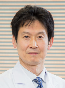 Yasuhisa Fujii合作论文数Department of Urology, Graduate School, Tokyo Medical and Dental University9
 Sjoerd Elias合作论文数Julius Center for Health Sciences and Primary Care, University Medical Center Utrecht9
 Bart De Keizer合作论文数Department of Nuclear Medicine, Erasmus Medical Center9
 Frederik Beek合作论文数8
 Marieke Haan合作论文数Dept Sociol, Univ Groningen8
 Marcel Nijland合作论文数8Jaap Stoker合作论文数8Tomohiro Yamashita合作论文数8
 M.B. Bierings合作论文数7Jan P Pennings合作论文数7Stefan J. Fransen合作论文数7
 Xaver U. Kahle合作论文数7Ryota Ishii合作论文数7




