Staphylococcus aureus infections often develop biofilms, abscesses, and intracellular reservoirs, which reduce antibiotic effectiveness. This reduced efficacy is partly due to bacterial metabolic inactivity. However, sugars can reactivate bacteria and restore their susceptibility. This study aims to identify effective combination of sitafloxacin and sugars and develop a carrier-free nanodrug delivery system to enhance treatment against metabolically inactive S. aureus. Sitafloxacin demonstrates the highest activity against stationary-phase USA300 S. aureus, achieving a ∼3 log10 bacterial reduction, which is more than tenfold enhanced when combined with glucose, fructose, or mannose. The antibacterial effects were confirmed in biofilm and abscess in vitro models, with sugars potentiating sitafloxacin in abscesses. Nanodrugs were synthesized by covalently linking sitafloxacin to sugars via Schiff-base bonds, allowing self-assembly into stable nanoparticles (∼100 nm). Antibiotic release reaches ∼35-40% over three days at physiological pH, indicating high stability. These nanodrugs reduce stationary and growing bacteria and in vitro abscesses by ∼2 log10 and biofilm bacteria by ∼4 log10, needing further refinement to achieve enhanced activity compared to free sitafloxacin. They accumulate in macrophages, reducing intracellular bacteria by 2 log10 without any cytotoxic effect. This novel nanosystem co-delivers antibiotic and sugar without inert carriers, demonstrating antimicrobial activity, biocompatibility, and delayed release, offering potential for targeting persistent bacterial infections.
To investigate, in an established rodent model, whether administration of an opioid (buprenorphine) increases the risk of developing an orthopaedic-device related infection (ODRI), and whether prolonged buprenorphine administration interferes with treatment outcome for established ODRI. The primary outcome was infection status; secondary outcomes included bone morphology and implant osseointegration assessed by quantitative microCT. The study comprised two arms. In the “risk study”, 20 rats received proximal tibia implants inoculated with 102 CFU of Staphylococcus epidermidis; 10 were treated with buprenorphine (0,1 mg/kg s.c., once daily for five days), and 10 received saline. Animals were euthanized on day 9. MicroCT scans were performed at surgery and euthanasia. Quantitative bacteriology of bone, soft tissues, and implants was performed post-mortem. In the “interference study”, 24 rats received implants inoculated with 106 CFU of S. epidermidis; 12 were treated with buprenorphine (0,1mg/kg s.c., day 7-18) and 12 received saline. Two buprenorphine-treated animals were excluded because of a deviation from the administration protocol. All animals received cefazolin and rifampin from day 7-21. Longitudinal MicroCT scans were acquired on day 0, 9, 20, and 28. Animals were euthanized on day 28. Three animals per group underwent histopathology; the remainder were assessed by bacteriology. Bacteriological analyses in both arms showed no significant differences between control and treated groups in infection burden or bacterial clearance. In the interference study, persistent infection occurred in 3/12 control animals versus 7/10 buprenorphine-treated animals (p = 0.0836). An exploratory comparison between the two study arms revealed reduced bone formation under high versus moderate contamination (p = 0.0056). No statistically significant effect of buprenorphine on bone morphology was detected under high contamination, whereas under moderate contamination buprenorphine was associated with reduced bone formation. No statistically significant increase in ODRI risk or antibiotic treatment response was detected in the buprenorphine-treated group. A non-significant trend toward higher infection persistence in buprenorphine-treated animals warrants further investigations. High bacterial load reduced bone formation, while buprenorphine did not impair short-term osseointegration or infection-related bone remodeling.
Musculoskeletal infection (MSKI) remains a major problem after trauma and elective orthopedic surgery. Chronic MSKI is related to the formation of biofilm, which impairs diagnosis and effective treatments. Therefore, to understand and communicate global standards and best practices, the 2025 International Consensus Meeting (ICM) on MSKI created a Biofilm Section to address crucial aspects of biofilm biology pertaining to its mechanisms of drug resistance and immune evasion, and potential approaches to overcome them. This featured a 2-year process, with final voting and discussion on May 8-10, 2025, in Istanbul, Turkey. This Consensus Article is the effort of the Biofilm Basic Mechanisms Workgroup, which interpreted the results on ICM questions related to (1) the infectious microenvironment; (2) appropriate inocula in preclinical research; (3) biofilm behavior in infected tissues; and (4) synergy within biofilms and with other comorbidities. Collectively, we find that this field has the necessary research tools to discover the pathophysiology of orthopedic implant-associated biofilm development and maturation, perform clinically relevant studies in animal models, and elucidate mechanisms that allow opportunistic infections in compromised tissues and patients with other health issues.
Introduction: Biofilm formation is one of the key elements making orthopaedic device-related infections (ODRIs) difficult to eradicate. Aminoglycosides such as gentamicin are frequently applied via local carriers, and systemic rifampin is added for its anti-biofilm activity. However, robust in vitro evidence of their ability to eradicate mature biofilm is limited. This study assessed whether gentamicin, alone or in combination with rifampin, can eradicate established Staphylococcus aureus biofilm in vitro. Methods: A clinical methicillin-susceptible S. aureus isolate was grown as a 5 d old biofilm on a peg lid microtiter plate. Three exposure regimens were tested: (i) continuous exposure to gentamicin (15-2000 mg L-1) for 28 d, (ii) intermittent 2 h exposures twice daily (at 15, 250 and 2000 mg L-1) for 28 d to reflect systemic twice-daily dosing and (iii) a 14 d burst release starting at 2000 mg L-1 with stepwise decline to model release from local carriers. Rifampin was either absent or added at 3.3 mg L-1, approximating peri-implant concentrations from preclinical pharmacokinetic studies. Biofilm viability was quantified as colony-forming units (CFUs) from sonicated pegs, and selected surviving isolates underwent susceptibility testing. Results: Across all regimens, concentration- and time-dependent decreases in CFU counts were observed, but no regimen resulted in bacterial counts falling below the lower limit of detection (LLOD). The addition of rifampin did not result in the sustained enhancement of biofilm killing, and, in some regimens, resulted in higher CFU counts. Isolates recovered from culture-positive pegs remained largely susceptible to gentamicin, whereas rifampin resistance arose sporadically. Conclusion: High-dose gentamicin exposures failed to eradicate 5 d old S. aureus biofilm in vitro, whatever the administration regimen. Rifampin co-administration did not alter the final outcome of biofilm persistence, despite its well-recognised anti-biofilm activity. These findings challenge the reliance on aminoglycoside-loaded carriers as curative strategies for ODRIs and suggest that persistent viability may reflect antibiotic tolerance that may not be overcome by antibiotics alone.
Staphylococcus epidermidis, a dominant human skin commensal that promotes microbial homeostasis from early life, can transition to an opportunistic pathogen under certain conditions, including invasive, biofilm-associated infections linked to medical devices. Neonatal exposure to skin commensals induces a lifelong immunological imprint in the skin, characterized by immunoregulatory responses. We therefore hypothesized that early life exposure to S. epidermidis influences immune responses to invasive biofilm-associated infections later in life. Using a murine model of biofilm-related S. epidermidis bone infection induced in adulthood, we show that mice previously colonized as neonates displayed substantially different immune responses to later-life invasive infection than those not colonized or those colonized as adults. Neonatal colonization led to greater numbers of NK cells and neutrophils than no colonization, along with reduced Tregs and Th1 cells, and consistent increase in immune checkpoint receptor PD-1+ Tregs, T effector, Th1 and Th2 cells across infected bone marrow, blood and spleen. These PD-1-related immune modulations were absent in the adult-colonized group, which had the highest numbers of Tregs, Th1 and Th2 cells of all three groups. These findings reveal that early exposure to commensal bacteria strongly impacts the response to invasive infection later in life. Notably, the response depends on the timing of previous exposure. Neonatal colonization drives T cell modulation, resembling neonatal immunity, while adult-colonization increases specific T cell abundance. These differences highlight the essential role of skin commensal colonization in shaping the quality of pathogen immunity to protect against invasive, biofilm-associated infection later in life.
Fracture-related infection (FRI) is a serious complication in orthopaedic trauma that can lead to delayed union, nonunion, and poor clinical outcomes. A better understanding of the host immune response may provide valuable insights into the pathophysiology of FRI and may help identify genomic elements that contribute to the infection. This observational study compared immune responses between patients with FRI and non-infected controls using bone/tissue biopsies and sonication fluid, and it explored the possibility of detecting bacterial and biofilm genes using transcriptome profiling with hybridization technology (nCounter® RNA hybridization technology). A total of 15 infected patients demonstrated significant upregulation of the innate immune pathway, including Toll-like receptor (TLR) signalling and the MyD88 cascade, suggesting an active immune response contributing to both infection control and bone resorption. Among the differentially expressed genes, PTGS2 (COX-2) showed the highest level of upregulation in the infection group. Bone biopsies showed enhanced chemokine (e.g. CXCL1, CXCL2, CCL4/L1/L2) signalling, with higher levels compared to tissue biopsies. Transcriptomic analysis identified bacterial transcripts in cases where conventional culture was negative, revealing potential cases of low-bacterial-load infections causing culture-negativity. Transcriptome profiling exposed distinct immune activation patterns in FRI and enabled the detection of pathogens missed by conventional culture. These findings call for larger, prospective studies to further explore the clinical utility of transcriptomics in understanding and managing FRI.
Introduction Surgical site infections (SSIs) are significant complications that can occur after spine surgery. Compared with systemic infections such as sepsis, or localised musculoskeletal infections such as periprosthetic joint infection, the immune responses in postoperative infection of the spine are poorly understood. A deeper understanding may provide clinically relevant diagnostic or therapeutic options.Methods This study examined changes in immune cell profiles in the blood of patients who developed infections after spinal surgery (Infection group, I, n = 17) compared to control patients without infections (Non-infection, NI, n = 20). The two groups were matched by age, body mass index (BMI), and the invasiveness of the surgical procedure. Peripheral blood mononuclear cells (PBMCs) were collected after surgery and subjected to high-dimensional mass cytometry.Results Cluster analysis identified 46 immune cell clusters, 30 of which exhibited significant differences between the I and NI patients. Natural killer cells as well as myeloid dendritic cells were decreased in I patients compared to NI patients (p=0.0269; and p=0.0267, respectively). Conversely, Th17, CD69+ and HLA-DR+ CD4+ T cells were significantly increased in I patients compared to NI patients (p=0.0422, p=0.0267; p=0.0267, respectively). ROC curve analysis of immune cell counts demonstrated potential for differentiating NI from I patients.Discussion This study reveals that patients with SSI exhibit a significantly altered immune cell profile, with greatest differences observed in increased HLA-DR+ CD4+ T cells and decreased numbers of several innate immune cells. The diagnostic potential of these markers may prove clinically relevant, and further research into the impact of activation and exhaustion may yield future therapeutic strategies.
Orthopedic device-related infection (ODRI) is a devastating complication, often requiring repeated revision surgeries and prolonged antibiotic treatment. Injectable biodegradable biomaterials that deliver and maintain high local concentrations of antibiotics to the surgical field may eliminate the need for additional surgeries while improving treatment outcomes. In this study, we introduce a Gel for Delivery of Antibiotics (GEDAI) for local administration of tobramycin (T), gentamicin (G) or gentamicin-vancomycin (GV). GEDAI was developed with translatability and ease of intraoperative handling as design goals. GEDAI displayed storage and loss moduli, shear thinning, and elastic recovery properties appropriate for intraoperative applications, with good adhesion to stainless steel and tissues but reduced adhesion to surgical gloves. In vitro assays showed no adverse effects of the GEDAI on viability of fibroblasts, macrophages and endothelial cells, or osteogenic differentiation of human bone marrow stromal cells (hBMSCs). Methicillin susceptible and resistant Staphylococcus aureus (MSSA and MRSA), growing in planktonic culture or biofilm, were significantly reduced after exposure to the antibiotic loaded-GEDAI compared to GEDAI without antibiotics (p < 0.05). In three distinct in vivo studies in sheep, GEDAI-T reached a peak local concentration above 2000 μg/mL within 2 h and substantially reduced bacterial counts in two models with implant retention (GEDAI-T or GEDAI-G), and completely eradicated infection in an implant exchange model (GEDA-GV). GEDAI directly targets two unmet needs in ODRI: achieving bactericidal local concentrations without systemic toxicity and providing a ready-to-use formulation. The antibiotic-loaded GEDAI formulations promise to improve treatment outcomes in challenging infection cases, potentially reducing healthcare costs and patient burden in postoperative infection management.
Despite advancements in surgical techniques, musculoskeletal infections (MSKI) remain severe complications following orthopedic surgery, imposing a substantial financial and personal burden on patients and healthcare systems globally. To establish the current state of knowledge in this field, International Consensus Meetings (ICM) were held in 2013, 2018, and 2025, including a Biofilm Section focused on establishing state-of-the-art basic science and translational research. The latest ICM utilized a 2-year-long Delphi process that commenced on May 31, 2023, and culminated in an in-person meeting involving voting on 30 questions by 47 delegates on May 8-10, 2025, in Istanbul, Turkey. Following the voting process, the Biofilm Section formed three workgroups (Biofilm Basic Science, Biofilm Treatment, and Research Priorities) to interpret the results and disseminate the findings in Consensus Articles that highlight priorities. The following is the summation of the Biofilm Treatment Workgroup, which aims to shape future pre-clinical MSKI research directions and grant funding with respect to: (1) elevating scientific rigor to ensure reproducibility and high-quality data in preclinical research; (2) transitioning mature therapeutic concepts into rigorous in vivo models to definitively prove their clinical feasibility; and (3) accelerating the development of novel molecular targets and advanced drug-delivery systems. Finally, the workgroup acknowledged a critical shift in the funding landscape. As government support faces future challenges, there is an urgent need for increased investment from industry and philanthropic partners. Such support is essential to develop effective treatments for serious orthopedic infections and to improve outcomes for patients facing life-altering illnesses.
Staphylococcus aureus is a major human pathogen that causes persistent infections characterised by the formation of aggregates such as surface-attached biofilm and staphylococcal abscess communities (SACs). Both consist of dense bacterial populations associated with self-produced matrices that impair immune cell and antibiotic access. Surface-attached biofilms have mostly protein- or polysaccharide-rich matrices, whereas SACs are spherical structures within tissue encased in a fibrin pseudocapsule and microcolony-associated meshwork. It remains unclear whether SAC are simply biofilms within tissue or if they display distinct features with unique genetic regulation. Here, we investigated genetic determinants of SAC and biofilm formation using four S. aureus deletion mutants targeting staphylokinase (Δsak), coagulase (Δcoa), the alternative sigma factor SigB (ΔrpoF), and sortase A (ΔsrtA). SACs were grown in collagen gels, whilst biofilms were grown on titanium discs, with bacterial viability, fibrin deposition, and biofilm biomass assessed by microscopy, image analysis, and crystal violet staining. Although deletion of coa or sak did not significantly impact either SAC or biofilm formation, biofilm surface coverage was markedly increased in ΔsrtA and biomass decreased in ΔrpoF; however, these mutations had no effect on SAC. These findings suggest that certain mutations can have different effects in the two experimental systems, and that the tested gene deletions were more important for surface-associated biofilm development than SAC formation.
Persistent bacteria (persisters) are phenotypic variants that emerge either randomly or in response to a range of adverse environmental conditions. Persistence represents a state whereby a subpopulation of microorganisms can spontaneously enter a "dormant" state in response to environmental factors, while simultaneously exhibiting elevated tolerance to antimicrobial agents. This review provides the current definition of bacterial persistence and summarizes the mechanisms of persisters formation as well as the various niches of bacterial persistence encountered in clinical practice. Strategies targeting persisters are outlined, including but not limited to direct killing, awakening of persistent bacteria, combined clearance, and inhibition of persistence formation, and we conclude by proposing challenges and solutions for addressing bacterial persistence in current clinical practice.
The management of fracture-related infection (FRI) with Debridement, Antibiotics, Irrigation, and Implant Retention (DAIR) is an appealing option, but its suitability is restricted to a relatively narrow proportion of patients. This study aimed to create a large animal model of DAIR after FRI and to evaluate outcomes after early (2 weeks) and delayed (5 weeks) DAIR. Additionally, intramedullary lavage (IML) of the intramedullary canal (IMC) is introduced as a novel technique to remove infected tissue. Our findings showed that DAIR failed to resolve infections in both early and delayed groups, whilst IML significantly reduced bacterial counts, leading to culture-negative results in the soft tissue and bone marrow. IML did not compromise long-term bone healing as revealed by an implant load sensor on the plate. In conclusion, DAIR was successfully achieved in a new large animal model with minimal losses. The IML method improves treatment efficacy, potentially broadening the range of patients suitable for DAIR.
Staphylococci are the most common cause of orthopaedic device-related infections (ODRIs), with Staphylococcus aureus responsible for a third or more of cases. This prospective clinical and laboratory study investigated the association of genomic and phenotypic variation with treatment outcomes in ODRI isolates. Eighty-six invasive S. aureus isolates were collected from patients with ODRI, and clinical outcome was assessed after a follow-up examination of 24 months. Each patient was then considered to have been ‘cured’ or ‘not cured’ based on predefined clinical criteria. Whole-genome sequencing and molecular characterization identified isolates belonging to globally circulating community- and hospital-acquired lineages. Most isolates were phenotypically susceptible to methicillin and lacked the staphylococcal cassette chromosome mec cassette [methicillin-susceptible S. aureus (MSSA); 94%] but contained several virulence genes, including toxins and biofilm genes. Whilst recognizing the role of the host immune response, we identified genetic variance, which could be associated with the infection severity or clinical outcome. Whilst this and several other studies reinforce the role antibiotic resistance [e.g. methicillin-resistant S. aureus (MRSA) infection] has on treatment failure, it is important not to overlook MSSA that can cause equally destructive infections and lead to poor patient outcomes.
BackgroundFracture-related infections (FRI), particularly those caused by antibiotic resistant Staphylococcus aureus, present significant clinical challenges due to the formation of biofilm on the implanted device, and reduced options for conventional antibiotic treatment. Bacteriophage (phage) therapy (PT) offers a targeted approach to managing such infections, however, evidence for pharmacokinetics and optimal route of administration is limited for FRI. This study aimed to evaluate safety, phage distribution kinetics, phage neutralization, and antibacterial efficacy after intravenous or local administration in a sheep model.MethodsThe study was conducted in two phases: Phase 1 assessed the safety and distribution of two successive rounds of intravenous and local phage administration in non-infected sheep, while Phase 2 evaluated the therapeutic efficacy of intravenous versus local phage administration in combination with intravenous vancomycin in treating MRSA-induced FRI (tibial osteotomy with plate fixation). The specific pathogen and phage used in the sheep were both taken from a human FRI patient treated with PT. Phage neutralization and phage distribution were the primary outcomes measured in both phases of the sheep study.ResultsBoth intravenous and local phage administration were well-tolerated in non-infected sheep. Phages were cleared rapidly from circulation after intravenous administration, with no phage detected after 240 minutes. Phage neutralization increased during PT, peaking at 99.9% in non-inoculated sheep by the end of the second phage treatment (day 50). In infected sheep, phage neutralization levels reached a maximum of 99.9% earlier (day 13), with no significant differences between intravenous and local administration. The bacterial load was not significantly changed by PT, either IV or locally applied.ConclusionsPT is a safe adjunct to antibiotic treatment for FRI, however, phage neutralization developed rapidly and was accelerated in infected hosts. Further research is required to optimize phage selection, dosing, and delivery methods to enhance its therapeutic potential as an adjunct to conventional antibiotic therapy, particularly in the face of challenges such as rapid clearance and phage neutralization.
Intracellular Staphylococcus aureus persisters are a dormant bacterial subpopulation responsible for chronic and recurrent infections due to their ability to evade antibiotic treatment within host cells. However, effective strategies for eliminating these intracellular pathogens remain limited. Herein, we proposed a versatile poly(amino acid)-based platform, F(AM), for the effective eradication of intracellular Staphylococcus aureus persisters via on-site antibiotic delivery. The F(AM) platform exhibited dual-targeting capability toward macrophages and Staphylococcus aureus persisters, efficiently penetrating cellular barriers and achieving precise antibiotic delivery at intracellular bacterial niches. Sitafloxacin, rifampicin, and polymyxin B were identified from a panel of 11 antibiotic candidates and individually loaded into the F(AM) platform. The resulting nanoparticles markedly improved intracellular drug accumulation, protected antibiotics from degradation within the adverse intracellular environment, and overcame microenvironment-induced bacterial metabolic shifts. Compared with free antibiotics, the drug-loaded F(AM) nanoparticles notably improved their intracellular bactericidal activity. Collectively, this study highlights F(AM) as a robust and versatile platform for overcoming intracellular barriers and restoring antibiotic efficacy, offering a valuable tool for antipersister strategies and intracellular pharmacokinetic investigations.
Osteomyelitis (OM) is a progressive, inflammatory infection of bone caused predominately by Staphylococcus aureus. Herein, we engineered an antibiotic-eluting collagen-hydroxyapatite scaffold capable of eliminating infection and facilitating bone healing. An iterative freeze-drying and chemical crosslinking approach was leveraged to modify antibiotic release kinetics, resulting in a layered dual-release system whereby an initial rapid release of antibiotic to clear infection was followed by a sustained controlled release to prevent reoccurrence of infection. We observed that the presence of microbial collagenase accelerated antibiotic release from the crosslinked layer of the scaffold, indicating that the material is responsive to microbial activity. As exemplar drugs, vancomycin and gentamicin-eluting scaffolds were demonstrated to be bactericidal, and supported osteogenesis in vitro. In a pilot murine model of OM, vancomycin-eluting scaffolds were observed to reduce S. aureus infection within the tibia. Finally, in a rabbit model of chronic OM, gentamicin-eluting scaffolds both facilitated radial bone defect healing and eliminated S. aureus infection. These results show that antibiotic-eluting collagen-hydroxyapatite scaffolds are a one-stage therapy for OM, which when implanted into infected bone defects simultaneously eradicate infection and facilitate bone tissue healing.
Staphylococcus aureus has multiple mechanisms to evade the host's immune system and antibiotic treatment. One such mechanism is the invasion of the osteocyte lacuno-canalicular network (OLCN), which may be particularly important in recurrence of infection after debridement and antibiotic therapy. The aim of this study was to develop an ex vivo model to facilitate further study of S. aureus invasion of the OLCN and early-stage testing of antibacterial strategies against bacteria in this niche. The diameter of the canaliculi of non-infected human, sheep, and mouse bones was measured microscopically on Schmorl's picrothionin stained sections, showing a large overlap in canalicular diameter. S. aureus successfully invaded the OLCN in all species in vitro as revealed by presence in osteocyte lacunae in Brown and Brenn-stained sections and by scanning electron microscopy. Murine bones were then selected for further experiments, and titanium pins with either a wild-type or ΔPBP4 mutant S. aureus USA300 were placed trans-cortically and incubated for 2 weeks in tryptic soy broth. Wild-type S. aureus readily invaded the osteocyte lacunae in mouse bones while the ΔPBP4 showed a significantly lower invasion of the OLCN (p = 0.0005). Bone specimens were then treated with gentamicin, sitafloxacin, R14 bacteriophages, or left untreated. Gentamicin (p = 0.0027) and sitafloxacin (p = 0.0280) significantly reduced the proportion of S. aureus-occupied lacunae, whilst bacteriophage treatment had no effect. This study shows that S. aureus is able to invade the OLCN in an ex vivo model. This ex vivo model can be used for future early-stage studies before proceeding to in vivo studies.
AimBacteriophages are remerging as alternative and adjunctive therapy for fracture-related infection (FRI). However, current administration protocols involve prolonged retention of a percutaneous draining tube with potential risk of developing superinfection. In this study, we applied a cocktail of in vitro evolved biofilm-targeting phages for Methicillin-resistant Staphylococcus aureus (MRSA) in a hydrogel platform co-delivering vancomycin. In vitro synergy and antibiofilm activity was assessed and a subsequent in vivo study was performed in a mouse FRI model with MRSA.MethodTwo evolved bacteriophages (MRSA-R14 and COL-R23) with improved antibiofilm activity against a clinical isolate (MRSA3) were tested in combination with vancomycin and a carboxymethylcellulose (CMC) hydrogel in vitro and in vivo. MRSA3 bacterial biofilms were formed on sterile 4 mm sintered porous glass beads at 37 °C for 24 h. Biofilms were exposed to i-phage cocktail (107 PFU/ml), ii-vancomycin at concentrations of 0.5, 1, 10 and 100 times the MIC, or iii-combination of phage cocktail and vancomycin. Recovered biofilm cells, were quantified by colony counting. The stability and release profiles of phage cocktail and vancomycin in co-delivery hydrogel were assessed in vitro for 8 days and 72 hrs, respectively, and subsequently tested in the treatment of 5-day-old MRSA3 infection of a femoral plate osteotomy in mice.ResultsIn vitro: The cocktail of evolved phages (107 PFU/ml, 1:1) combined with 0.5 MIC vancomycin achieved 99.72% reduction in MRSA3 biofilm in vitro compared to the growth control. This combination was stable in the co-delivery hydrogel over 8 days. The release profile showed that 57% of phages and 80% of vancomycin were released after 72hrs, which was identical to the performance for gels loaded with phage or antibiotic alone. In the in vivo study, the bacterial load from animals that received co-delivery hydrogel and systemic vancomycin was significantly reduced compared to controls, animals that received systemic vancomycin and animals that received co-delivery hydrogel alone (p<0.05).ConclusionsOur study demonstrates the potential of using evolved phages in combination with vancomycin and hydrogel delivery systems for the treatment of MRSA-related infections. Further research in this area may lead to the development of specific therapies for biofilm-related infection.
Rifampicin is a key antibiotic in the treatment of staphylococcal biofilm infections. In this pilot study, we found that patients who received rifampicin for treatment of an orthopaedic-device-related infection (ODRI) were colonized with rifampicin-resistant staphylococci during treatment and this persisted for up to 2 months after cessation of treatment.
INTRODUCTION:With the introduction and continuous improvement in operative fracture fixation, even the most severe bone fractures can be treated with a high rate of successful healing. However, healing complications can occur and when healing fails over prolonged time, the outcome is termed a fracture non-union. Non-union is generally believed to develop due to inadequate fixation, underlying host-related factors, or infection. Despite the advancements in fracture fixation and infection management, there is still a clear need for earlier diagnosis, improved prediction of healing outcomes and innovation in the treatment of non-union. AREAS COVERED:This review provides a detailed description of non-union from a clinical perspective, including the state of the art in diagnosis, treatment, and currently available biomaterials and orthobiologics.Subsequently, recent translational development from the biological, mechanical, and infection research fields are presented, including the latest in smart implants, osteoinductive materials, and in silico modeling. EXPERT OPINION:The first challenge for future innovations is to refine and to identify new clinical factors for the proper definition, diagnosis, and treatment of non-union. However, integration of in vitro, in vivo, and in silico research will enable a comprehensive understanding of non-union causes and correlations, leading to the development of more effective treatments.
 Richards R Geoff合作论文数AO Research Institute Davos/ECM Journal116
 Stephan Zeiter合作论文数AO Research Institute55
 Mario Morgenstern合作论文数41
 Willem-Jan Metsemakers合作论文数32
 David Eglin合作论文数21
 Daniel Arens合作论文数20
 Claudia Siverino合作论文数AO Res Inst Davos18Wj. Metsemakers合作论文数16
 Keith Thompson合作论文数AO Research Institute Davos15
 Stephen L. Kates合作论文数Department of Orthopaedics, University of Rochester14
 D'Este Matteo合作论文数13
 Michiel H J Verhofstad合作论文数13Post Virginia合作论文数13
 Charalampos Zalavras合作论文数Keck School of Medicine, University of Southern California13
 Edward Thomas James Rochford合作论文数12Jan Puetzler合作论文数11Marco Chittò合作论文数11
 Jolien Onsea合作论文数11
 Edward M. Schwarz合作论文数Center for Musculoskeletal Research, University of Rochester Medical Center;Department of Orthopaedics, University of Rochester11
 Muthukrishnan Gowrishankar合作论文数College of Medicine, University of Central Florida11
 D.W. Grijpma (Dirk)合作论文数Faculty of Science and Technology, University of Twente11Richard Kuehl合作论文数11
 Martijn Riool合作论文数Swammerdam Institute for Life Sciences, University of Amsterdam10
 Andrew Foster合作论文数AO Research Institute Davos9
 Caroline Constant合作论文数9
 Liam O’Mahony合作论文数Univ Zurich, Swiss Inst Allergy & Asthma Res SIAF, Davos, Switzerland9
 Peter Wahl合作论文数9
 Niels Vanvelk合作论文数8Marloes I Hofstee合作论文数8
 Tanja Schmid合作论文数8
 Sebastian Zaat合作论文数8
 Andrej Trampuz合作论文数Centrum fur Muskuloskeletale Chirurgie, Charite - University Medicine Berlin8Alexandra H. C. Poulsson合作论文数7Dirk Nehrbass合作论文数7
 Baixing Chen (陈柏行)合作论文数7
 William Obremskey合作论文数Div Orthopaed Trauma, Vanderbilt Orthopaed Inst7
 Olivier Guillaume合作论文数7Boot Willemijn合作论文数7
 Xing Wang (王兴)合作论文数College of Life Science and Technology, Beijing University of Chemical Technology7
 Stefaan Nijs合作论文数Department of Traumatology, UZ Leuven7
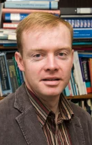 Michael Tunney合作论文数School Of Pharmacy, Queen's University Belfast;Faculty of Medicine, Health & Life Sciences, Queen's University Belfast7
 Martin McNally合作论文数Nuffield Orthopaedic Centre, Oxford University Hospitals;Royal College of Surgeons, The University of Edinburgh7
 Stuart Elborn合作论文数Centre for Experimental Medicine, Institute for Health Sciences, School of Medicine, Dentistry and Biomedical Sciences, Queen's University Belfast7
 Michael J. Raschke合作论文数Klinik für Unfall-, Hand- und Wiederherstellungschirurgie;, Universitatsklinikum Munster7Christoph Erichsen合作论文数6
 Guofeng Li合作论文数6
 Rob Lavigne合作论文数Department of Biosystems, KU Leuven;Faculty of Bioscience Engineering, KU Leuven;Leuven Plant Institute, KU Leuven6
 Vincent a Stadelmann合作论文数Laboratory of Biomechanical Orthopedics;Ecole Polytechnique Fédérale de Lausanne;Laboratory of Biomechanical Orthopedics, Ecole Polytechnique Fédérale de Lausanne6




