OBJECTIVES:Experiential avoidance is the unwillingness to come into contact with aversive internal experiences. Trauma exposure is associated with greater experiential avoidance and insomnia symptoms. Experiential avoidance may perpetuate insomnia symptoms in patients with posttraumatic stress disorder (PTSD). We examined the relationship between experiential avoidance and insomnia symptoms among veterans with PTSD (based on the Clinician-Administered PTSD Scale for DSM-5). METHOD:The sample included 93 veterans (M = 54.7 years; 86.0% male) who attributed their sleep disturbance onset to experiences of trauma on the CAPS-5. Experiential avoidance, insomnia, sleep disturbance, daytime sleepiness, and daytime consequences were measured with the Brief Experiential Avoidance Questionnaire (BEAQ), Insomnia Severity Index (ISI), Pittsburgh Sleep Quality Index (PSQI), Epworth Sleepiness Scale (ESS), and International Classification of Sleep Disorders (ICSD) items. We conducted multiple linear regressions with age, sex, and BEAQ as the independent variables and sleep variables as the dependent variables. RESULTS:There were significant positive associations between the BEAQ and the ISI, PSQI daily disturbance factor, ESS, and ICSD daytime consequences. CONCLUSIONS:Greater experiential avoidance was associated with worse insomnia symptoms and consequences, particularly daytime dysfunction. Experiential avoidance may be an overlooked, but relevant treatment target for patients with comorbid insomnia and PTSD.
Objective:Polygenic risk scores (PRS) for posttraumatic stress disorder (PTSD) often account for a low amount of variance. Ancestry-related differences in PRS scale and variance limit cross-group comparisons. This methodological challenge further complicates gene-by-environment (GxE) analyses, given that socioenvironmental exposures are inequitably distributed across ethnoracial groups. We constructed an ancestry-calibrated polygenic risk score (AC-PRS) for PTSD in the largest longitudinal study of trauma survivors to date and investigated GxE interactions. Method:Recent trauma survivors (N=1,801) provided a blood specimen for genotyping. Six PTSD trajectories were previously identified from PTSD Checklist for DSM-5 (PCL-5) scores at 2-weeks, 8-weeks, 3-months, and 6-months post-trauma. Greenspace (normalized difference vegetation index [NDVI) and socioeconomic disadvantage (area deprivation index [ADI]) were derived from residential addresses. Logistic regressions examined interactions between newly developed AC-PRS and neighborhood factors on trajectories after adjusting for sociodemographic and trauma-related covariates. Secondary linear models considered GxE interactions on 6-month PCL-5 scores. Results:AC-PRS performed well across ethnoracial groups, explaining significant variability in PTSD trajectories (R2=.053). ADI moderated the association between AC-PRS and the likelihood of assignment in a high nonremitting trajectory of PTSD symptoms and severity of symptoms at 6-months (ps < .05). There were no NDVI x AC-PRS interactions in any models. Conclusions:AC-PRS captures genetic risk for PTSD in admixed trauma survivors, demonstrating good discrimination between nonremitting and resilient courses of PTSD. However, neighborhood disadvantage may modify utility of PRS for PTSD, warranting careful consideration when applying these scores across contexts.
Abstract We identified “Sleep Bursts” (SBs), as a novel phenomenon of brief (1-2 sec), often periodic bursts in cranial forces occurring during human sleep. Our goal was to characterize SBs in normal subjects, then compare SB in a cohort of subjects with neurodegenerative disease (NDD). We recorded 32 cognitively healthy subjects (23 -87 years) and 13 subjects with NDD (51 – 84 years). SBs occurred in all 45 subjects. SBs occurred at 0.57 SB/min (once per 105 seconds) in controls and 0.40 SB/min (once per 150 seconds) in NDD (p = 0.0043). SB occurred with equal rates across all sleep stages in both groups. When occurring periodically, SBs had modal intervals (3.75 bursts/min (0.0625 Hz) - 2.67 bursts/min (0.044 Hz)). EEG power increased in the delta range 1-2 seconds before and following the SB. EEG delta power during a SB was significantly lower in all NDD subjects across sleep stages compared to controls. The relatively low frequency of SB events and synchronization with EEG power has no parallel in human sleep; we hypothesize that SBs may represent a brain-generated pulsatile component of brain glymphatic drainage.
Background Mental health conditions are leading causes of disability, driven substantially by inadequate treatment access in low-income and middle-income countries. WHO identifies psychosocial and pharmacological treatments as essential services. We aimed to compare the effectiveness of first-line treatments with interpersonal psychotherapy (IPT) versus fluoxetine delivered by non-specialists, for major depressive episode and post-traumatic stress disorder (PTSD) among public-sector primary care patients in Kenya, and to assess first-line and second-line treatment sequences. Methods We conducted a sequential, multiple-assignment, randomised trial (SMART) at Kisumu County Referral Hospital (Kisumu, Kenya) Eligible participants were outpatients aged 18 years or older, positive for major depression, PTSD, or both, on the Mini International Neuropsychiatric Interview 7.0.2, and able to attend IPT and fluoxetine treatment appointments. We randomly assigned (1:1) participants to first-line treatment (stage 1) with IPT (12 weekly sessions delivered by non-specialists) or fluoxetine (6 months, 20 mg starting dose) prescribed by a non-specialist (nurse or clinical officer). In stage 2, participants not in remission from both major depressive episodes (MDE) and PTSD were randomly assigned to second-line treatment consisting of switch (IPT to fluoxetine or vice versa), or combination (IPT and fluoxetine). The primary outcome was remission of MDE and PTSD at end of stage 1 (ie, after first-line treatment; 3 months for IPT and 6 months for fluoxetine) and end of stage 2 (ie, after second-line, crossover treatment; up to 12 months) using thresholds on the Beck Depression Inventory 2 and PTSD Checklist 5. We used intention-to-treat analyses to investigate first-line treatment superiority and treatment sequences, using relative risk (RR) and ratios of RRs for interactions. The completed trial is registered at ClinicalTrials.gov, NCT03466346. Findings Between Sept 1, 2020, and Oct 15, 2021, we screened 3864 participants for eligibility. Of these, we randomly assigned 2162 (56·0%) participants in stage 1 to IPT (1082 [50·0%]) or fluoxetine (1080 [50·0%]). 1958 (90·6%) were female and 204 (9·4%) were male. Treatment adherence was 98·7% for IPT and participants assigned to fluoxetine received a mean of 126 (SD 60; 70%) of the 180 doses for fluoxetine. At the end of treatment (stage 1), 127 (12·9%) of 986 IPT participants and 89 (10·1%) of 877 fluoxetine participants had MDE (RR 0·15 [95% CI 0·13–0·18] for IPT and 0·011 [0·090–0·13] for fluoxetine), favouring fluoxetine (ratio of RRs 0·73 [95% CI 0·57–0·94]; p=0·015). At the end of treatment (stage 1), 173 (17·5%) IPT participants and 108 (12·3%) fluoxetine participants had PTSD (RR 0·20 [0·18–0·23] for IPT and 0·14 [0·12–0·16] for fluoxetine), favouring fluoxetine (ratio of RRs 0·68 [0·54–0·84] p=0·0005). For stage 2, 104 people who had initially received IPT received second-line fluoxetine, 99 people who had initially received IPT received second-line fluoxetine and IPT, 68 people who had initially received fluoxetine received second-line fluoxetine, and 61 people who had initially received fluoxetine received second-line fluoxetine and IPT. As a result, stage 2 comparisons were underpowered. In stage 2, there were no between-sequence remission differences (between-group risk ratio for major depression in those who received first-line fluoxetine 1·18 [95% CI 0·99–1·39]; p=0·091; between-group RR for PTSD in those who received first-line fluoxetine 1·19 [0·96–1·46]; p=0·14; between-group RR for major depression in those who received first-line IPT 0·96 [0·86–1·07]; p=0·56; between-group risk ratio for PTSD in those who received first-line IPT 0·90 [0·77–1·06]; p=0·25). There were 43 adverse events in the fluoxetine group—the most common were gastrointestinal distress (16 [1·5%] of 1080), sweating (five [0·5%]), dizziness (three [0·3%]), anxiety or insomnia (three [0·3%]), sedation (two [0·2%])—one in the combined treatment group (thoughts of self-harm or suicide), none in the IPT group, and no deaths. Interpretation Although fluoxetine was superior as first-line treatment for remission of major depressive episode and PTSD, no differences were observed between the groups after treatment crossover at the end of second-line treatment. Our results suggest that non-specialist mental health services that adhere to global standards can be successfully delivered via primary care in a lower-middle-income east-African setting. Funding US National Institute of Mental Health and Global Alliance for Chronic Disease.
BackgroundAdverse posttraumatic neuropsychiatric sequelae are common after trauma. Early identification of individuals at risk for these outcomes could enable the deployment of preventive interventions to survivors at greatest risk. Smartphone keystroke biomarkers show promise in identifying individuals with neuropsychiatric symptoms; however, to our knowledge, no research has examined whether they can be used to identify symptoms in the aftermath of trauma. ObjectiveThis study evaluates whether passively collected keystroke data from smartphone use in daily life could identify individuals with high symptom levels, as well as worsening or recovery of symptoms, after trauma exposure. MethodsData from a diverse cohort of individuals presenting to 27 emergency departments after trauma were analyzed. Inclusion criteria were presenting to the emergency department within 72 hours of trauma, age 18-75, and the ability to speak and read English. Exclusion criteria were solid organ injury, significant hemorrhage, operative intervention, or likely admission for over 72 hours. Participants installed an app that passively collected keystroke data during use of any app on their smartphone, beginning in the emergency department. Participants also completed serial symptom assessments over 8 weeks after trauma exposure. ResultsA total of 3445 patients met study criteria, provided informed consent, and completed assessments in the emergency department. Of these, 1072 (mean age 40, SD 13; 616/1072, 57.46%, women; 565/1072, 52.71% non–Hispanic Black) installed the app on their Android smartphone and completed the 8-week assessment and were therefore included in analyses. Keystroke biomarkers related to typing speed, identified using bivariate linear mixed models controlling for false discovery rates, were associated with elevated pain, reexperiencing, and mental fatigue (absolute values of rs=0.22-0.25, Ps=.02). Separate change-of-operation and scrolling keystroke biomarkers were associated with increased reexperiencing symptoms (r=0.18, P=.047) and mental fatigue (rs=0.18-0.19, Ps=.031-.047). Further, changes in specific keystroke biomarkers were associated with worsening or recovery of pain (rs=0.07-0.10, Ps=.02), somatic symptoms (rs=0.02, Ps=.02), mental fatigue (rs=0.02-0.04, Ps=.02), sleep disturbance (absolute rs=0.07-0.09, Ps=.02), reexperiencing (rs=0.02-0.04, Ps=.02), and hyperarousal (rs=0.02-0.04, Ps=.02). ConclusionsIn general, slower typing and scrolling speeds were associated with higher symptom levels, with small to medium effect sizes. Keystroke data passively collected via smartphone use may help identify individuals with significant or changing posttraumatic symptoms. Future research should continue to explore these keystroke biomarkers and whether they can be leveraged to connect vulnerable trauma survivors to appropriate services. Overall, these results add to the literature, indicating that passively collected keystroke data may help identify individuals with neuropsychiatric symptoms or changes and are, to our knowledge, the first to test whether keystroke biomarkers are useful in the aftermath of trauma. This represents a critical period during which preventive interventions could be deployed to reduce the long-term burden of trauma-related sequelae.
Stress is increasingly recognized as an important, modifiable factor for Alzheimer's disease (AD), yet its roles in initiation, progression, and outcomes remain incompletely elucidated. Epidemiologic studies link chronic stress, early-life adversity, and trauma to increased AD risk, while experimental models have uncovered mechanisms by which stress hormones directly drive core AD pathological processes, including amyloid beta and tau aggregation, neuroinflammation, and neurodegeneration. Complicating the relationship, brain structures that regulate the stress response are themselves selectively vulnerable to early degeneration in AD. As these circuits degenerate, interpreting changes in stress biomarkers becomes more challenging, with physiological measures potentially decoupling from perceived stress. Here, we review evidence connecting stress to AD pathophysiology as both a risk factor and a driver, examine how the degeneration of stress neuroendocrine systems accelerates disease progression, and discuss implications for intervention and clinical trial design.
Importance:Prior work demonstrates that earlier trauma exposure increases the risk of posttraumatic stress disorder (PTSD) after a subsequent trauma, and cross-sectional work has also identified sexual violence (SV) as the form of traumatic stressor with the greatest conditional risk for PTSD. However, little work explores these findings longitudinally with a comparison group with no history of SV. Objective:To examine the differences in PTSD symptoms after a new incident of trauma exposure in survivors with and without an earlier history of SV. Design, Setting, and Participants:In this case-control study, data from a longitudinal cohort sample of patients enrolled in the Advancing Understanding of Recovery After Trauma (AURORA) study were examined. Participants were admitted to emergency departments due to a traumatic injury (primarily motor vehicle crash), and they were followed up for 1 year after emergency department (ED) admission. They were recruited within 72 hours of ED admission and then assessed at 5 time points (2 weeks, 8 weeks, 3 months, 6 months, 12 months) after their injury. Participants were a diverse and heterogeneous sample of individuals admitted to EDs across the US. Study data were evaluated from January 2025 to March 2026. Exposure:Earlier history of SV as assessed at ED admission using the Childhood Trauma Questionnaire and the Life Events Checklist. Main Outcomes and Measures:Symptoms of PTSD as assessed by the PTSD Checklist for DSM-5. Results:The study sample included 2423 individuals (mean [SD] age, 36 [13] years; 1566 female [64.6%]), 1270 whom (52%) reported an earlier history of SV. At every time point, participants with a history of SV demonstrated significantly elevated symptoms of PTSD (β = 4.62; 95% CI, 3.09-6.21; P < .001) compared with those without a history of SV, even after controlling for earlier nonsexual trauma exposure. Within the group with prior SV, the PTSD symptoms that remained the highest across time points were hypervigilance symptoms (criterion E), although negative alterations in cognition and mood (criterion D) were the symptom cluster that recovered most slowly. Conclusions and Relevance:Results of this case-control study indicate that having a history of SV was associated with elevated symptoms of PTSD and slower symptom recovery after a subsequent nonsexual trauma. These results raise important considerations for clinicians, regardless of whether or not they self-identify as working specifically with survivors of SV.
Chronic pain following traumatic stress exposure (TSE) is common. Increasing evidence suggests inflammatory and immune mechanisms are activated following TSE, play a role in the transition from acute to chronic pain, and may differ by sex. In this study, we tested the hypothesis that elevated levels of the inflammatory marker C-reactive protein (CRP) would be associated with acute and chronic pain in a sex-specific manner. We utilized blood-plasma samples and pain questionnaire data from men (n=116) and women (n=269) enrolled in AURORA, a multi-site emergency department (ED)-based longitudinal study of TSE survivors. CRP levels were measured by ELISA using plasma samples collected in the ED ('peritraumatic CRP') and six-months following TSE. Multivariate models were used to assess the relationship between CRP and pain. Men and women reported similar acute pain levels in the ED; however, pain resolved more rapidly in men over time. Peritraumatic CRP was not associated with acute pain severity in either men or women. However, six-month CRP levels were positively associated with six-month pain severity in both men (r=0.19, p=0.089, non-significant) and women (r=0.21, p=0.0015). In men only, higher peritraumatic CRP predicted lower chronic pain severity (β=-0.36, p=0.024) whereas no association was observed in women (β=0.04, p=0.628). Among men with elevated peritraumatic CRP, decreases in CRP over time were associated with decreases in pain over time (r=-0.38, p=0.0197). These findings suggest sex-specific relationships between CRP and chronic pain following TSE and highlight inflammation as a potential mechanism contributing to differential pain recovery trajectories in men and women. PERSPECTIVE: Longitudinal CRP levels following traumatic stress exposure showed sex-dependent associations with chronic pain outcomes. Unexpectedly, higher early CRP predicted lower chronic pain severity in men, suggesting inflammatory responses may differentially influence pain recovery trajectories in men and women.
OBJECTIVE:The Posttraumatic Stress Related Functioning Inventory (PRFI) is a 33-item, self-report measure that assesses functioning associated with symptoms of posttraumatic stress disorder (PTSD) across occupational, social relationships, and lifestyle domains (McCaslin et al., 2016). The purpose of the current investigation was to examine the psychometric properties of the DSM-5 version of the PRFI (PRFI-5). METHOD:The data were drawn from a completed randomized, controlled trial that compared Individual Placement and Support (IPS) supported employment with a stepwise vocational rehabilitation involving transitional work (TW) among veterans with PTSD. Veterans completed the PRFI-5 and related measures (e.g., quality of life, PTSD symptom severity, self-esteem, and suicidality; N = 450). Psychometric analyses included a review of item and scale descriptives, evaluation of the scale's reliability and cohesiveness, assessment of the scale factor structure, and its convergent and discriminant validity. RESULTS:Broadly, results were similar to those of the previous version of the PRFI and support the PRFI-5 as a reliable (αs = .70-.97) and valid measure of posttraumatic stress-related functional impairment. The measure can provide a high-level summary of functioning (e.g., total scale score) yet also can be used to provide a detailed clinical analysis of psychosocial functioning across several domains (i.e., work/school, relationships, lifestyle). CONCLUSIONS:This study examines a pragmatic measure of PTSD-related psychosocial functioning and highlights the importance of incorporating a measure of psychosocial functioning routinely alongside symptom measures as a component of psychological assessment and practice. (PsycInfo Database Record (c) 2026 APA, all rights reserved).
Socioenvironmental factors may be associated with patterns of biological age and modify individual vulnerability to trauma- and stress-related psychiatric conditions. We examined whether neighborhood socioeconomic disadvantage is associated with accelerated epigenetic aging in the early aftermath of trauma. Next, we tested whether individual psychological resources, previously shown to attenuate stress-related accelerated aging, moderated the association between neighborhood disadvantage and epigenetic aging. Participants provided blood samples approximately 2 weeks post-trauma (N = 203; 64% female) and completed sociodemographic and psychometric assessments, including a measure of individual psychological resources (Connor-Davidson Resilience Scale [CD-RISC]). Epigenetic aging was assessed using the Horvath, Hannum, GrimAge, PhenoAge, and DunedinPACE clocks. Neighborhood disadvantage (national area deprivation index [ADI]) was derived from participants' home addresses. Linear models evaluated the relationship between ADI and epigenetic aging as well as a subsequent CD-RISC x ADI interaction after covarying for chronological age, income, sex, lifetime trauma, lifetime tobacco and alcohol use, PTSD symptoms, and five estimated cell-type proportions. Higher ADI was associated with accelerated aging in GrimAge, PhenoAge, and DunedinPACE models (pscorrected <0.05) after adjusting for covariates. There was an ADI x CD-RISC interaction on GrimAge, such that the effect of neighborhood disadvantage was not significant among individuals with higher levels of individual psychological resources. Findings underscore a link between neighborhood disadvantage and accelerated biological aging and highlight several modifiable factors at both the neighborhood and individual levels associated with epigenetic age.
BACKGROUND:Systemic inflammation is hypothesized to contribute to post-traumatic stress disorder (PTSD) vulnerability. Few studies have examined inflammation shortly after trauma as a predictor of later PTSD symptoms. We examined whether inflammation from the emergency department (ED) post-trauma is associated with PTSD symptom severity over the following 6 months. METHODS:Our sample included 742 AURORA participants, a longitudinal cohort of patients in 29 EDs across the United States after a traumatic stressor, followed up to 6 months. Plasma cytokines were assessed from a study blood draw in the ED: an inflammatory index (standardized sum of generally pro-inflammatory markers interleukin [IL]-6, IL-8, tumor necrosis factor alpha [TNF-α], interferon gamma [IFN-γ]), and generally anti-inflammatory IL-10. PTSD symptoms were self-reported at 2 weeks, 8 weeks, 3 months, and 6 months post-ED. Covariate-adjusted repeated-measures regressions estimated associations between inflammation and PTSD symptoms, overall and sex-stratified. RESULTS:Among 742 participants (age m = 40.0 [13.7]; 479 [64.6%] female), PTSD symptoms were elevated then modestly decreased over follow-up. Higher ED inflammation was associated with higher PTSD symptoms across follow-up (standardized symptoms β = 0.05, 95% CI: 0.01-0.09), adjusted for potential confounders. Higher pro-inflammatory index levels and IL-6, IL-8, and TNF-α were associated with higher PTSD symptoms in males only, while higher IL-10 was associated with higher PTSD symptoms in females only. CONCLUSIONS:Pro-inflammatory levels shortly after traumatic stress are associated with heightened PTSD symptoms, particularly among males. Inflammatory markers may prove useful additions to prediction models for PTSD following trauma, with attention to sex differences.
QuestionIn veterans with chronic pain, does the Department of Veterans Affairs (VA) whole health team approach reduce pain interference more than cognitive behavioral therapy and more than usual care?FindingsIn this randomized clinical trial of 764 VA patients, the whole health team approach reduced pain interference (range, 0-10; higher scores indicate worse pain) more than cognitive behavioral therapy (mean difference, -0.58 points) and more than usual care (mean difference, -0.77 points).MeaningThese results support the whole health team approach to improve pain interference in patients with chronic pain. ImportanceThe US Department of Veterans Affairs (VA) Whole Health approach was congressionally mandated in 2016 for patients with chronic pain receiving care in VA hospitals, but no randomized clinical trials have tested its benefits.ObjectiveTo evaluate the effectiveness of a whole health team intervention in VA patients with chronic pain compared with cognitive behavioral therapy and with usual care, and to evaluate the effectiveness of cognitive behavioral therapy compared with usual care in reducing long-term pain interference.Design, Setting, and ParticipantsThis randomized clinical trial involving 6 VA health systems in the US enrolled participants between September 18, 2020, and January 19, 2024. Final follow-up occurred on January 27, 2025. Analyses took place between April 1, 2025, and February 3, 2026. Participants were patients with chronic pain receiving VA primary care.InterventionsPatients with chronic pain were randomized (11:11:2) to receive a whole health team intervention (n = 343), cognitive behavioral therapy for chronic pain delivered in group sessions (n = 339), or usual care (n = 82) for 12 months. The whole health team included a primary physician or nurse practitioner, a second clinician providing nonpharmacological or integrative pain care, and a coach. The team provided interdisciplinary, individualized care consistent with the VA Whole Health model to attain personal health goals aligned with patients' personal values and life goals.Main Outcomes and MeasuresThe primary outcome was the Brief Pain Inventory interference (BPI-I) subscale score (range, 0-10 points; higher scores indicate worse interference from pain; minimal clinically important difference, 1.0) at 12 months.ResultsOf 764 randomized patients (mean [SD] age, 60.5 [12.3] years; 66.5% were men), 632 (82.7%) completed 12-month follow-up. At 12 months, the whole health group had significantly improved pain interference scores (from 6.6 to 4.9) compared with the cognitive behavioral therapy (from 6.4 to 5.5) (mean difference, -0.58 [97% CI, -1.11 to -0.05]; P = .02) and usual care (from 6.4 to 5.7) (mean difference, -0.77 [99% CI, -1.40 to -0.15]; P = .002) groups. At 12 months, cognitive behavioral therapy did not improve pain interference scores significantly more than usual care (mean difference, -0.19 [99% CI, -0.89 to 0.50]; P = .46). The most common adverse event was suicidal ideation, which occurred in 15.9% of patients in the cognitive behavioral therapy group, 13.7% in the whole health team group, and 13.4% in the usual care group.Conclusions and RelevanceThese results support use of the whole health team approach to attain a statistically significant but small improvement in pain interference in VA patients with chronic pain.Trial RegistrationClinicalTrials.gov Identifier: NCT04330365 This randomized clinical trial examines the effectiveness of a whole health team intervention in veterans with chronic pain compared with cognitive behavioral therapy and usual care in reducing long-term pain interference.
Abstract Background Growing evidence suggests that sleep plays an important role in PTSD outcomes, potentially due to its influence on emotional memory consolidation, though these mechanisms remain unknown. This study sought to test the hypotheses that sleep neurophysiology, PTSD status, and sex moderates the degree to which the late positive potential (LPP) mediates memory accuracy for affective visual stimuli. Methods N = 39 participants (18 female) viewed 75 negative and 75 neutral IAPS images while EEG was recorded. After viewing the images, participants took a two-hour long nap which was followed by a memory assessment. Memory accuracy was measured using d’ = Z(hit rate) – Z(false alarm rate), where hit rate refers to the proportion of images seen during the memory assessment that are correctly identified as being previously seen, false alarm rate refers to the proportion of images seen during the memory assessment that are incorrectly identified as being previously seen, and Z() is the inverse cumulative distribution function of the standard normal distribution function. Results The early (300 – 1000 ms) and late (1000 – 1500 ms) LPP mediated enhanced discrimination accuracy for emotional compared to neural stimuli (d’) ( ps < 0.001). The association between the late LPP and d’ was moderated by sleep such that the association was stronger when participants spent proportionately more time in N3 and REM ( p = 0.02). The differences in reactivity between emotional and neutral images for both the early and late LPP were attenuated in PTSD+ individuals vs. controls ( ps < 0.001). Despite mediation results showing greater d’ for emotional compared to neutral stimuli, women showed overall worse memory accuracy for negative compared to neutral stimuli ( p < 0.001) whereas men showed no difference ( p = 0.64). Conclusions N3 and REM sleep play a critical role for memory of stimuli that produce large and sustained neural responses. PTSD is marked by a diminished ability to distinguish between negative and neutral information. More research is critical to understand sex effects on emotional memory.
OBJECTIVE:Preventing posttraumatic psychopathology (e.g., depression or posttraumatic stress disorder [PTSD]) following an acute traumatic event requires attention to modifiable factors that may protect against such psychopathology. In this study, we aimed to identify the relative contributions of resiliency factors across multiple domains to posttraumatic psychopathology and to delineate subcomponents that may be most influential. METHOD:This study leveraged prospective data from 2,043 trauma-exposed individuals recruited from emergency departments in the Advancing Understanding of RecOvery afteR traumA Study. We first used structural equation modeling to examine higher level regulatory and interpersonal strength domains following acute trauma exposure and their relative associations with symptoms of depression or PTSD at 3 months posttrauma. We then tested which specific factors within these domains were associated with 3-month symptoms. RESULTS:Both regulatory and interpersonal strength domains were linked to fewer symptoms of depression and PTSD 3 months later, though relational strengths were more strongly associated than regulatory strengths when modeled together. Within interpersonal strengths, higher levels of emotional support and supportive networks, but not social engagement, were associated with lower depressive and PTSD symptoms. Within regulatory strengths, trait resilience was associated with lower depressive and PTSD symptoms, and self-efficacy showed mixed associations, whereas mindfulness (measured as nonreactively observing internal experiences) was associated with higher symptoms. CONCLUSIONS:Findings suggest enhancing interpersonal resiliency, above and beyond regulatory strengths, may be crucial in the aftermath of acute trauma, with emotional support and supportive networks as especially strong potential buffers against posttraumatic psychopathology. (PsycInfo Database Record (c) 2026 APA, all rights reserved).
BACKGROUND:Sleep phenotypes differ in progressive supranuclear palsy (PSP) and Alzheimer's disease (AD). The human intermediate nucleus (IntN), a putative ventrolateral preoptic analog, promotes non-rapid eye movement (NREM) sleep, but its disease-specific vulnerability is unclear. METHODS:Post mortem hypothalami (n = 30; baseline [Braak stage I-II] n = 6; PSP n = 9; Braak III-IV n = 4; Braak V-VI n = 11) underwent marker-guided IntN delineation, galanin/phospho-tau (T231) immunohistochemistry, and stereology. RESULTS:Advanced PSP showed profound IntN degeneration (84% neuron reduction vs baseline). In AD neuropathologic change, IntN neuronal loss and phospho-tau burden were greater in higher Braak stages and preferentially affected galanin-positive neurons (≈77% reduction in late AD); phospho-tau burden was higher in galanin-positive neurons. CONCLUSIONS:The IntN is a disease-sensitive node, with near-ablation in PSP and progressive, preferential loss of detectable galaninergic neurons in AD. Although longitudinal observations and premortem sleep/wake measurements were not available for this sample, these findings are consistent with disease-level differences in NREM/ slow-wave sleep disturbance reported in PSP and AD.
Residential segregation is associated with differential exposure to air pollution. Hippocampus structure and function are highly susceptible to pollutants and associated with posttraumatic stress disorder (PTSD) development. Therefore, we investigated associations between residential segregation, air pollutants, hippocampal neurobiology, and PTSD in recent trauma survivors. Participants (N = 278; 34% non-Hispanic white, 46% Non-Hispanic Black, 16% Hispanic) completed multimodal neuroimaging two weeks after trauma. Yearly averages of air pollutants (PM2.5 and NO2) and racial/economic segregation (Index of Concentration at the Extremes) were derived from each participant's address. Linear models assessed if air pollutants mediated associations between segregation and hippocampal volume, threat reactivity, or parahippocampal cingulum fractional anisotropy (FA) after covarying for age, sex, income, and 2-week PTSD symptoms. Further models evaluated if pollutants or segregation prospectively predicted PTSD symptoms six months post-trauma. We found that non-Hispanic Black participants lived in neighborhoods with significantly greater segregation and air pollution compared to Hispanic and non-Hispanic white participants (ps < 0.001). PM2.5 concentration was positively correlated with threat reactivity (r(276) = 0.16, p < 0.006), while NO2 concentration was positively correlated with hippocampus volume (r(276) = 0.17, p < 0.005) and negatively correlated with white matter tract FA (r(276) = -0.18, p < 0.003). There was a significant indirect effect of NO2 between segregation and FA values (β = 0.08, 95% CI[0.01, 0.15]), and an indirect effect of PM2.5 between segregation and threat reactivity (β = -0.08, 95% CI[-0.14, -0.01]). There was no direct effect of segregation on hippocampal features. Pollutants and segregation were not associated with PTSD symptoms. In conclusion, residential segregation is associated with greater air pollution exposure, which is in turn associated with variability in hippocampal features among recent trauma survivors. Further research is needed to assess relationships between other environmental factors and trauma and stress-related disorders.
Background: Traumatic Brain Injury (TBI) is a major public health concern, and accurate classification is essential for effective treatment and improved patient outcomes. Sleep/wake behavior has emerged as a potential biomarker for TBI classification, yet the optimal time window in which to identify sleep/wake changes after TBI remains unclear. Methods: We evaluated daily longitudinal sleep/wake data from a prospective cohort of more than 2,000 emergency department patients with and without blood biomarker-documented TBI (Glial Fibrillary Acidic Protein - GFAP $ > 268 \frac{pg}{ml}$). We utilized a deep learning model to identify the impact of time from trauma and duration of data collection on the model's ability to distinguish between TBI-positive (TBI+) and TBI-negative (TBI-) cases. Results: Our analysis showed that sleep/wake data from the first 7 days after TBI most accurately identified TBI. Sleep-wake data from the first 7, 14, and 21 days after trauma achieved sensitivity/specificity of 81%/25%, 40%/66%, and 45%/58%, respectively. F1 scores of deep learning models developed from the first 7, 14, and 21 days were 22%, 21%, and 20%, respectively. Conclusions: The results suggest that early sleep/wake data has promise for assisting with TBI identification. Significance: In the future, the incorporation of sleep/wake derived biomarkers into TBI identification tools could assist in the identification of individuals with potential TBI for further screening and intervention.
 Thomas J. Metzler合作论文数129
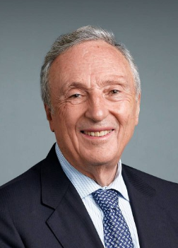 Charles R. Marmar合作论文数Department of Psychiatry, Grossman School of Medicine, New York University101
 Aoife O'Donovan合作论文数Mental Health Service, San Francisco Veterans Affairs Healthcare System79
 Sabra S Inslicht合作论文数Veterans Affairs Medical Center73Xinming An合作论文数Univ N Carolina, Sch Med, Inst Trauma Recovery, Chapel Hill, NC 27515 USA68
 Samuel McLean合作论文数Department of Psychiatry, School of Medicine, University of North Carolina at Chapel Hill;Department of Emergency Medicine, School of Medicine, University of North Carolina at Chapel Hill;Institute for Trauma Recovery, School of Medicine, University of North Carolina at Chapel Hill68
 Sarah Linnstaedt合作论文数Univ N Carolina64
 Francesca L Beaudoin合作论文数Center for Epidemiologic Research,School of Public Health, Brown University;Department of Epidemiology,School of Public Health, Brown University61
 Shira Maguen合作论文数61Beth E Cohen合作论文数59
 Phyllis L Hendry合作论文数Dept Emergency Med, Univ Florida57Pearson Claire合作论文数57Christopher Lewandowski合作论文数56
 Stacey Lynn House合作论文数56
 Ronald C. Kessler合作论文数Department of Health Care Policy, Harvard Medical School;Department of Mental Health, Bloomberg School of Public Health, Johns Hopkins University;Department of Sociology, Survey Research Center, University of Michigan56Sheikh Sophia合作论文数Emory University55
 Steven E. Bruce合作论文数55Niels K. Rathlev合作论文数Department of Emergency Medicine, Boston Medical Center55
 Scott L. Rauch合作论文数McLean Hospital;Harvard Medical School54Robert M. Domeier合作论文数54
 Leon Dahomey Sanchez合作论文数Department of Emergency Medicine, Beth Israel Deaconess Medical Center54
 Karestan Koenen合作论文数Department of Social and Behavioral Sciences, Harvard T.H. Chan School of Public Health53
 Anne Richards合作论文数53
 Kerry J. Ressler合作论文数Division of Depression and Anxiety Disorders, McLean Hospital, Harvard Medical School;Learning and Memory Study Section, Center for Scientific Review, National Institutes of Health;Department of Psychiatry and Behavioral Sciences, Emory University52
 Tanja Jovanovic合作论文数Institute of Microbiology and Immunology, School of Medicine, Belgrade, Yugoslavia52
 David Peak合作论文数51
 Gari Clifford合作论文数Department of Biomedical Informatics, Emory University School of Medicine;Department of Biomedical Engineering, Georgia Institute of Technology;LifeBell AI51
 Mark J. Seamon合作论文数Surgery Faculty, Perelman School of Medicine, University of Pennsylvania50
 Lea Grinberg合作论文数Laboratory Medicine and Pathology , Neurosciences, Mayo Clinic Florida50
 Christopher Jones合作论文数Department of Medicine, Duke University School of Medicine49
 Harte Steven E合作论文数49Christine M. Walsh合作论文数47Brian J. O’Neil合作论文数46
 Jose L. Pascual合作论文数Department of Neurosurgery, Perelman School of Medicine, University of Pennsylvania45Alan B. Storrow合作论文数44
 Laura Germine合作论文数Department of Psychology, Harvard University44
 Jennifer L Stevens合作论文数Department of Psychiatry and Behavioral Sciences, Emory University School of Medicine;Department of Psychiatry and Behavioral Sciences, Emory University School of Medicine43
 Brittany Punches合作论文数Univ Cincinnati, Coll Nursing, POB 210038, Cincinnati, OH 45221 USA41
 John P Haran合作论文数40
 Robert A Swor合作论文数Corewell Health;William Beaumont University Hospital39Lauren A. Hudak合作论文数38Paulina B. Sergot合作论文数36
 Roland C Merchant合作论文数Sch Publ Hlth, Brown Univ35
 Michael Weiner合作论文数School of Medicine, University of California, San Francisco35Karen H. Seal合作论文数34
 Jutta Joormann合作论文数Department of Psychology, Yale University31Leslie M Yack合作论文数30Shannon E. Mccaslin合作论文数Department of Psychiatry, University of California30
 Clare Henn-Haase合作论文数30




