ObjectivesThe aim of this study was to analyze the impact of deep brain stimulation (DBS) of the posteromedial hypothalamus (pHyp) on seizure frequency in patients with drug-resistant epilepsy (DRE) associated with intractable aggressive behavior (IAB).MethodsData were collected retrospectively from nine patients, who received bilateral stereotactic pHyp-DBS for the treatment of medically intractable aggressive behavior, focusing on five patients who also had DRE. All patients were treated at the Colombian Center and Foundation of Epilepsy and Neurological DiseasesFIRE (Chapter of the International Bureau for Epilepsy), in Cartagena de Indias, Colombia from 2010 to 2014. Each case was evaluated previously by the institutional ethical committee, assessing the impact of aggressive behavior on the patient's family and social life, the humanitarian aspects of preserving the safety and physical integrity of caregivers, and the need to prevent self-harm. Epilepsy improvement was measured by a monthly seizure reduction percentage, comparing preoperative state and outcome. Additional response to epilepsy was defined by reduction of the antiepileptic drugs (AEDs). Aggressive behavior response was measured using the Overt Aggression Scale (OAS).ResultsAll the patients with DRE associated with IAB presented a significant decrease of the rate of epileptic seizures after up to 4years follow-up, achieving a general 89.6% average seizure reduction from the state before the surgery. Aggressiveness was significantly controlled, with evident improvement in the OAS, enhancing the quality of life of patients and families.SignificanceIn well-selected patients, DBS of the pHyp seems to be a safe and effective procedure for treatment of DRE associated with refractory aggressive behavior. Larger and prospective series are needed to define the pHyp as a target for DRE in different contexts.
Holmes tremor (HT) is a difficult-to-treat, very disabling symptomatic condition which characteristically appears weeks to years after a brain lesion. It features a unique combination of rest, action, and postural tremors. Pharmacotherapy is mostly not effective. Chronic deep brain stimulation (DBS) of ventralis intermedius nucleus (Vim) of thalamus has been described as being the best surgical approach in singular case series; various authors observe, however, cases with partial responses only; therefore, alternatives are still needed. We report ten patients with HT unresponsive to best medical therapy who underwent DBS in our center from March 2002 to June 2012. Based in our previous experience dealing with cases of unsatisfactory Vim intraoperative tremor control and in order to optimize surgical results, presurgical target planning included two Nuclei: Vim and posteroventral Globus pallidus internus (GPi) (Espinoza et al. 2010; Espinoza et al. Stereotact Funct Neurosurg 90(suppl 1):1–202, p 61, 2012). Definitive chosen target was decided after single-cell microelectrode recording, intraoperative test stimulation, thresholds for stimulation-induced adverse effects and best clinical response compared to baseline status. Fahn-Tolosa-Marin tremor rating scale (FTM-TRS) was used to evaluate outcome. The electrode was implanted in the nucleus with the best tremor suppression achievement; on the other hand, GPi DBS was initially decided if one of the following conditions was present: (a) If Vim nucleus anatomy was grossly altered; (b) when intraoperative tremor control was unsatisfactory despite Vim high-intensity stimulation; or (c) if unaffordable side effects or even tremor worsening occurred during intraoperative macrostimulation. Seven patients received definitive Gpi DBS implantation, while three patients received Vim DBS. In all observed cases, we observed an improvement on the TRS. In two cases where Vim thalamic anatomy was altered by the pathological insult GPI was planned from the beginning, and same was true in two additional cases where the Gpi nucleus showed major alterations allowing only Vim planning. Over all cases, the average improvement in tremor was of 2.55 points on the TRS or a 64 % increase in measured results; with a minimum of 1 point (25 %) improvement in one case and a maximum of 4 points (100 % improvement) also in one case. All the results were sustained at 2 years follow-up. One case with predominant resting component, implanted in the GPi, achieved the maximum possible tremor reduction (from 4 to 0 points, meaning 100 % tremor reduction); in the nine resting cases, the average reduction was of 3 points (or 75 %). DBS demonstrated in this case series adequate tremor control in 10 patients unresponsive to medical therapy. Presurgical planning of two targets allowed choosing best optimal response. Gpi stimulation could be considered as an alternative target for cases in which thalamic anatomy is considerably altered or Vim intraoperative stimulation does not produce satisfactory results.
Background: Magnetic resonance imaging (MRI) is the standard neuroimaging method to diag-nose neoplastic brain lesions, as well as to perform stereotactic biopsy surgical planning. MRI has the advantage of providing structural anatomical details with high sensitivity, though histological specificity is limited. Although combining MRI with other imaging modalities, such as positron-emission tomography (PET), has proven to increment specificity, exact correlation between PET threshold uptake ratios (URs) and histological diagnosis and grading has not yet been described. Objectives: The aim of this study was to correlate exactly the histopathological criteria of the biopsy site to its PET uptake value with high spatial resolution (mm 3 ), and to analyze the diagnostic value of PET using the amino acid O -(2-[ 18 F]fluoroethyl)- l -tyrosine ( 18 F-FET) PET in patients with newly diagnosed brain lesions in comparison to histological findings obtained from stereotactic serial biopsy. 18 stereotactic newly stereotactic computed tomography series, and 18 F-FET PET images were semiautomatically fused, and hot-spot detection performed serial 27 Comparisons between FET URs along the biopsy and the were made with Pearson product-moment correlation coefficients. Analysis of variance was performed to test for significant differences in maximum UR between different tumor grades. Results: A total of 363 biopsy specimens were taken from 23 patients by stereotactic serial biopsies. Histological examination revealed eight patients (35%) with an LGG: one with a World Health Organization (WHO)-I lesion and seven with a WHO-II lesion. Thirteen (57%) patients revealed an HGG (two with a WHO-III and three with a WHO-IV tumor), and two patients (9%) showed a process that was neither HGG nor LGG (group X or no-grade group). The correlation matrix between histological findings and the UR revealed five strong correlations. Low cell density in tissue samples was found to have a significant negative correlation with the measured cortical uptake rate ( r =- 0.43, P = 0.02), as well as moderate cell density ( r =- 0.48, P = 0.02). Pathological patterns of proliferation ( r = 0.37, P = 0.04), GFAP ( r = 0.37, P = 0.04), and Olig2 ( r = 0.36, P = 0.05) showed a significant positive correlation with cortical URs. Analysis of variance tests showed a significant difference between the LGG and the HGG groups ( F = 8.27, P , 0.002), but no significant differences when differentiating between the X group and the HGG ( P = 0.2)/LGG ( P = 0.8) groups, nor between the no-grade group and the WHO-I group. Conclusion: 18 F-FET PET is a valuable tool, as it allows the differentiation of HGGs from LGGs. Its use is not limited to preoperative evaluation; it may also refine biopsy targeting and improve tumor delimitation for radiotherapy. Histology is still necessary, and remains the gold standard for definitive diagnosis of brain lesions. at of highest the target chosen by PET the PET-targeted predominantly
BACKGROUND:Adult brainstem gliomas are rare primary brain tumors (<2% of gliomas). The goal of this study was to analyze clinical, prognostic and therapeutic factors in a large series of histologically proven brainstem gliomas.METHODS:Between 1997 and 2007, 104 patients with a histologically proven brainstem glioma were retrospectively analyzed. Data about clinical course of disease, neuropathological findings and therapeutic approaches were analyzed.RESULTS:The median age at diagnosis was 41 years (range 18-89 years), median KPS before any operative procedure was 80 (range 20-100) and median survival for the whole cohort was 18.8 months. Histopathological examinations revealed 16 grade I, 31 grade II, 42 grade III and 14 grade IV gliomas. Grading was not possible in 1 patient. Therapeutic concepts differed according to the histopathology of the disease. Median overall survival for grade II tumors was 26.4 months, for grade III tumors 12.9 months and for grade IV tumors 9.8 months. On multivariate analysis the relative risk to die increased with a KPS ≤ 70 by factor 6.7, with grade III/IV gliomas by the factor 1.8 and for age ≥ 40 by the factor 1.7. External beam radiation reduced the risk to die by factor 0.4.CONCLUSION:Adult brainstem gliomas present with a wide variety of neurological symptoms and postoperative radiation remains the cornerstone of therapy with no proven benefit of adding chemotherapy. Low KPS, age ≥ 40 and higher tumor grade have a negative impact on overall survival.
Introduction: In this study, we assessed the outcomes of patients with dystonia who underwent surgery treatment following the same algorithm.Patients and methods: Eighty consecutive patients with dystonia were submitted to neurosurgical management by means of intrathecal pump implantation, pallidotomy or deep brain stimulation (GPi or VIM). These patients included 48 patients with primary dystonia and 32 patients with secondary dystonia. Burke-Fahn-Marsden Dystonia Rating Scale (BFMDRS) was used to access pre- and postoperative outcomes. Patients were followed from 12 to 114 months.Results: Mean improvement in BFMDRS score among patients with PrD was 87.54% and 42.21% for SeD. Hemidystonic patients in both groups (PrD, SeD) showed a mean improvement in BFMDRS of 71.05% with GPiDBS. Patients with SeD due to previous perinatal insults showed a mean improvement in BFMDRS of 41.9%, with better results in purely dyskinetic patients (mean improvement of 61.2%).Conclusion: Use of the proposed algorithm facilitated surgical decision planning, which translated in improved diagnostic rates, earlier interventions, appropriate management plans, and outcomes for both groups (PrD, SeD). Therefore, neuroimaging findings had a positive prognostic significance in the response to treatment in patients with primary dystonia compared with patients with secondary dystonia or distortion of basal ganglia anatomy. However, further studies in this line are warranted. (C) 2014 Elsevier B.V. All rights reserved.
In a retrospective single-centre study, we analysed data of 1,378 patients (55 % male, 45 % female) who underwent interstitial radiotherapy with 1,596 implanted Iodine-125 seeds in the Department of Stereotactic and Functional Neurosurgery in Freiburg from January 1990 to December 2011. The medical prerequisites and physical parameters of the treatment with Iodine-125 seeds are given. The method used in Freiburg relying on temporary Iodine-125 seed implants is described in detail and analysed. The survival rates and the peri-operative risk are evaluated. We conclude that interstitial radiosurgery with Iodine-125 seeds is a safe and useful tool, offering a wide range of treatment options for benign and also malignant intracranial lesions, especially if they are small, deep-seated, in eloquent areas, or not suitable for micro-surgery.
OBJECTIVE:The aim of this study was to explore the impact of automated hotspot detection on surgical planning of (18)FET PET-guided stereotactic serial biopsy.METHOD:Imaging of ten patients with brain lesions detected by MRI and showing increased (18)FET uptake on PET who were retrospectively and randomly assigned to compose the study. Stereotactic biopsy plans (PET-guided and MR-guided) were performed by two neurosurgeons for each patient, independently and blinded. For PET-guided plans, biopsy target was achieved by means of an automated hotspot detection system. MR-guided plans targeted contrast enhancement areas or hyperintense areas in T2-weighted sequences. FET uptake ratio (UR) was determined in the biopsy trajectory across the lesion. Highest UR (HUR) from both planning techniques was compared.RESULTS:Each single HUR obtained through PET-guided technique was higher than correspondent values from MR-guided technique. Mean HUR of 2.41 (SE ± 0.23) for PET-guided plans and 1.85 (±0.16) for MR-guided plans were respectively obtained. This difference was statistically significant (p = 0.002).CONCLUSION:The use of an automated hotspot detection system was able to provide higher FET HUR along stereotactic biopsy trajectories in comparison to those from MR-guided plans. The use of specially designed computational tools may refine surgical planning by improving biopsy targeting.
The prognosis for glioma remains dismal, and little is known about the final disease phase. To obtain information about this period, we surveyed caregivers of patients who were registered in the German Glioma Network and who died from the disease.A questionnaire with 15 items, focusing on medical, logistic, and mental health support and symptom control during the final 4 weeks, was sent to caregivers. For some of the questions, a scale from 1 (inadequate) to 10 (excellent) was used.Of 1655 questionnaires, 605 were returned (36.6) and evaluated. We found that 67.9 of the patients were taken care of at home for the last 4 weeks; 47.7 died at home, 22.6 died in hospitals, and 19.3 died in hospice facilities. Medical support was provided by general practitioners in 72.3 of cases, by physicians affiliated with a nursing home or hospice in 29.9, and by general oncologists in 17. Specialized neuro-oncologists were involved in 6. The caregivers ranked the medical support with a mean of 7.2 (using a 10-point scale), nursing service with 8.1, and mental health support with 5.5. In 22.9 of cases, no support for the caregivers themselves was offered by medical institutions.Although these data reflect the caregivers subjective views, they are useful in understanding and improving current patterns of care. While patients and their caregivers are supported mainly by neuro-oncologists for most of the disease phase, the end-of-life phase is managed predominantly by general practitioners and specialists in palliative care. Close cooperation between these specialties is necessary to meet the specific needs of glioma patients.
Purpose: To report on iodine-125 (1125) interstitial irradiation in the treatment of low grade brain stem gliomas in adults.Patients and methods: Ten patients with well-circumscribed lesions of the brainstem and histological confirmation of low grade gliorna treated with stereotactically implanted 1-125 seed in our department between 1995 and 2012 were retrospectively analyzed.Results: In 9 patients the lesion was treated with one I-125 seed and in one patient, 2 spatial separated lesions were implanted, therefore a total of 11 I-125 seeds were implanted. The mean volume of the 11 lesions was 2.76 ml (range: 0.5-7.2 ml), mean activity of the seeds was 6.23 mCi (range: 1.5-11.1 mCi), mean duration of irradiation was 28.5 days (range: 21-41 days) and mean effective dose rate was 9.16 cGy/h (range: 6.2-12 cGy/h). The 30 days perioperative morbidity and mortality rate was 0%. Median follow up was 72.5 month (range 5-168 months). Six of ten patients were free of progression until last follow up.Conclusion: In our experience at the University Clinic in Freiburg Germany, interstitial radiosurgery based on MRI is a safe and effective method to diagnose and treat low grade gliomas of the brain stem. Furthermore randomized studies are needed to confirm the therapeutic impact of this method in comparison to external beam radiation of brain stem gliomas. (C) 2013 Elsevier B.V. All rights reserved.
Here we show that activation of the canonical WNT/β-catenin pathway increases the expression of stem cell genes and promotes the migratory and invasive capacity of glioblastoma. Modulation of WNT signaling alters the expression of epithelial-to-mesenchymal transition activators, suggesting a role of this process in the regulation of glioma motility. Using immunohistochemistry in patient-derived glioblastoma samples we showed higher numbers of cells with intranuclear signal for β-catenin in the infiltrating edge of tumor compared to central tumor parenchyma. These findings suggest that canonical WNT/β-catenin pathway is a critical regulator of GBM invasion and may represent a potential therapeutic target.
PURPOSE:The aim of the study was the comparison of C-11 methionine (MET) and C-11 choline (CHO) in the positron emission tomography (PET) imaging of brain metastases in correlation to the histopathology findings in stereotactic biopsy.METHODS:In all, 8 patients underwent MET and CHO PET and magnetic resonance imaging, in 7 of these, the metastases were previously treated by radiation therapy. Histopathologic diagnosis was made for each patient by frame-based stereotactic serial biopsy. Standardized uptake values of tumor uptake and lesion-to-normal brain tissue ratios (LNRs) of the lesions were determined. LNRs for each tracer were compared with the histopathology findings and follow-up.RESULTS:In 6 patients, biopsy revealed viable metastases, 1 patient suffered from tumor recurrence in follow-up and 1 patient was tumor free in biopsy and follow-up. In the last mentioned patient, LNR was the lowest determined in all patients for CHO, but not for MET. Mean standardized uptake values of the lesions were in median 1.8 for MET and 1.1 for CHO, median LNRs were 1.5 for MET and 6.6 for CHO. LNRs for CHO were significant higher than those for MET (P = 0.007).CONCLUSIONS:In the direct comparison to the well-established amino acid tracer MET, CHO seems to be promising for the imaging of brain metastases because of significantly higher LNRs in tumor tissue compared with MET without evidence for a lower specificity of CHO uptake.
BACKGROUND:The prognosis for patients with recurrent glioblastoma is still poor with a median survival between 3 and 6 months. Reports about the application of carmustine (BCNU), one of the standard chemotherapeutic drugs in the treatment of newly diagnosed glioblastoma, in the recurrent situation are rare.METHODS:We performed a retrospective analysis of 35 patients with recurrent or progressive glioblastoma treated with 80 mg/m2 BCNU on days 1 on 3 intravenously at our department for efficacy, toxicity and prognostic factors. Progression free survival and overall survival were estimated by the Kaplan-Meier method. The influence of age, Karnofsky performance status (KPS), tumor burden, pretreatment with temozolomide (TMZ), type of surgery for initial diagnosis and number of previous relapses on outcome was analyzed in a proportional hazards regression model.RESULTS:The median age of the group was 53 years, median KPS was 70. Median progression free survival was 11 weeks (95% confidence interval [CI]: 8-15), median overall survival 22 weeks (95% CI: 18-27). The rate of adverse events, especially hematological toxicity, is relatively high, and in 3 patients treatment had to be terminated due to adverse events (one pulmonary embolism, one pulmonary fibrosis, and one severe bone marrow suppression). No influence of age, KPS, tumor burden, pre-treatment with TMZ and number of previous relapses on outcome could be demonstrated, while gross total resection prior to recurrence showed a borderline statistically significant negative impact on PFS and OS. These data compare well with historical survival figures. However prospective randomized studies are needed to evaluate BCNU efficacy against newer drugs like bevacizumab or the intensified temozolomide regime (one week on/one week off).CONCLUSION:In summary, BCNU treatment appears to be a valuable therapeutic option for recurrent glioblastomas, where no other validated radio- and/or chemotherapy are available.
The use of amino acid PET tracers like F-18 fluorethyltyrosine (FET) in the diagnostic workup of brain tumors has gained widespread acceptance. We report a case of a man who initially presented with focal neurologic symptoms. CT scan revealed a lesion suspicious of an ischemia or a low-grade glioma. The lesion showed high amino acid uptake in the FET PET examination, but without evidence of tumor tissue in the following stereotactic biopsy. A second FET PET examination 7 weeks later showed normal tracer uptake. Follow-up contrast enhanced CT finally confirmed a vascular embolic event as cause for the false-positive PET result.
The use of amino acid PET tracers like F-18 fluorethyltyrosine (FET) in the diagnostic workup of brain tumors has gained widespread acceptance. We report a case of a man who initially presented with focal neurologic symptoms. CT scan revealed a lesion suspicious of an ischemia or a low-grade glioma. The lesion showed high amino acid uptake in the FET PET examination, but without evidence of tumor tissue in the following stereotactic biopsy. A second FET PET examination 7 weeks later showed normal tracer uptake. Follow-up contrast enhanced CT finally confirmed a vascular embolic event as cause for the false-positive PET result.
 Guido Nikkhah合作论文数11
 Michael Trippel合作论文数9
 William Omar Contreras Lopez合作论文数Department of Neurology of School of Medicine, University of São Paulo5
 Soroush Doostkam合作论文数4
 Rottenburger Christof合作论文数4Marcus O Pinsker合作论文数3
 Philipp T. Meyer合作论文数3
 Furlanetti Luciano L合作论文数Laboratory of Stereotaxy and Interventional Neurosciences, University Freiburg-Medical Center3
 Marco Prinz合作论文数Institute of Neuropathology, Medical Center - University of Freiburg3C. Anastasopoulos合作论文数2Gabriel J Arango合作论文数2
 Stephan Meckel合作论文数2Martinez Jairo Alberto Espinoza合作论文数2
 Jaroslaw Maciaczyk合作论文数2
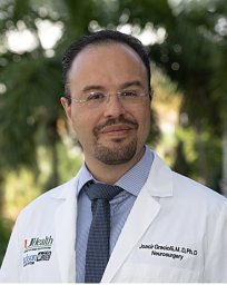 Joacir Graciolli Cordeiro合作论文数Department of Neurosurgery, Hospital Nossa Senhora das Graças2
 Erich Talamoni Fonoff合作论文数Divisão De Neurocirurgia Funcional, Instituto de Psiquiatri, Faculdade de Medicina, Universidade de São Paulo2
 Markus Loeffler合作论文数Institute for Medical Informatics, Statistics and Epidemiology, University of Leipzig2
 Michael Mix合作论文数Division of Nuclear Medicine;Albert Ludwigs University;Medical Center;Medical Center, Albert Ludwigs University2j luebke合作论文数1Jorge Humberto Marin合作论文数1Robert D Kirch合作论文数1Thomas Kelly合作论文数1Esther Perea-Castro合作论文数1Martín Torres Zambrano合作论文数1
 Andres Vargas-Toscano合作论文数Dept Neurosurg, Heinrich Heine Univ Dusseldorf1Bogiel Tomasz合作论文数1Heese Oliver合作论文数1Joerg-Christian Tonn合作论文数1
 Farsin Hamzei合作论文数1
 Travis S Tierney合作论文数1
 Ulf Kahlert合作论文数Div Neuropathol, Johns Hopkins Univ Hosp1Fernandes da Silva Fabio Eduardo合作论文数1Albicker Ulrich合作论文数1
 Gabriel Alcala-Cerra合作论文数Universidad de Cartagena1
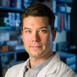 Orr Brent A合作论文数Department of Pathology, Johns Hopkins University School of Medicine1Aanyo Kuzeawu合作论文数1Piroth Tobias合作论文数1Maciaczyk Donata合作论文数1Nguyen-Thanh T合作论文数1Vogeler Eva合作论文数1Hentschel Michael合作论文数1Uwe Schlegel合作论文数1Oliver Schnell合作论文数1
 Irina Mader合作论文数1
 Juan Carlos Benedetti-Isaac合作论文数1
 Gabriele Schackert合作论文数Carl Gustav Carus University Hospital1Manfred Westphal合作论文数1
 Michael Weller合作论文数Department of Neurology, University Hospital Zurich1
 Johannes Schramm合作论文数Department of Neurosurgery, Medical School, University of Bonn1




