Hydrocephalus is a common complication following aneurysmal subarachnoid hemorrhage (aSAH), associated with increased morbidity and mortality. While its immediate negative impact on cognitive function is well-known, the longitudinal effects, especially in lower-grade aSAH patients, remain unclear. This study aimed to assess these effects. Within the prospective, multicenter “MoCA-DCI study” (ClinicalTrials.gov NCT03032471), patients with a GCS of 13–15 < 72 h post-aSAH underwent serial neuropsychological assessments using the Montreal Cognitive Assessment (MoCA) at baseline (< 72 h post-aSAH), around discharge (14–28 days post-aSAH), and at 3-month follow-up. Standardized MoCA scores were compared to evaluate cognitive outcomes, and the likelihood of a clinically meaningful decline (≥ 2 points) was assessed in patients with and without hydrocephalus. We included 112 patients, mean age 53.9 years (SD 13.9), 66.1
Introduction:Despite advances in cerebral aneurysm treatment, information on detailed outcomes remains limited. We compared complications and multidimensional outcomes in alert aneurysmal subarachnoid hemorrhage (aSAH) patients (GCS ≥13) who received clipping or coiling after individualized decisions were made through multidisciplinary board discussions. Research question:Are there significant differences in multidimensional outcomes between clipping and coiling treatments for alert aSAH patients when treatment selection is individualized? Material and methods:Within the prospective MoCA-DCI study (ClinicalTrials.gov identifier: NCT03032471), patients with a GCS of 13-15 72h post-aSAH in six neurovascular centers underwent neuropsychological (Montreal Cognitive Assessment; MoCA), neurological (National Institutes of Health Stroke Scale; NIHSS), headache (visual analog scale; VAS), disability (modified Rankin Scale; mRS) and health-related quality of life (EuroQol Five Dimensions; EQ5D) assessments within 72h, 14 days and three months after aSAH. We compared these multidimensional outcomes and complications for clipped and coiled patients. Results:Of 126 patients (mean age 53.8 years; 63.9 % female), 84 were coiled and 42 clipped. MoCA scores for clipped vs coiled patients were 23(7) vs 23(8; p = 0.250), 25(8) vs 28(5; p = 0.346), and 27(4) vs 28(5; p = 0.481). Normal cognition (MoCA ≥26) was achieved within 72h by 28.6 % of coiled and 40.5 % of clipped patients (p = 0.179). Complication rates were similar, though surgical patients had higher intraprocedural rupture rates (p = 0.027). Clipped patients reported higher headaches at three months (p = 0.013), while NIHSS, mRS and EQ5D showed no differences. Discussion and conclusion:After interdisciplinary selection of the most appropriate treatment, similar favorable outcomes can be achieved in alert aSAH patients.
The vertebral arteries represent in the adult the main blood supply of the posterior cranial fossa, even if they appear relatively late during the embryological life. We reviewed all the most important steps of the vertebral artery's embryological development and the most common variants that can occur in the adult. The aim of this review is to summarize the main events of the development of this artery and to give an embryological explication for the most common variants of this artery.
The ophthalmic artery has one of the most fascinating embryologic developments among the craniofacial arteries. Most of the ophthalmic artery orbital branches develop from the formation and regression of the stapedial artery and share their origin with dural branches of the ophthalmic artery. The concomitant embryologic development of the ophthalmic artery and middle meningeal artery explains adequately the important varieties of anastomosis between these 2 arteries. It also explains the presence of many dural branches from the ophthalmic artery. In this review, we focused on dural branches of the ophthalmic artery with the description of rare variations possible, in particular the ophthalmic artery origin of the middle meningeal artery and the ophthalmic artery origin of the marginal tentorial artery.
BACKGROUND:Thoracic pial arteriovenous fistulas (pAVFs) are rare vascular malformations that usually consist of a single dilated pial artery connecting directly to an enlarged draining vein. Multiple shunting point thoracic pAVFs are even rarer entities causing progressive myelopathy.METHOD:We present our surgical technique to identify and exclude multiple shunting point thoracic pAVF with appropriate pre-operative planning. This surgical technique is illustrated by an intraoperative video.CONCLUSION:Double injection pre-operative angiography represents a helpful tool to plan the surgery. Intraoperative exposure with pedicle removal and the use of micro-Doppler improve the identification and the exclusion of a multiple shunting thoracic pAVF.
OBJECTIVE:Acute hydrocephalus is a frequent complication after aneurysmal subarachnoid hemorrhage (aSAH). Among patients needing CSF diversion, some cannot be weaned. Little is known about the comparative neurological, neuropsychological, and health-related quality-of-life (HRQOL) outcomes in patients with successful and unsuccessful CSF weaning. The authors aimed to assess outcomes of patients by comparing those with successful and unsuccessful CSF weaning; the latter was defined as occurring in patients with permanent CSF diversion at 3 months post-aSAH.METHODS:The authors included prospectively recruited alert (i.e., Glasgow Coma Scale score 13-15) patients with aSAH in this retrospective study from six Swiss neurovascular centers. Patients underwent serial neurological (National Institutes of Health Stroke Scale), neuropsychological (Montreal Cognitive Assessment), disability (modified Rankin Scale), and HRQOL (EuroQol-5D) examinations at < 72 hours, 14-28 days, and 3 months post-aSAH.RESULTS:Of 126 included patients, 54 (42.9%) developed acute hydrocephalus needing CSF diversion, of whom 37 (68.5%) could be successfully weaned and 17 (31.5%) required permanent CSF diversion. Patients with unsuccessful weaning were older (64.5 vs 50.8 years, p = 0.003) and had a higher rate of intraventricular hemorrhage (52.9% vs 24.3%, p = 0.04). Patients who succeed in restoration of physiological CSF dynamics improve on average by 2 points on the Montreal Cognitive Assessment between 48-72 hours and 14-28 days, whereas those in whom weaning fails worsen by 4 points (adjusted coefficient 6.80, 95% CI 1.57-12.04, p = 0.01). They show better neuropsychological recovery between 48-72 hours and 3 months, compared to patients in whom weaning fails (adjusted coefficient 7.60, 95% CI 3.09-12.11, p = 0.02). Patients who receive permanent CSF diversion (ventriculoperitoneal shunt) show significant neuropsychological improvement thereafter, catching up the delay in neuropsychological improvement between 14-28 days and 3 months post-aSAH. Neurological, disability, and HRQOL outcomes at 3 months were similar.CONCLUSIONS:These results show a temporary but clinically meaningful cognitive benefit in the first weeks after aSAH in successfully weaned patients. The resolution of this difference over time may be due to the positive effects of permanent CSF diversion and underlines its importance. Patients who do not show progressive neuropsychological improvement after weaning should be considered for repeat CT imaging to rule out chronic (untreated) hydrocephalus.
BACKGROUND AND PURPOSE:Internal carotid artery (ICA) agenesis is a rare anatomical variant that can involve different segments of the ICA. Although many authors proposed their own classifications of this variant basing on different criteria, none of these allows to include all the cases described in the literature. The aim of the authors is to propose a new classification that allows to include all cases of ICA agenesis described until now.MATERIALS AND METHODS:The study is based on the review of all the cases of ICA agenesis described in the literature and of the classifications already proposed. After the analysis of these cases, we looked for the limits of each classification to elaborate a new more complete one.RESULTS:We found 228 cases of ICA agenesis. Among them 40 were not includible in the existing classifications. For this reason, we proposed a new classification based on the type of flow compensation and on the embryological events that determine the different variants. The flow is compensated in Type I by the Circle of Willis; in Type II by the non-regression of an embryonic artery; in Type III by the presence of an arterio-arterial anastomosis and in Type IV by external-internal carotid arteries anastomoses.CONCLUSION:After the literature review, we proposed a new comprehensive classification based on the detailed analysis of arterial embryology. Even if all the embryological details that determine this complex variant are not completely understood yet, this classification allows to include all the cases described in literature.
Spinal pial arteriovenous fistulas (spAVFs) are rare, high-flow vascular malformations consisting of a single dilated pial artery connecting directly to an enlarged draining vein. This article describes the anatomic, clinical, and diagnostic features of spAVFs, to help clinicians to select the best treatment option. Both surgical and endovascular treatments are effective; microsurgery has a higher success rate. To maximize the exclusion rate, a multidisciplinary approach and adequate preoperative study are mandatory.
 Gabriele Ciccio合作论文数58
 Michel Piotin合作论文数50
 Raphael Blanc合作论文数Basecamp Vascular47S Smajda合作论文数44Sara Bonasia合作论文数42
 Fahed Robert合作论文数Department of Interventional Neuroradiology, Hôpital de la Pitié-Salpétrière30Hocine Redjem合作论文数Fondation Ophtalmologique Rothschild22
 Bruno Bartolini合作论文数19
 Philippe Bijlenga合作论文数University of Geneva Medical Center, University of Geneva19h redjem合作论文数18
 Martin N. Stienen合作论文数Klinik für Neurochirurgie, Kantonsspital St. Gallen;Ostschweizer Wirbelsäulenzentrum18
 Maduri Rodolfo合作论文数Cattedra ed U.O. di Neurochirurgia, Università degli Studi di Catanzaro Magna Græcia15
 Michael Reinert合作论文数University of Bern15Fung Christian合作论文数14
 Roy Thomas Daniel合作论文数UNIL;CHUV14Michel W. Bojanowski合作论文数14
 Alessio Chiappini合作论文数Department of Neurosurgery, University Hospital Basel13
 Luca Regli合作论文数Department of Neurosurgery, University Hospital Zurich;Universität Zürich13
 Johannes Goldberg合作论文数12
 Schatlo Bawarjan合作论文数Neurochirurgische Klinik, Universitätsklinikum Göttingen12
 David Bervini合作论文数12
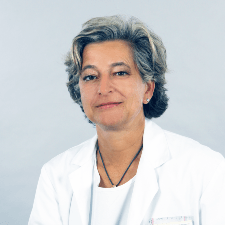 Emanuela Keller合作论文数Universitätsspital Zürich11
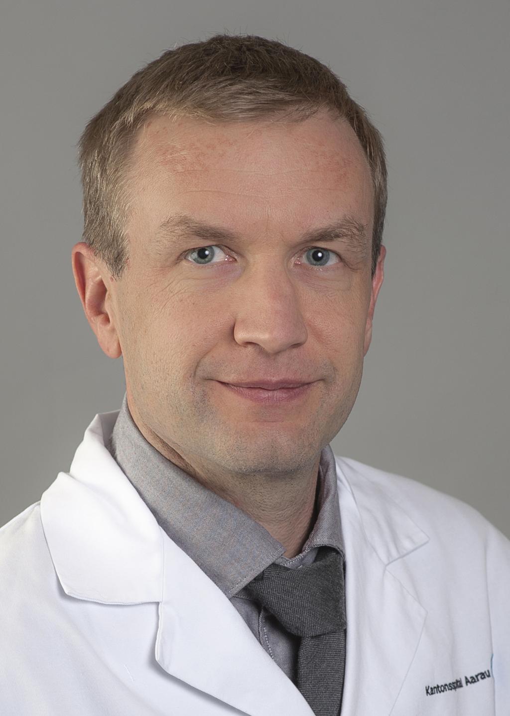 Serge Marbacher合作论文数Department of Neurochirurgie, Kantonsspital Aarau10Maldaner Nicolai合作论文数Department of Neurosurgery, Charité - Universitätsmedizin Berlin10Gilboa Boaz合作论文数10
 Burkhardt Jan-Karl合作论文数Dept Neurol Surg, Univ Calif San Francisco9Schaller Karl合作论文数Dept Neurosurg, Univ Hosp Geneva9
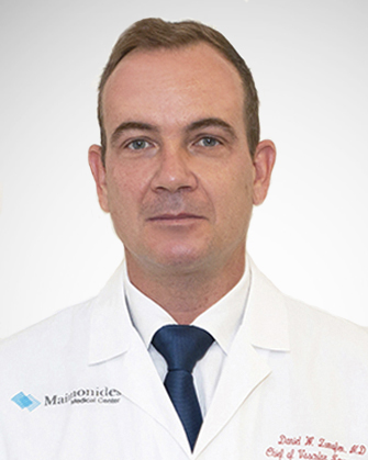 Daniel W. Zumofen合作论文数Maimonides Medical Center9
 Martin Alexander Seule合作论文数9
 Oliver Bozinov合作论文数Dept of Neurosurgery, University Hospital of Zurich9
 Lorenzo Bertulli合作论文数7
 Michel Roethlisberger合作论文数7Donato D'Alonzo合作论文数7
 Adrien May合作论文数Serv Neurochirurg, Hop Nord Marseille7
 Javier Fandino合作论文数Department for BioMedical Research, Neurochirurgie Hirslanden7Klemens Gutbrod合作论文数7Mikael Mazighi合作论文数6Stefania Rossi合作论文数6Silvia Pistocchi合作论文数6C Chaalala合作论文数6
 Daniele Starnoni合作论文数6
 Jean-Philippe Desilles合作论文数French Institute of Health and Medical Research6
 Menno R Germans合作论文数6
 Peter Brugger合作论文数Neuropsychology Unit, Kliniken Valens;Klinik für Psychiatrie, University of Zurich6Severin Früh合作论文数5D. Botta合作论文数5Simon Escalard合作论文数Hospital de Egas Moniz, Centro Hospitalar Lisboa Ocidental;Stroke Centre (, Lausanne University Hospital;School of Health Sciences, University of Thessaly;Boston Medical Center, Boston University School of Medicine;Comprehensive Stroke Centre, Charles University Faculty of Medicine and University Hospital;2nd Department of Neurology (A.C., M.A.K., M.N.),, Institute of Psychiatry and Neurology;Neurology Department (J.D., R.L.),, Leuven University Hospital;Hospitals and Affiliated Stroke Network (O.Y.M.),, Alexandria University;Department of Neurology, University Hospital of Zurich;Stroke Center (C.W.C., G.B.),, Neurocenter of Southern Switzerland;Bern University Hospital, University of Bern;Stroke Centre (E.C.),, Geneva University Hospital;Department of Neuroradiology, Geneva University Hospital;University Hospital Basel, University of Basel;Stroke Centre (M.B.),, Kantonsspital Lucerne;Stroke Centre (N.P., S.W.),, Hirslanden Hospital;Hospital de Egas Moniz, Centro Hospitalar Lisboa Ocidental;Department of Neurology, Centro Hospitalar Universitário de Coimbra;Department of Neuroradiology (E.M.),, Centro Hospitalar Universitário de Coimbra;Hospital de São José, Centro Hospitalar Universitário Lisboa Central;Hospital de Santa Maria, Centro Hospitalar Universitário Lisboa Norte;Hospital de Santa Maria, Centro Hospitalar Universitário Lisboa Norte;Department of Neurology (P.C., E.A.),, Centro Hospitalar Universitário São João;Department of Neuroradiology (L.A.),, Centro Hospitalar Universitário São João;Departments of Neurology (J.N.A., J.F.-P.), and Neuroradiology (T.M.),, Hospital de Braga;Department of Neurology (L.P., M.R.),, Hospital Garcia de Orta;Department of Neuroradiology (A.P.A., M.R.),, Centro Hospitalar de Vila Nova de Gaia/Espinho;Department of Neurology (M.R.),, Centro Hospitalar de Vila Nova de Gaia/Espinho;Department of Neurology (A.P.-F, L.R.),, Unidade Local de Saúde de Matosinhos;Department of Neurology (R.V., S.M.),, Centro Hospitalar Universitário do Porto;Stroke Unit (M.C., C.Z.),, Azienda Ospedaliera Universitaria Integrata;Department of Neurology and Stroke Centre, Maggiore Hospital;Department of Neurology (M.S., S.L.G.),, ASST Papa Giovanni XXIII;Department of Clinical and Experimental Sciences (A.P.),, University of Brescia;Department of Neurology and Stroke Unit (D.S.),, Azienda Socio Sanitaria Territoriale;Stroke Unit, Azienda Unità Sanitaria-IRCCS di Reggio Emilia;Neuroradiology Unit (R.P.),, Azienda Unità Sanitaria-IRCCS di Reggio Emilia;San Gerardo Hospital, University of Milano Bicocca;Stroke Unit (G.S.),, Fondazione IRCCS Ca' Granda Ospedale Maggiore Policlinico;Department of Neurology (G.F.),, Policlinico Universitario Agostino Gemelli;Emergency Neurology and Stroke Unit (S.M.),, IRCCS Humanitas Clinical and Research Center;Department of Neurology (C.S., S.E.),, Hôpital Fondation Ade Rothschild;Department of Interventional Neuroradiology (M.P., B.M.),, Hôpital Fondation Ade Rothschild;Department of Interventional Neuroradiology (G.C., F.V.),, Centre Hospitalier Régional Universitaire;Stroke Unit, Centre Hospitalier Universitaire;Department of Interventional and Diagnostic Neuroradiology (J.-S.L., I.S.),, Bordeaux University Hospital;Department of Diagnostic and Interventional Neuroradiology (F.F, G.B., N.-O.G.),, University Medical Center-Hamburg-Eppendorf;University Hospital Frankfurt, Goethe University;Berlin Institute of Health, Charité-Universitätsmedizin Berlin;Department of Neuroradiology (E.S.),, Charité-Universitätsmedizin Berlin;Department of Neurology (, St. John's Hospital;Departments of Neurology, Medical University of Innsbruck;Department of Neurology (J.S., Amsterdam University Medical Centers;Medical Centre, Leiden University;Universitair Ziekenhuis Brussel, Vrije Universiteit Brussel;Department of Neurology (M.P.R, A.G.),, Europe Hospitals;Department of Neurology, Centre Hospitalier Universitaire de Charleroi;Instituto de Investigación Hospital 12 de Octubre (i+12), Hospital Universitario de Octubre;Ramon y Cajal Institute for Health Research (IRYCIS),, Hospital Universitario Ramón y Cajal;La Paz University Hospital, Universidad Autónoma de Madrid;Department of Neurology (S.P.-S., J.M.),, Hospital Universitario Virgen Macarena;Stroke Centre (F.D-.O.),, Hospital General Universitario Gregorio Marañón;Stroke Unit (B.F.-P., J.M.-N.),, Germans Trias Hospital;Comprehensive Stroke Centre, Hospital Clinic from Barcelona;Department of Neurology, Complejo Hospitalario Universitario de Albacete;Department of Neurology, and Interventional Neuroradiology Unit, Hospital Universitario Miguel Servet;Department of Neurology, Hospital Universitario Miguel Servet;Stroke and Geriatric Medicine (T.H.),, Aintree University Hospital;University College London, University College London;Queen Square Institute of Neurology, University College London (D.W.),;Akershus University Hospital, University of Oslo;Sahlgrenska Academy at University of Gothenburg, Sahlgrenska University Hospital;at the University of Gothenburg, Sahlgrenska University Hospital;Faculty of Medicine and University Hospital, Charles University;St. Anne´s University Hospital, Masaryk University;Faculty of Medicine, University of Ostrava;Department of Neurology (R.H, S.V.),, České Budějovice Hospital;Department of Neurology (O.S., M.P.),, Jihlava Hospital;Neurocenter (L.J., Z.E., M.J.),, Regional Hospital Liberec;Cerebrovascular Centre (M.K., M.P., P.M.),, Na Homolce Hospital;Department of Neurology (H.P.),, Karviná Miners Hospital Inc.;Cerebrovascular Centre (A.T, P.J, A.O.),, University Hospital in Motol;Cerebrovascular Centre (M.S., R.H, P.M., L.T.),, Central Military Hospital;Cerebrovascular Centre (J.F., M.S.),, General University Hospital;1th Department of Neurology (H.S.-J, A.B.), Institute of Psychiatry and Neurology, Warsaw, Poland;University Hospital, Jagiellonian University;Medical College, Rzeszow University;Department of Neurology and Stroke (M.B, St. John Paul II Western Hospital;Department of Neurology (, Central Clinical Hospital of the Ministry of the Interior and Administration;Departments of Neurology (M.N.-K., K.O., P.U.), and Radiology (M.G.),, Wroclaw Medical University;Department of Neurosurgery and Neurology (M.S.),, Nicolaus Copernicus University in Torun Ludwik Rydygier Collegium Medicum;Stroke Intervention Centre (I.R., Jan Biziel University Hospital;Institute of Medical Sciences, University of Opole;Clinic of Neurology (, Military Institute of Medicine;Department of Neurology (J.Z.),, University of Warmia and Mazury;Department of Radiology (C.W.),, Provincial Specialist Hospital;University Emergency Hospital Bucharest, University of Medicine and Pharmacy "Carol Davila",;Department of Radiology, University Emergency Hospital Bucharest;Elias University Emergency Hospital, University of Medicine and Pharmacy "Carol Davila",;Department of Neurology (A.O.),, Eskisehir Osmangazi University;Affiliated Saudi German Hospital, Ain Shams University;Neuropsychiatry Department (H.A.),, Tanta University;Department of Neurology (J.A.-H.),, Ibn Sina Hospital;Department of Neurology (I.I.I.),, Jaber Al-Ahmad Hospital;School of Medicine, Zanjan University of Medical Sciences;Neurology Department, Hillel Yaffe Medical Center;Department of Neurosurgery (P.J., Thomas Jefferson University Hospital;Departments of Radiology (G.A.M., P.G.N.),, Grady Memorial Hospital;Department of Neurology (A.C.),, Henry Ford Hospital;Comprehensive Stroke Centre and Department of Neurosciences (, Michigan State University;Department of Neurology (K.N., S.O.),, University of Arkansas for Medical Sciences;Department of Neurology (M.K.),, Upstate University Hospital;Medical Centre, University of Kansas;Endovascular Neurological Surgery and Neurology (P.K., I.B, M.O., M.B.),, The State University of New Jersey;Detroit Medical Center, Wayne State University;Stroke Clinic (V.C.-N, A.A.),, Instituto Nacional de Neurologia y Neurocirugia Manuel Velasco Suarez;Department of Neurology (P.A.),, Fundación Valle del Lili;Centro de Investigaciones Clínicas (N.L., A.A.),, Fundación Valle del Lili;Department of Neurology (M.A.V.),, Hospital Nacional Edgardo Rebagliati Martins;Hospital General San Juan de Dios (J.D.B.G.),;Department of Neurology (R.C., R.T.M.),, Hospital Nossa Senhora da Conceição Hospital;Stroke Unit, Ramos Mejía Hospital (S.D.S.),;St. Luke's Medical Center (P.M.Y.), Global City, Philippines;Department of Neurology (S.N., A.G.),, Grant Medical College and Sir JJ Hospital;Department of Neurology (K.-D.S.),, National Health Insurance Service Ilsan Hospital;School of Biomedical Engineering and Imaging Sciences (G.G.),, King's College London;Department of Clinical Therapeutics (G.G.),, National and Kapodistrian University of Athens;University of Thessaly, Lausanne University Hospital5Valérie Beaud合作论文数5




