Astrocytes perform multifarious roles in the formation, regulation, and function of synapses in the brain, but the mechanisms involved are incompletely understood. Interestingly, astrocytes abundantly express neuroligins, postsynaptic adhesion molecules that function as synaptic organizers by binding to presynaptic neurexins. Here, we examined the function of neuroligins in astrocytes with a rigorous genetic approach that uses the conditional deletion of all major neuroligins (Nlgn1–3) in astrocytes in vivo in mice and complemented this approach by a genetic deletion of neuroligins in glial cells that are co-cultured with human neurons. Our results show that early postnatal deletion of neuroligins from astrocytes in vivo has no detectable effect on cortical or hippocampal excitatory or inhibitory synapses, and does not alter the cytoarchitecture of astrocytes when evaluated in young adult mice. Moreover, deletion of astrocytic neuroligins in co-cultures of human neurons produced no detectable consequences for the formation and function of synapses. Thus, astrocytic neuroligins are unlikely to fundamentally shape synapse formation or astrocyte morphogenesis, but likely perform other important roles that remain to be discovered.
In transfected cells, adhesion G protein-coupled receptors (GPCRs) are activated by tethered agonists that are embedded in their canonical autoproteolytic GAIN domain. It is unknown, however, whether a tethered agonist-dependent activation mechanism generally mediates the physiological functions of adhesion GPCRs. Here, we show that G protein signaling by BAI3 (Adgrb3), a brain-specific adhesion GPCR, is essential for its functions in controlling axon and dendrite growth and promoting synapse formation. Moreover, our signal transduction assays confirm that constitutive exposure of BAI3's tethered agonist massively stimulates (~5-fold) its GPCR activity. However, the constitutive exposure of BAI3's tethered agonist, produced by deletion of its extracellular domains, blocked instead of activating BAI3's functions in regulating axonal and dendritic growth and promoting synapse formation. Moreover, inactivating mutations of BAI3's tethered agonist or deletion of BAI3's constituent GAIN domain did not detectably impair BAI3's physiological functions. Thus, the GPCR activity of BAI3 is functionally required, whereas tethered agonist-mediated stimulation of its GPCR activity is not.
Extensive experiments document that SPARCL1, a secreted protein that is produced primarily by astrocytes in brain and endothelia throughout the body and that is also known as Hevin, enhances synapse formation. However, the mode of action of SPARCL1 at synapses remains unclear owing to divergent results in the literature. Here, we use cultured neurons from newborn male and female mouse embryos to show that the C-terminal follistatin-like and Ca2+-binding domains of SPARCL1, which account for only 35% of the total SPARCL1 sequence, are sufficient to potently enhance synapse numbers. SPARCL1 acts at nanomolar concentrations at which SPARCL1 does not robustly bind to neurexins, neuroligins or neurexin/neuroligin complexes but avidly interacts with all teneurins. Strikingly, the follistatin-like domain of SPARCL1 on its own strongly binds to teneurins but is unable to stimulate synapse formation. Only when combined with the SPARCL1 Ca2+-binding domain does the follistatin-like domain induce synapses, suggesting that SPARCL1 enhances synapse numbers by binding to teneurins via its C-terminal follistatin-like domain and by activating synapse formation via its Ca2+-binding domain.
Developing therapies for Alzheimer's disease (AD) is a pressing need. A key feature of AD is synapse loss. ApoE4, amyloid-β (Aβ)-peptides, phospho-tau, and neuroinflammation are thought to induce AD pathogenesis, but their molecular mechanisms are incompletely understood. Free Aβ-peptides whose levels decrease in AD are synaptogenic, whereas aggregating Aβ-peptides that accumulate in AD are synaptotoxic. It is unclear whether AD is driven primarily by the loss of free Aβ, the increase of aggregated Aβ, or both, nor do we know how ApoE4 affects synapses. Therefore, understanding the molecular pathways mediating synapse loss in AD is a priority in developing new therapies. ANN NEUROL 2026.
Adhesion G protein-coupled receptors (aGPCRs) constitute a structurally and functionally distinct group within the superfamily of GPCRs. In 2015, the International Union of Pharmacology invited the Adhesion GPCR Consortium to publish a comprehensive review about aGPCRs and establish a unified nomenclature. Since then, substantial progress has been made in delineating the biological roles, molecular architecture, biochemical properties, expression profiles, ligand repertoire, and activation and signaling strategies of aGPCRs. Commensurate with these advances, their relevance to human pathophysiology has become increasingly apparent. In a coordinated effort, the Adhesion GPCR Consortium has reviewed recent progress in this field and provides a comprehensive assessment of the current understanding of aGPCR biology, including a focus on human and mammalian aGPCRs, their evolutionary origins, methodological approaches, and model systems for their investigation, as well as emerging approaches for their therapeutic targeting. SIGNIFICANCE STATEMENT: Adhesion G protein-coupled receptors are versatile cell-surface proteins that integrate structural, biochemical, and physiological functions, with major roles in health and disease. This review summarizes current knowledge of their molecular features, functions in diverse model systems, and emerging opportunities for therapeutic targeting, providing a comprehensive resource that connects basic biology with translational applications across multiple scientific disciplines.
Rapid delivery of glutamate receptors to the postsynaptic membrane via vesicle fusion is a central component of synaptic plasticity. However, it is unknown how this process supports specific neural computations during behavior. To bridge this gap, we combined hippocampal CA1 deletion of the t-SNARE protein Syntaxin3 (Stx3), a component of the postsynaptic membrane fusion machinery, with population in vivo calcium imaging. This approach revealed that Stx3 is necessary for neural responses to novelty and for forming stable representations of rewarded locations. By contrast, CA1 Stx3 is dispensable for maintaining aspects of the neural code that exist presynaptic to CA1, such as representations of context and space. Thus, altering the Stx3-dependent postsynaptic membrane fusion machinery identified computations that specifically require synaptic restructuring via membrane trafficking in CA1 and distinguished them from neural representations that could be inherited from upstream brain regions or learned through other mechanisms.
This Perspective is motivated by the mounting distrust of science by the public fueled by integrity issues in scientific papers (1) and by the intense questioning of our own laboratory’s work by PubPeer, a popular website for finding mistakes in papers (2). Science is primarily communicated via scientific papers whose integrity is being increasingly doubted because of often contradictory conclusions and large number of retractions. In this Perspective, I aim to discuss some of the underlying causes for the current science integrity problems and to offer potential solutions. My main proposition is that to regain the trust of the public in science, we scientists as a community need to reform science communication and science publishing and we need to manage and rectify science integrity issues more directly.
Presynaptic neurexins are key regulators of synapse properties that arguably represent the best-studied synaptic adhesion molecules. Despite thousands of papers, however, no direct comparison of overall neurexin functions in different types of synapses is available. A decade ago, we provided such an analysis but we recently retracted this paper because four images contained microduplications that, although without discernible impact on the paper's conclusions, could not be corrected. As a result, the scientific community lost access to primary data that established the fundamental principle that neurexins perform profound but distinct functions in different types of synapses. In the present study, we have therefore reanalyzed the original raw data and expanded their conclusions with new experiments to document in a single study the basic contributions of neurexins to different synapses. Using triple conditional knockout mice that target all neurexins except for Neurexin-1γ and applying neuron-specific manipulations combined with slice electrophysiology, two-photon Ca2+ imaging and immunohistochemistry, we focussed on excitatory climbing-fiber synapses in the cerebellum and on inhibitory synapses formed by parvalbumin- or somatostatin-positive neurons in the cerebellum, hippocampus, and medial prefrontal cortex. Our results show that pan-neurexin deletions produce dramatically different phenotypes in synapses, ranging from modest to massive impairments in synapse assembly (climbing-fiber and parvalbumin-positive synapses) to severe but selective decreases in presynaptic action potential-induced Ca2+-transients (somatostatin-positive synapses). Thus, neurexins perform powerful but distinct context-dependent roles in different synapses that shape the brain's circuits. Significance Statement:Neurexins are abundant presynaptic adhesion molecules expressed from three genes in more than a thousand splice variants. Although neurexins are well studied, no analysis that compares neurexin deletions in multiple types of synapses is currently available. Here, we provide such an analysis by examining triple conditional knockout mice that delete all neurexins except for Neurexin-1γ. Using neuron-specific manipulations combined with slice electrophysiology, two-photon Ca2+ imaging and immunohistochemistry, we show that pan-neurexin deletions produce distinct phenotypes in different synapses, ranging from impairments in synapse assembly (climbing-fiber and parvalbumin-positive cortical synapses) to severe selective decreases in presynaptic action potential-induced Ca2+- transients (somatostatin-positive cortical synapses). Our data reveal context-dependent distinct functions of neurexins in different synapses whose properties shape the input-output relations of neural circuits.
The ability to avoid confusion between similar episodic memories enables organismal survival and fitness. This evolutionarily conserved differentiation process of memories as distinct representations is known as pattern separation. A central role for the entorhinal cortex→dentate gyrus (EC→DG) circuit in pattern separation memory is well established, but the molecular mechanisms that enable this circuit to mediate pattern separation memory are incompletely understood. We previously found that a trans-synaptic protein complex formed by presynaptic Cerebellin-4 and postsynaptic Neogenin-1 is selectively required for long-term potentiation (LTP) in the EC→DG circuit. We now demonstrate that this complex is essential for normal pattern separation memory, suggesting a role for this form of LTP in pattern separation memory. Deletion of either presynaptic Cerebellin-4 in the entorhinal cortex or of postsynaptic Neogenin-1 in the dentate gyrus impaired pattern separation but did not affect pattern completion memory. Thus, we describe a specific memory function for a defined molecular complex at an identified synapse, providing direct support for the hypothesis that synaptic plasticity contributes to the encoding of memory.
Microglia are the immune cells of the central nervous system and are thought to be key players in both physiological and disease conditions. Several microglial features are poorly conserved between mice and human, such as the function of the neurodegeneration-associated immune receptor Trem2. Induced pluripotent stem cell (iPSC)-derived microglia offer a powerful opportunity to generate and study human microglia. However, human iPSC-derived microglia often exhibit activated phenotypes in vitro, and assessing their impact on other brain cell types remains challenging due to limitations in current co-culture systems. Here, we developed fully defined brain microtissues, composed of human iPSC-derived neurons, astrocytes, and microglia, co-cultured in 2D or 3D formats. Our microtissues are stable and self-sufficient over time, requiring no exogenous cytokines or growth factors. All three cell types exhibit morphologies characteristic of their in vivo environment and show functional properties. Co-cultured microglia develop more homeostatic phenotypes compared to microglia exposed to exogenous cytokines. Hence, these tri-cultures provide a unique approach to investigate cell-cell interactions between brain cell types. We found that astrocytes and not neurons are sufficient for microglial survival and maturation, and that astrocyte-derived M-CSF is essential for microglial survival. Single-cell and single-nucleus RNA sequencing analyses nominated a network of reciprocal communication between cell types. Brain microtissues faithfully recapitulated pathogenic α-synuclein seeding and aggregation, suggesting their usefulness as human cell models to study not only normal but also pathological cell biological processes.
The adhesion-GPCR Brain-specific Angiogenesis Inhibitor-3 (BAI3) plays a crucial role in organizing synapses in the brain. However, how BAI3 engages one of its ligands, the C1q-like proteins (C1qls), remains largely unexplored. Here, we present the single-particle cryo-electron microscopy (cryo-EM) structure of the C1ql3-BAI3 complex at 2.8 Å resolution. The structure reveals a hexameric configuration, where C1ql3 forms a central homotrimer that effectively captures three BAI3 molecules. These BAI3 molecules fit snugly into the grooves between the trimeric C1q domains of the C1qls, employing calcium ion (Ca 2+ )-mediated interactions that differ from previously characterized structures of C1q-like domain-mediated complexes. Furthermore, we conducted mutant analysis and cell surface staining, which confirmed the essential contact residues involved in this interaction. This unique binding mechanism not only enhances our understanding of the C1ql-BAI3-mediated synaptic organization but also sheds light on the functional dynamics of BAI3 in the brain.
Neuroligins are postsynaptic cell-adhesion molecules that regulate synaptic function with a remarkable isoform specificity. Although Nlgn1 and Nlgn2 are highly homologous and biochemically interact with the same extra- and intracellular proteins, Nlgn1 selectively functions in excitatory synapses whereas Nlgn2 functions in inhibitory synapses. How this excitatory/inhibitory (E/I) specificity arises is unknown. Using a comprehensive structure-function analysis, we here expressed wild-type and mutant neuroligins in functional rescue experiments in cultured hippocampal neurons lacking all endogenous neuroligins. Electrophysiology confirmed that Nlgn1 and Nlgn2 selectively restored excitatory and inhibitory synaptic transmission, respectively, in neuroligin-deficient neurons, aligned with their synaptic localizations. Chimeric Nlgn1-Nlgn2 constructs reveal that the extracellular neuroligin domains confer synapse specificity, whereas their intracellular sequences are exchangeable. However, the cytoplasmic sequences of Nlgn2, including its Gephyrin-binding motif that is identically present in the Nlgn1, is essential for its synaptic function whereas they are dispensable for Nlgn1. These results demonstrate that although the excitatory vs. inhibitory synapse specificity of Nlgn1 and Nlgn2 are both determined by their extracellular sequences, these neuroligins enable normal synaptic connections via distinct intracellular mechanisms.
BAI1, BAI2, and BAI3 (for ‘Brain-specific Angiogenesis Inhibitor-1, -2, and -3’) are adhesion-GPCRs implicated in neuronal development. The precise roles of individual BAIs remain unclear. BAIs interact with two sets of ligands, secreted C1ql proteins and membrane-bound RTN4R proteins (a.k.a. NoGo receptors), but which of these ligands regulate specific functions of BAIs is incompletely understood. To address these key questions, we here systematically examine the functions of the three BAIs in neuronal development using hippocampal neuron-glia cultures, genetic knockouts, and rescue experiments. In a direct comparison, we demonstrate that deletions of BAI1 or BAI3, but not of BAI2, increase axonal and dendritic arborizations but decrease excitatory synapse formation, while inhibitory synapse formation remains unaffected. Since biochemical and cellular assays reveal that only BAI3 binds to both RTN4Rs and C1qls, we analyzed the role of these two ligands in controlling BAI3 functions using rescue experiments. We find that RTN4R-binding to BAI3 is essential for restricting axonal and dendritic arborizations and for enabling excitatory synapse formation, whereas C1ql-binding to BAI3 is only required for synapse organization as monitored in hippocampal neuron-glia cultures. Thus, BAI1 and BAI3 perform diverse functions that shape multiple facets of neuronal development and that require their interaction with RTN4Rs.
Whether amyloid-β (Aβ) peptides are synaptogenic or synaptotoxic remains a pivotal open question in Alzheimer's disease research. Here, we chronically treated human neurons with precisely controlled concentrations of chemically defined synthetic Aβ40, Aβ42, and Aβ42arctic peptides that exhibit distinct aggregation propensities. Remarkably, chronic exposure of human neurons to free Aβ40 at higher concentrations or to free Aβ42 at lower concentrations potently promoted synapse formation. In contrast, aggregated Aβ42 or Aβ42arctic at higher concentrations were neurotoxic and synaptotoxic. The synaptotoxic effects of Aβ peptides manifested as an initial contraction of the synaptic vesicle cluster followed by synapse loss. Aβ40 and Aβ42 peptides with scrambled or inverted sequences were inactive. Thus, our experiments reveal that Aβ peptides exhibit an aggregation-dependent functional dichotomy that renders them either synaptogenic or synaptotoxic, thereby providing insight into how Aβ peptides straddle a thin line between physiological synapse organization and pathological synapse disruption. Among others, our data suggest that Alzheimer's disease therapies might aim to shift the balance of Aβ peptides from the aggregated to the free state instead of suppressing all Aβ peptides.
 Marius Wernig合作论文数Chemical and Systems Biology, Stanford University/Department of Pathology, Stanford University53
 Jose Rizo-Rey合作论文数Rizo Lab, Department of Biophysics, University of Texas Southwestern Medical Center;Department of Pharmacology, University of Texas Southwestern Medical Center;Department of Biochemistry, University of Texas Southwestern Medical Center44
 Robert Malenka合作论文数Wu Tsai Neurosciences Institute, Stanford University;Department of Psychiatry and Behavioral Sciences, Stanford University School of Medicine43
 Reinhard Jahn合作论文数Department of Neurobiology, Max Planck Institute for Multidisciplinary Sciences35
 Xinran Liu (欣然劉)合作论文数28
 Zhiping P. Pang合作论文数rutgers university28Christian Rosenmund合作论文数27
 Pascal S. Kaeser合作论文数Department of Molecular and Cellular Physiology and Howard Hughes Medical Institute, Stanford University School of Medicine24
 Nils Brose合作论文数Department of Molecular Neurobiology, Max Planck Institute for Multidisciplinary Sciences20
 Manu Sharma合作论文数Neuroscience, Weill Cornell Graduate School of Medical Sciences, Cornell University18Soham Chanda合作论文数18
 Katsuhiko Tabuchi合作论文数Howard Hughes Medical Institute, The University of Texas Southwestern Medical Center18Jacqueline Burre合作论文数Neuroscience, Weill Cornell Graduate School of Medical Sciences, Cornell University17Xu Wei合作论文数17
 Axel Brunger合作论文数Department of Molecular and Cellular Physiology, Stanford University17Weiping Han合作论文数A*STAR16Anton Maximov合作论文数16Martin Geppert合作论文数16Taulant Bacaj合作论文数16
 Xiaofei Yang (阳小飞)合作论文数School of Biomedical Engineering, South-Central Minzu University15Robert E. Hammer合作论文数15Irina Dulubova合作论文数15
 Kif Liakath-Ali合作论文数Stanford Univ, Dept Mol & Cellular Physiol, Stanford, CA 94305 USA14
 Richard (Rick) Sando合作论文数Department of Pharmacology, School of Medicine, Vanderbilt University;Vanderbilt Brain Institute14
 Diana R. Tomchick合作论文数Department of Biochemistry, The University of Texas Southwestern Medical Center14
 Jaewon Ko合作论文数14Ok-Ho Shin合作论文数14
 Stephan Maxeiner合作论文数13
 Peng Zhou合作论文数Peking University, Peking University13
 Justin Trotter合作论文数Johnnie B Byrd Sr Alzheimer's Center & Research Institute, University of South Florida Tampa13
 Pablo E. Castillo合作论文数Department of Psychiatry and Behavioral Sciences, Albert Einstein College of Medicine;Dominick P. Purpura Department of Neuroscience, Albert Einstein College of Medicine13Craig M. Powell合作论文数13
 Demet Arac-Ozkan合作论文数The Neuroscience Institute, The University of Chicago12
 Christopher Patzke合作论文数Department of Molecular and Cellular Physiology, Stanford University School of Medicine, 265 Campus
DriveUnited States 12
Christopher Patzke合作论文数Department of Molecular and Cellular Physiology, Stanford University School of Medicine, 265 Campus
DriveUnited States 12 Stephen Quake合作论文数School of Engineering, Stanford University;Department of Bioengineering, Stanford University;Department of Applied Physics, Stanford University;Department of Physics, Stanford University;Cornell University12Markus Missler合作论文数12
 Sung-Jin Lee合作论文数Dept Mol & Cellular Physiol, Stanford Univ11
 Nan Yang合作论文数11
 Gokce Ozgun合作论文数Department of Molecular and Cellular Physiology and Howard Hughes Medical Institute, Stanford University Medical School11
 Cheen Euong Ang合作论文数Department of Pathology, Stanford University;Stanford Institute for Stem Cell Biology and Regenerative Medicine, Stanford University;Stanford University, Stanford University11
 Frank Schmitz合作论文数11
 Bo Zhou合作论文数11
 Antony Boucard合作论文数Department of Molecular and Cellular Physiology, Neurology and Psychiatry and Behavioral Sciences, Stanford University11
 Rafael Fernandez-Chacon合作论文数Departmento Fisiología Médica y Biofísica, Universidad de Sevilla11
 Jacqueline Blundell合作论文数Memorial University11
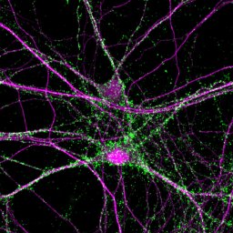 Jin-Zhao Wang (王今朝)合作论文数Sch Life Sci, South China Normal Univ10
 Samuele Marro合作论文数10
 Mischa Machius合作论文数10Mark R. Etherton合作论文数Department of Neuroscience, The University of Texas Southwestern Medical Center10




