Dendritic cells (DCs) play pivotal roles in initiating and shaping both innate and adaptive immune responses. The spatiotemporal expression of transcription factor networks and activation of specific signal transduction pathways determine the specification, distribution and differentiation of DC subsets. Even though pioneering studies have established indispensable roles for specific catalytic subunits (p110δ and p110γ) in immune cells, functions of the regulatory subunits, particularly of Class I PI3K, within the hematopoietic system remain incompletely understood. In the study presented here, we deleted the key regulatory subunits—p85α and p85β of the Class I A PI3K in hematopoietic cells and studied its impact on DC differentiation. Our studies identify that a deficiency of p85 causes increased differentiation of conventional DC (cDC) 2 and plasmacytoid DC (pDC) subsets in the spleen. On the other hand, DC numbers in the bone marrow (BM), thymus and lymph nodes were decreased in p85 mutant mice. Analysis of DC-specific progenitors and precursors indicated increased numbers in the BM and spleen of p85 deficient mice. In-vitro differentiation studies demonstrated augmented DC-differentiation capacities of p85 deficient BM cells in the presence of GM-CSF and Flt3L. BM chimera studies established that p85 deficiency affects DC development through cell intrinsic mechanisms. Molecular studies revealed increased proliferation of DCs and common DC progenitors (CDPs) in the absence of p85 and altered signal transduction pathways in p85 mutant DC subsets in response to Flt3L. In essence, data presented here, for the first time, unequivocally establish that the P85α subunit of class I A PI3Ks has an indispensable role in the development and maintenance of DCs.
Basophils are one of the major players in the pathophysiology of allergy related immune disorders. In spite of their key roles in autoimmune and inflammatory disorders in humans, molecular mechanisms that control basophils remain largely unknown. Therefore we have been involved in elucidating the biochemical and molecular mechanisms through which development and functions of basophils are controlled in the immune system. In this study, we utilized the Itch deficient mouse as a model system for understanding the biology of Basophils. Itch is an E3 ubiquitin ligase that belongs to the HECT family. Mice deficient for itch develop severe autoimmune defects (‘itchy’ phenotype) and patients with ITCH mutations show characteristic clinical features, including multisystem autoimmune disease. Our preliminary analysis indicated that itch deficiency leads to Basophilia, hyperactivation of basophils, elevated IL4 expression and Th2 priming by basophils, in a cell intrinsic manner. Strikingly, depleting basophils in itch deficient mice resulted in rescue of the ‘itchy’ phenotype. Our mechanistic studies demonstrated that notch signals are elevated in itch deficient basophils, due to reduced ubiquitylation of notch. Interestingly, ablation of notch signals in itch deficient mice resulted in normal frequencies of basophils and rescue of the ‘itchy’ phenotype. At this annual meeting, we would be discussing the precise involvement of the signaling pathways and molecular mechanisms in the restriction of pathogenic basophils.
Species-dependent variation in proteins that aid or limit virus replication determines the ability of lentiviruses to jump between host species. Identifying and overcoming these differences facilitates the development of animal models for HIV-1, including models based on chimeric SIVs that express HIV-1 envelope (Env) glycoproteins, (SHIVs) and simian-tropic HIV-1 (stHIV) strains. Here, we demonstrate that the inherently poor ability of most HIV-1 Env proteins to use macaque CD4 as a receptor is improved during adaptation by virus passage in macaques. We identify a single amino acid, A281, in HIV-1 Env that consistently changes during adaptation in macaques and affects the ability of HIV-1 Env to use macaque CD4. Importantly, mutations at A281 do not markedly affect HIV-1 Env neutralization properties. Our findings should facilitate the design of HIV-1 Env proteins for use in non-human primate models and thus expedite the development of clinically relevant reagents for testing interventions against HIV-1.
The APOBEC3 family of cytidine deaminases cause lethal hypermutation of retroviruses via deamination of newly reverse-transcribed viral DNA. Their ability to bind RNA is essential for virion infiltration and antiviral activity, yet the mechanisms of viral RNA recognition are unknown. By screening naturally occurring, polymorphic, non-human primate APOBEC3H variants for biological and crystallization properties, we obtained a 2.24-Å crystal structure of pig-tailed macaque APOBEC3H with bound RNA. Here, we report that APOBEC3H forms a dimer around a short RNA duplex and, despite the bound RNA, has potent cytidine deaminase activity. The structure reveals an unusual RNA-binding mode in which two APOBEC3H molecules at opposite ends of a seven-base-pair duplex interact extensively with both RNA strands, but form no protein-protein contacts. CLIP-seq analysis revealed that APOBEC3H preferentially binds to sequences in the viral genome predicted to contain duplexes, a property that may facilitate both virion incorporation and catalytic activity.
Hematopoietic stem cells (HSCs) are capable of both self-renewing throughout the lifetime of an organism and differentiating into all lineages of the blood system. A proper balance between quiescence and proliferation is critical for the self-renewal and functions of HSCs. The choice of HSCs to remain quiescent or to enter proliferation has been tightly regulated by a variety of cell intrinsic and extrinsic pathways. Identifying molecular players that control HSC quiescence and proliferation may lead to new treatment strategies and therapeutic interventions for hematologic disorders. To identify the functions of the slicer endonuclease Argonaute (Ago) 2 in the physiology of HSCs, we generated Ago2Hem-KO mice, that are deficient for Ago2 in HSCs and in their progeny. Analysis of Ago2Hem-KO mice indicated that a loss of Ago2 results in reduced HSC pool size and altered frequencies of hematopoietic progenitors. Ago2 deficient HSCs exhibit defective multilineage differentiation capacities and diminished repopulation abilities, in a cell intrinsic manner. Interestingly, Ago2 mutant HSCs remain largely quiescent and show reduced entry into cell cycle. Genome-wide transcriptome studies and gene set enrichment analysis revealed that Ago2 deficient HSCs downregulate the “HSC signature” and upregulate the “lineage signature.” Moreover, our analysis on transcription factors (TFs) identified that a loss of Ago2 is sufficient to alter the “molecular signature” and “TF networks” that control the quiescent and proliferative states of HSCs. In essence, our study identified Ago2 as a key determinant of quiescence exit in HSCs.
A balance between quiescence and proliferation is critical for proper maintenance of the hematopoietic stem cell (HSC) pool. Although a lot is known about hematopoiesis, molecular mechanisms that control HSC quiescence remain largely unknown. The ubiquitin-editing enzyme A20 functions as a central regulator of inflammation and adaptive immunity. Here, we show that a deficiency of A20 in the hematopoietic system causes anemia, lymphopenia, and postnatal lethality. Lack of A20 in HSCs results in diminished pool size, impaired radioprotection, defective repopulation, and loss of quiescence. A20-deficient HSCs display increased IFN-γ signaling, caused by augmented NF-κB activation. Strikingly, deletion of both IFN-γ and A20 in hematopoietic cells results in partial rescue of the HSC phenotype. We anticipate that our experiments will facilitate the understanding of mechanisms through which A20-mediated inflammatory signals control HSC quiescence and functions.
 Chozhavendan Rathinam合作论文数Bill and Melinda Gates Foundation4
 Alice Raymond合作论文数Rockefeller Univ, Lab Retrovirol, New York, NY 10065 USA3
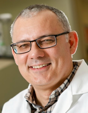 Paul Bieniasz合作论文数The Rockefeller University;Howard Hughes Medical Institute3Theodora Hatziioannou合作论文数3Masahiro Marshall Nakagawa合作论文数2
 Brandon Keele合作论文数AIDS and Cancer Virus Program, Frederick National Laboratory, National Cancer Institute, National Institutes of Health, U.S. Department of Health and Human Services2
 Gregory Q Del Prete合作论文数2Swanstrom Adrienne E合作论文数2
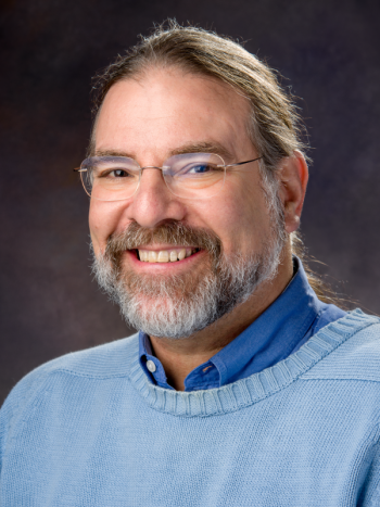 Jeffrey Lifson合作论文数Frederick National Laboratory for Cancer Research2
 Jacob Estes合作论文数2
 Vineet Narain Kewalramani合作论文数NCI, HIV Drug Resistance Program2
 Jennifer Bohn合作论文数Univ Michigan, Dept Biol Chem, Ann Arbor, MI 48109 USA1
 Celia Crane LaBranche合作论文数Department of Surgery, School of Medicine, Duke University1
 Laura Pasqualucci合作论文数Institute for Cancer Genetics, Vagelos College of Physicians & Surgeons, Columbia University Irving Medical Center1
 Ashley York合作论文数Microbiology, nrmicro nature1
 William Clay Brown合作论文数1
 Janet Smith合作论文数Center for Structural Biology, Life Sciences Institute, University of Michigan;University of Michigan Medical School;College of Literature, Science, and the Arts, University of Michigan1
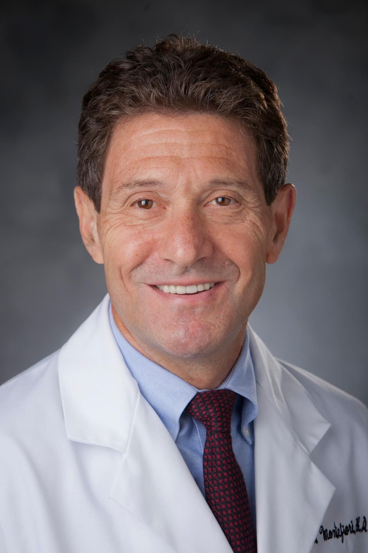 David Charles Montefiori合作论文数Laboratory for HIV and COVID-19 Vaccine Research and Development, Department of Surgery, Division of Surgical Sciences, Duke University School of Medicine;Duke Human Vaccine Institute, Duke University1Jonathan Mandelbaum合作论文数1




