PURPOSE: To determine cardiac dose received by patients treated with high dose rate interstitial brachytherapy. Patients with early -stage, node negative breast cancer can be treated using multi -catheter interstitial brachytherapy accelerated partial breast irradiation (MIB-APBI), with the benefit of reduced treatment volumes and favorable toxicity. METHODS AND MATERIALS: We conducted a retrospective review of left -sided breast cancer patients treated using MIB-APBI at our institution since 2014. The mean heart dose (MHD) was calculated using the Oncentra 3.2 planning system. The minimum distance between the planning target volume (PTVeval) and heart contour was measured manually. FINDINGS: 81 patients were included. The upper outer quadrant was the most common site. The MHD was 97.8 cGy (EQD2a/b = 2) (range 22-229 cGy). MHD significantly correlated with the closest distance between PTVeval and heart contour (correlation coefficient -0.823, p < 0.001); size of PTVeval (cc) and quadrant location did not. CONCLUSIONS: Appropriately selected women with early -stage, low -risk, left -sided breast cancer who received MIB-APBI had acceptable MHD. There was a strong correlation between the distance of PTVeval and MHD. Quadrant breast tumor is in cannot be used as a surrogate for MHD in brachytherapy. Our findings contribute to the growing evidence of the utility and safety of MIB-APBI.
With nearly two decades of expertise in the field of radiotherapy physics, she possesses a specialized focus on motion management, including surface-guided radiotherapy and breathing adaptive treatments.Her doctoral studies were focused on polymer gel dosimetry for 3D verification of dynamic radiotherapy.In the early 2000s, she obtained certification as a medical physicist from both the Swedish and Danish National Boards of Health and Welfare.Gaining international experience in Belfast, Sydney, and Copenhagen, she received in 2015 a research grant for promising young clinical researcher in Sweden.Throughout her clinical career, she has assumed a leading role in introducing motion management techniques in Sweden, such as breathing-adapted treatment and surfaceguided radiotherapy.Under her leadership, collaborative efforts involving industry, university hospital resulted in the world-leading clinical implementation of motion management technology.In 2018, she attained a career development position as an Associate University Lecturer at Lund University within the Department of Medical Radiation Physics.Since then, she has been able to dedicate even more time to activities aligned with her passions, including research, supervising doctoral and master's level students,
In the clinic, routine quality assurance tests ensure the proper functioning of each individual aspect of a radiation therapy treatment delivery. However, these tests may not guarantee an accurate treatment delivery. In this work, we present the design and application of a head phantom (using 3D printing technology) developed for end-to-end quality assurance of a stereotactic radiation therapy treatment for brain metastases, coupled with inserts for ion chamber, and film and gel dosimeters. The head phantom was subjected to the entire clinical workflow, with each stage of the process being performed by the appropriate clinical personnel to ensure that this quality assurance test mimics the clinical scenario.
A segment of a spine was 3D-printed based on real patient anatomy, using metal-doped high density plastic to radiographically mimic bone. This spine was submerged in a water tank to create an anthropomorphic phantom. The spine print incorporated a slot for Gafchromic EBT3 film dosimeters and fiducials for alignment of measured and calculated dose distributions. Spine SBRT treatment plans were generated for both 6 MV and 10FFF energies based on oncologist-drawn contours transferred from real anatomy. Plans were delivered under image guidance using our clinical procedures, to evaluate the dosimetric accuracy of our planning system in high density inhomogeneities and the geometric accuracy of delivery. Results show that the Acuros XB algorithm (dose-to-water) agrees well with film measurements throughout the measured region, including within the bone substitute material. Alignment of the steep dose gradients in planned and measured doses was within 0.5 mm in the ANT-POST direction and within 0.9 mm in the SUP-INF direction, both within machine tolerances. Our results give us confidence in our ability to plan and accurately deliver spinal SBRT treatments.
High-dose-rate brachytherapy is an accepted standard-of-care treatment for prostate cancer. In this procedure, catheters are inserted using three-dimensional (3D) transrectal ultrasound image-guidance. Their positions are manually segmented for treatment planning and delivery. The transverse ultrasound sweep, which is subject to tip and depth error for catheter localization, is a commonly used ultrasound imaging option available for image acquisition. We propose a two-step pipeline that uses a deep-learning network and curve fitting to automatically localize and model catheters in transversely reconstructed 3D ultrasound images. In the first step, a 3D U-Net was trained to automatically segment all catheters in a 3D ultrasound image. Following this step, curve fitting was implemented to detect the shapes of individual catheters using polynomial fitting. Of the 343 catheters (from 20 patients) in the testing data, the pipeline detected 320 (93.29%) with 7 false positives (2.04%) and 13 false negatives (3.79%). The average distance± one standard deviation between the ground truth and predictions for each catheter shaft was 1.9 ± 0.3 mm. The average difference of each catheter tip was 3.0 ± 0.4 mm. The proposed pipeline provides a method for reducing time spent on verification of catheter positions, minimizing uncertainties, and improving clinical workflow during the procedure. Reducing human variability in catheter placement predictions may increase the accuracy of tracking and radiation dose modelling.
PURPOSE: High-dose-rate brachytherapy (HDR-BT) is an important treatment modality for prostate cancer that maximizes radiation dose to cancerous tissue while sparing surrounding organs. Currently, treatment planning during HDR-BT is manually completed by medical physicists, a time-consuming and observer dependent process. We propose using deep learning through a U-Net architecture to automatically segment catheters in HDR prostate brachytherapy treatment planning. METHODS: 3D Ultrasound data along with the corresponding manual contours were obtained from 49 patients undergoing HDR prostate brachytherapy. The dataset was preprocessed and then exported for training and evaluation. The resulting model was assessed both quantitatively with binary segmentation metrics and qualitatively through 3D reconstructions. RESULTS: The output segmentations demonstrated consistency on different patient datasets and good visual agreement with ground truth images. The average execution time per patient is under 30.0 s, a significant improvement from manual contouring, which may require upwards of an hour. CONCLUSION: We trained and evaluated a 3D U-Net model for automatic catheter segmentation on 3D transrectal ultrasound images generated through HDR prostate brachytherapy. Deep learning methods such as the 3D U-Net used in this scenario appear to be a promising method for automatic catheter segmentation in prostate brachytherapy.
Breast conservation therapy with a partial mastectomy followed by adjuvant radiotherapy has become the standard approach for women diagnosed with an early stage breast cancer. Accelerated partial breast irradiation using multi-catheter interstitial brachytherapy can be used to deliver RT. Electromagnetic reconstruction (EMR) is a new technology that has been recently developed to determine the catheter placement post implantation. In this technique, a position sensor is pulled through a catheter. The path of the sensor is then reconstructed to delineate the path of the catheter. EMR is a promising technology that can potentially be used to validate the position of the catheters post implantation. We have previously shown that our EMR technology can accurately reconstruct catheter paths in breast phantoms. We performed a clinical feasibility study in patients to assess the accuracy of EMR in reconstructing the catheter paths in patients. Eligible patients for breast brachytherapy were enrolled in a prospective, single-center pilot study. All patients underwent breast brachytherapy as per the standard of care. A spatial tracking machine is positioned next to the patient's bed, which creates an external electromagnetic field around the patient's breast. A reference electromagnetic sensor is clamped on the patient's sternum. An electromagnetic sensor was guided through each of the implanted catheters, and the positions in real time were recorded. Using 3D slicer (an open source platform, we reconstructed the paths of the catheters. The paths generated manually on the planning CT scan were registered over the reconstructed paths. The accuracy of the EMR workflow was determined by measuring the mean + standard deviation between the EM generated reconstruction to the path segmented on the planning CT scan. A total of 38 catheters were implanted in 3 patients between July 2019-September 2019. We found that the mean distance between the CT generated catheter paths and the paths created by EMR was 1.7 + 0.3 mm. Reconstruction of the catheter paths in a patient typically took less than 5 minutes. Our results are promising and suggest that EM reconstruction can be accurate in identifying the catheter paths for interstitial breast brachytherapy. We will expand the patient cohort to confirm this data.
Voxelizing three-dimensional surfaces into binary image volumes is a frequently performed operation in medical applications. In radiation therapy (RT), dose-volume histograms (DVHs) calculated within such surfaces are used to assess the quality of an RT treatment plan in both clinical and research settings. To calculate a DVH, the 3D surfaces need to be voxelized into binary volumes. The voxelization parameters may considerably influence the output DVH. An effective way to improve the quality of the voxelized volume (i.e., increasing similarity between that and the original structure) is to apply oversampling to increase the resolution of the output binary volume. However, increasing the oversampling factor raises computational and storage demand. This paper introduces a fuzzy inference system that determines an optimal oversampling factor based on relative structure size and complexity, finding the balance between voxelization accuracy and computation time. The proposed algorithm was used to automatically calculate oversampling factor in four RT studies: two phantoms and two real patients. The results show that the method is able to find the optimal oversampling factor in most cases, and the calculated DVHs show good match to those calculated using manual overall oversampling of two. The algorithm can potentially be adopted by RT treatment planning systems based on the open-source implementation to maintain high DVH quality, enabling the planning system to find the optimal treatment plan faster and more reliably.
To evaluate a novel navigation system for breast brachytherapy, based on ultrasound (US)-guided catheter needle implantations followed by electromagnetic (EM) tracking of catheter paths. Breast phantoms were produced, containing US–visible tumors. Ultrasound was used to localize the tumor pose and volume within the phantom, followed by planning an optimal catheter pattern through the tumor using navigation software. An electromagnetic (EM)-tracked catheter needle was used to insert the catheters in the desired pattern. The inserted catheters were visualized on a post-implant CT, serving as ground truth. Electromagnetic (EM) tracking and reconstruction of the inserted catheter paths were performed by pulling a flexible EM guidewire through each catheter, performed in two clinical brachytherapy suites. The accuracy of EM catheter tracking was evaluated by calculating the Hausdorff distance between the EM-tracked and CT-based catheter paths. The accuracy and clinical feasibility of EM catheter tracking were also evaluated in three breast cancer patients, performed in a separate experiment room. A total of 71 catheter needles were implanted into 12 phantoms using US guidance and EM navigation, in an average ± SD time of 8.1 ± 2.9 min. The accuracy of EM catheter tracking was dependent on the brachytherapy suite: 2.0 ± 1.2 mm in suite 1 and 0.6 ± 0.2 mm in suite 2. EM catheter tracking was successfully performed in three breast brachytherapy patients. Catheter tracking typically took less than 5 min and had an average accuracy of 1.7 ± 0.3 mm. Our preliminary results show a potential role for US guidance and EM needle navigation for implantation of catheters for breast brachytherapy. EM catheter tracking can accurately assess the implant geometry in breast brachytherapy patients. This methodology has the potential to evaluate catheter positions directly after the implantation and during the several fractions of the treatment.
The accuracy of EclipseTM-calculated head and neck VMAT plan dose delivery was assessed in the situation when there is a gap between placed bolus on a head and neck thermoplastic mask and the body surface. GafchromicEBT3 film dosimetry was used for dose delivery validation. The results from 3%, 3 mm 2D gamma comparison indicate better than 95% agreement between measured film dose and calculated plan dose for gap widths up to 25 mm between placed bolus and body surface.
As part of a recent commissioning process for an ultrasound-guided prostate HDR technique at our centre, a representative dose delivery was validated using Gafchromic EBT3 film dosimetry. Agreement between Oncentra Prostate-calculated plan dose and measured film dose was within 5-10% over most of the 2D film dose planes, except in the regions within 5 mm of the catheter axes. Given the uncertainties associated with the measurement, this result was deemed to be clinically acceptable.
A dosimetric evaluation of a respiratory gated VMAT SABR technique was performed at two different beam energies. Dynamic ion chamber, EBT3 film and Fricke-xylenol orange-gelatin (FXG) gel measurements were acquired using a motion phantom with custom inserts for each dosimeter. Ion chamber and gel dosimeter measurements show good agreement between the measured and calculated plan dose within the planning target volume (PTV). Lower agreement than expected was observed between calculated plan dose and measured film dose, particularly for the 10 MV flattening filter free beam plan, a result that warrants further investigation.
Three dimensional dosimetry is being used in an increasingly wide variety of clinical applications as more gel and radiochromic plastic dosimeters become available. However, accessible 3D dosimetry analysis tools have not kept pace. 3D dosimetry data analysis is time consuming and laborious, creating a barrier to entry for busy clinical environments. To help in the adoption of 3D dosimetry, we have produced a streamlined, open-source dosimetry analysis system by developing a custom extension in 3D Slicer, called the Gel Dosimetry Analysis slicelet, which enables rapid and accurate data analysis. To assist those interested in adopting 3D dosimetry in their clinic or those unfamiliar with what is involved in a 3D dosimeter experiment, we first present the workflow of a typical gel dosimetry experiment. This is followed by the results of experiments used to validate, step-wise, each component of our software. Overall, our software has made a full 3D gel dosimeter analysis roughly 20 times faster than previous analysis systems.
 L John Schreiner合作论文数Queen's University34
 k m alexander合作论文数Kingston General Hospital20Martin Korzeniowski合作论文数7
 Gabor Fichtinger合作论文数School of Computing, Queen’s University;Department of Pathology and Molecular Medicine, Faculty of Health Sciences, Queen's University7Michael Sayer合作论文数Department of Physics, Engineering Physics & Astronomy, Queen's University7
 Johnson Darko合作论文数Cancer Center of Southeastern Ontario, Kingston General Hospital;Department of Oncology, Queens University;Kingston General Hospital, Queens University6Kerr Andrew合作论文数6
 Kim B. Mcauley合作论文数Department of Chemical Engineering, Faculty of Engineering and Applied Science, Queen’s University6mary ann ruggiero合作论文数5
 Kurtis H Dekker合作论文数Dept Med Biophys, Univ Western Ontario5
 Abdullah Nasr合作论文数5
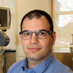 Tamas Ungi合作论文数School of Computing, Faculty of Arts and Science, Queen's University;Department of Surgery, School of Medicine, Queen's University;Centre for Health Innovation, Queen's University5M Lukacs合作论文数4C. Joshi合作论文数4Oliver Holmes合作论文数4
 Csaba Pinter合作论文数4G Salomons合作论文数3Logan Montgomery合作论文数3Harry Brastianos合作论文数3
 Chandra P. Joshi合作论文数Biophysics Group, Freie Universität Berlin3T Halsall合作论文数2
 Aquila Akingbade合作论文数2Nicole Kitner合作论文数2
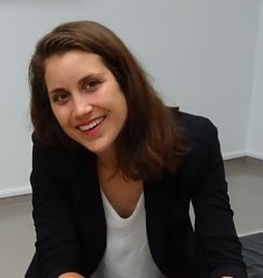 Natasja Janssen合作论文数Lab Percutaneous Surg, Queens Univ2Samantha Sigurdson合作论文数2
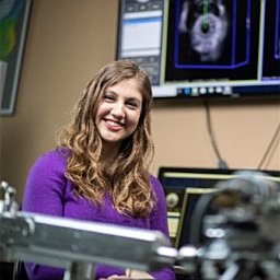 Jessica R. Rodgers合作论文数Sch Comp, Queens Univ2Stephane Thibodeau合作论文数2
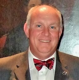 Conrad Falkson合作论文数Queens University2
 Giles Santyr合作论文数Department of Medical Biophysics, Temerty Faculty of Medicine, University of Toronto2Nkongchu Ken合作论文数2
 Sherrit Stewart合作论文数NASA Jet Propulsion Laboratory2
 Moraes Fábio Ynoe de合作论文数2Lalonde Michel合作论文数2Lourdes Botelho Garcia合作论文数2
 Parvin Mousavi合作论文数School of Computing, Queen's University;Medical Informatics Laboratory, Queen's University2B. Leclerc合作论文数2
 Andras Lasso合作论文数School of Computing, Queen's University2Guofeng Pang合作论文数1N B Campbell合作论文数1Lockwood, G.R.合作论文数1Mary Westerland合作论文数1C Joshi合作论文数1Paul DeJean合作论文数1David Barrow合作论文数1Andrew Kerr合作论文数1Lester M. Cohenb合作论文数1Thomas Vaughan合作论文数1




