Several studies suggest links between cancer and tricyclic antidepressant use. A case–control study using the General Practice Research Database examined whether previous tricyclic usage was associated with reduced incidence of brain (with glioma as a sub-category), breast, colorectal, lung and prostate cancers. Conditional logistic regression adjusted for age, gender, general practice, depression, smoking, body mass index, alcohol use and non-steroidal anti-inflammatory drug use. A total of 31 953 cancers were identified, each matched with up to two controls. We found a statistically significant reduction in tricyclic prescriptions compared with controls in glioma (odds ratio (OR) =0.59, 95% confidence interval (CI)=0.42–0.81) and colorectal cancer patients (OR=0.84, CI=0.75–0.94). These effects were dose-dependent (P-values for trend, glioma=0.0005, colorectal=0.001) and time-dependant (P-values for trend glioma=0.0005, colorectal=0.0086). The effects were cancer-type specific, with lung, breast and prostate cancers largely unaffected by antidepressant use. The biologically plausible, specific and dose- and time-dependant inverse association that we have found suggests that tricyclics may have potential for prevention of both colorectal cancer and glioma.
The in vitro effects of antidepressant drugs on mitochondrial function were investigated in a CHOβ2SPAP cell line used previously to determine the effects of antidepressants on gene transcription (Abdel-Razaq et al., Biochem Pharmacol 73:1995–2003, 2007) and in rat heart isolated mitochondria. Apoptotic effects of clomipramine (CLOM), desipramine (DMI) and of norfluoxetine (NORF, the active metabolite of fluoxetine), on cellular viability were indicated by morphological changes and concentration-dependent increases in caspase-3 activity in CHO cells after 18 h exposure to CLOM, DMI and NORF. However, tianeptine (TIAN) was without effect. CLOM and NORF both reduced integrated mitochondrial function as shown by marked reductions in membrane potential (MMP) in mitochondria isolated from rat hearts. DMI also showed a similar but smaller effect, whereas, TIAN did not elicit any significant change in MMP. Moreover, micromolar concentrations of CLOM, DMI and NORF caused significant inhibitions of the activities of mitochondrial complexes (I, II/III and IV). The inhibitory effects on complex IV activity were most marked. TIAN inhibited only complex I activity at concentrations in excess of 20 μM. The observed inhibitory effects of antidepressants on the mitochondrial complexes were accompanied by a significant decrease in the mitochondrial state-3 respiration at concentrations above 10 μM. The results demonstrate that the apoptotic cell death observed in antidepressant-treated cells could be due to disruption of mitochondrial function resulting from multiple inhibition of mitochondrial enzyme complexes. The possibility that antimitochondrial actions of antidepressants could provide a potentially protective pre-conditioning effect is discussed.
The effects of the antidepressant drugs clomipramine (CLOM), desipramine (DMI), tianeptine (TIAN) and of norfluoxetine (NORF, the active metabolite of fluoxetine), were investigated in CHO cells expressing human beta(2) adrenoceptors and a secreted placental alkaline phosphatase (SPAP) reporter gene to determine their actions on cyclic AMP-driven gene transcription.After 18 h of exposure, CLOM, DMI and NORF, but not TIAN, hadbiphasic effects on 1 mu M wisoprenaline-stimulated SPAP fsproduction with concentrations between 10 nM and mu M enhancing the maximal (E-max) SPAP response, without changing EC50 values, but higher concentrations produced marked inhibitory effects.At nanomolar concentrations, CLOM and DMI increased expression of phospho-CREB (cyclic AMP response element binding protein). NORF was less effective but did significantly increase phospho-CREB at a concentration of 200 nM. TIAN had no effect. None of the antidepressants had any effect on CREB expression, nor on the accumulation of cyclic AMP.After prolonged exposure (7-21 days) to a low concentration (200 nM) of the antidepressants, the enhanced E-max values for SPAP production evident after 18 h were not maintained but CLOM and DMI induced a significant leftward shift in the isoprenaline EC50, after a 7-day period of treatment and this was sustained at the 21 day time point. TIAN did not produce any significant changes.The results demonstrate that, in vitro, some but not all antidepressants can modify gene transcription via monoamine and cyclic AMP-independent mechanisms. The in vivo adaptive responses to TIAN probably involve alterations in different gene sets to those affected by other antidepressants. (c) 2007 Elsevier Inc. All rights reserved.
The present study utilised an in vitro cell model of the cAMP signalling pathway to investigate the actions of desipramine (DMI) and other psychoactive agents on cAMP-driven gene transcription. The model comprised CHO beta(2) SPAP cells; Chinese hamster ovary cells expressing human beta(2) adrenoceptors and a secreted placental alkaline phosphatase (SPAP) reporter gene with multiple cAMP response elements (CREs) in its promoter region. SPAP assays showed DMI to inhibit isoprenaline or forskolin-enhanced gene transcription in a time and concentration-dependent manner (IC50 = 16.6 +/- 2.0 mu M after 18 h). This effect of DMI was not dependent upon activity at the levels of the beta(2) receptor, cAMP accumulation or phosphorylation of the transcription factor, cAMP response element binding protein (CREB). The inhibitory effects were maintained in the presence of DMI for at least 3 weeks and were mimicked by exposure to norfluoxetine (the major metabolite of fluoxetine; IC50 = 7.2 +/- 1.8 mu M) and the neuroleptics, chlorpromazine and clozapine, all at a concentration of 10 mu M. Amphetamine (10 mu M, 18 h) enhanced SPAP gene transcription. Ca2+ imaging experiments ruled out an inhibitory effect of DMI on Ca2+ influx as concluded by previous studies.The results suggest a molecular target for DMI that lies downstream of CREB phosphorylation. Whether the inhibitory action of DMI is common to naturally expressed CRE-driven genes involved in adaptive responses to antidepressants in vivo remains to be determined. (c) 2005 Elsevier Inc. All rights reserved.
Mitochondria have been suggested to be a potential intracellular target for cancer chemotherapy. In this report, we demonstrate the ability of the tricyclic antidepressant chlorimipramine to kill human glioma cells in vitro by a molecular mechanism resulting in an increase in caspase 3 activity following inhibition of glioma oxygen consumption. Studies with isolated rat mitochondria showed that chlorimipramine specifically inhibited mitochondrial complex III activity, which causes decreased mitochondrial membrane potential as well as mitochondrial swelling and vacuolation. The use of chlorimipramine in human as an effective, non-toxic cancer therapeutic having a strong selectivity between cancer cells and normal cells on the basis of their mitochondrial function is discussed.
Oligodendroglia play an important role in axonal conduction in the CNS and are sensitive to oxidative toxicity induced by glutamate in the absence of ionotropic glutamate receptors. In this study, oligodendrocyte signalling cascades were examined, in response to glutamate-induced oxidative injury and to excitotoxicity. Rat cortical oligodendrocytes, differentiated in culture, were highly vulnerable to glutamate-induced cell death. Competitive inhibition of cystine uptake and increased oxidative stress appeared responsible for this death, and caused an accumulation of intracellular peroxides as well as chromatin fragmentation and condensation. Glutamate receptor subtype agonists (quisqualate, ibotenate) known to inhibit cystine uptake were cytotoxic, but not NMDA itself; moreover, glutamate receptor antagonists were not protective. Oligodendrocytes were also vulnerable to overactivation of glutamate receptors, as kainic acid and AMPA proved to be toxic. AMPA toxicity required the presence of cyclothiazide, suggesting rapid desensitization of AMPA receptors. Glutamate-induced oxidative stress and kainate/AMPA receptor stimulation activated the mitogen-activated protein kinase (MAP kinase) pathway, as well as the transcription factor ELK. However, MAP kinase kinase inhibitors only protected against injury from glutamate-induced oxidative stress. Oligodendrocytes were sensitive to oxygen-glucose deprivation injury as well, in a MAP kinase dependent fashion. Glutamate toxicity may conceivably be operative in neuropathological conditions that disrupt neuronal/oligodendrocyte interactions in axons, e.g. multiple sclerosis and ischaemia-reperfusion injury.
Mitochondrial cytochrome oxidase is competitively and reversibly inhibited by inhibitors that bind to ferrous heme, such as carbon monoxide and nitric oxide. In the case of nitric oxide, nanomolar levels inhibit cytochrome oxidase by competing with oxygen at the enzyme's heme–copper active site. This raises the Km for cellular respiration into the physiological range. This effect is readily reversible and may be a physiological control mechanism. Here we show that a number of in vitro and in vivo conditions result in an irreversible increase in the oxygen Km. These include: treatment of the purified enzyme with peroxynitrite or high (μM) levels of nitric oxide; treatment of the endothelial-derived cell line, b.End5, with NO; activation of astrocytes by cytokines; reperfusion injury in the gerbil brain. Studies of cell respiration that fail to vary the oxygen concentration systematically are therefore likely to significantly underestimate the degree of irreversible damage to cytochrome oxidase.
Nitric oxide (NO) is hypothesized to play a role in the immunopathogenesis of multiple sclerosis (MS). Increased levels of NO metabolites have been found in patients with MS. Peroxynitrite, generated by the reaction of NO with superoxide at sites of inflammation, is a strong oxidant capable of damaging tissues and cells. Inducible NO synthase (iNOS) is up‐regulated in the CNS of animals with experimental allergic encephalomyelitis (EAE) and in patients with MS. In this study, Western blots of cerebrospinal fluid (CSF) from patients with MS demonstrated the presence of iNOS, which was absent in CSF from control subjects. There was also NOS activity present in both MS and control CSF. Total NOS activity was increased (by 24%) in the CSF from MS patients compared with matched controls. The addition of 0.1 mM ITU (a specific iNOS inhibitor) to the samples did not change the activity of the control samples but decreased the NOS activity in the MS samples to almost control levels. The addition of 1 mM L‐NMMA (a nonisoform specific NOS inhibitor), completely inhibited NOS activity in CSF from control and MS subjects. Nitrotyrosine immunostaining of CSF proteins was detectable in controls but was greatly increased in MS samples. There were also significant increases in CSF nitrate + nitrite and oxidant‐enhanced luminescence in MS samples compared with controls. Additionally, a significant decrease in reduced glutathione and significant increases in oxidized glutathione and S‐nitrosothiols were found in MS samples compared with controls. Parallel changes in NO metabolites were observed in the plasma of MS patients, compared with controls, and accompanied a significant increase of reduced glutathione. These data strongly support a role for nitrosative stress in the pathogenesis of MS and indicate that therapeutic strategies focussed on decreasing production of NO by iNOS and/or scavenging peroxynitrite may be useful in alleviating the neurological impairments that occur during MS relapse. © 2002 Wiley‐Liss, Inc.
Conference Abstract| June 01 2002 Controlled release of cytochrome c from functional brain mitochondria; implications for apoptotic mechanisms in neurdegenerative disease R. A. Clayton; R. A. Clayton 1Division of Neurochemistry, Institute of Neurology, Queen Square, London, WC1N 3BG Search for other works by this author on: This Site PubMed Google Scholar M. A. Sharpe; M. A. Sharpe 1Division of Neurochemistry, Institute of Neurology, Queen Square, London, WC1N 3BG Search for other works by this author on: This Site PubMed Google Scholar J. B. Clark; J. B. Clark 1Division of Neurochemistry, Institute of Neurology, Queen Square, London, WC1N 3BG Search for other works by this author on: This Site PubMed Google Scholar T. E. Bates T. E. Bates 1Division of Neurochemistry, Institute of Neurology, Queen Square, London, WC1N 3BG Search for other works by this author on: This Site PubMed Google Scholar Biochem Soc Trans (2002) 30 (3): A70. https://doi.org/10.1042/bst030a070a Views Icon Views Article contents Figures & tables Video Audio Supplementary Data Peer Review Share Icon Share Twitter LinkedIn Cite Icon Cite Get Permissions Citation R. A. Clayton, M. A. Sharpe, J. B. Clark, T. E. Bates; Controlled release of cytochrome c from functional brain mitochondria; implications for apoptotic mechanisms in neurdegenerative disease. Biochem Soc Trans 1 June 2002; 30 (3): A70. doi: https://doi.org/10.1042/bst030a070a Download citation file: Ris (Zotero) Reference Manager EasyBib Bookends Mendeley Papers EndNote RefWorks BibTex toolbar search Search Dropdown Menu toolbar search search input Search input auto suggest filter your search All ContentAll JournalsBiochemical Society Transactions Search Advanced Search This content is only available as a PDF. © 2002 Biochemical Society2002 Article PDF first page preview Close Modal You do not currently have access to this content.
Calcium overload of neural cell mitochondria plays a key role in excitotoxic and ischemic brain injury. This study tested the hypothesis that brain mitochondria consist of subpopulations with differential sensitivity to calcium-induced inner membrane permeability transition, and that this sensitivity is greatly reduced by physiological levels of adenine nucleotides. Isolated non-synaptosomal rat brain mitochondria were incubated in a potassium-based medium in the absence or presence of ATP or ADP. Measurements were made of medium and intramitochondrial free calcium, light scattering, mitochondrial ultrastructure, and the elemental composition of electron-opaque deposits within mitochondria treated with calcium. In the absence of adenine nucleotides, calcium induced a partial decrease in light scattering, accompanied by three distinct ultrastructural morphologies, including large-amplitude swelling, matrix vacuolization and a normal appearance. In the presence of ATP or ADP the mitochondrial calcium uptake capacity was greatly enhanced and calcium induced an increase rather than a decrease in mitochondrial light scattering. Approximately 10% of the mitochondria appeared damaged and the rest contained electron-dense precipitates that contained calcium, as determined by electron-energy loss spectroscopy. These results indicate that brain mitochondria are heterogeneous in their response to calcium. In the absence of adenine nucleotides, approximately 20% of the mitochondrial population exhibit morphological alterations consistent with activation of the permeability transition, but less than 10% exhibit evidence of osmotic swelling and membrane disruption in the presence of ATP or ADP.
It is becoming increasingly evident that the mitochondrial genome may play a key role in neurodegenerative diseases. Mitochondrial dysfunction is characteristic of several neurodegenerative disorders, and evidence for mitochondria being a site of damage in neurodegenerative disorders is partially based on decreases in respiratory chain complex activities in Parkinson's disease, Alzheimer's disease, and Huntington's disease. Such defects in respiratory complex activities, possibly associated with oxidant/antioxidant balance perturbation, are thought to underlie defects in energy metabolism and induce cellular degeneration. Efficient functioning of maintenance and repair process seems to be crucial for both survival and physical quality of life. This is accomplished by a complex network of the so-called longevity assurance processes, which are composed of genes termed vitagenes. A promising approach for the identification of critical gerontogenic processes is represented by the hormesis-like positive effect of stress. In the present review, we discuss the role of energy thresholds in brain mitochondria and their implications in neurodegeneration. We then review the evidence for the role of oxidative stress in modulating the effects of mitochondrial DNA mutations on brain age-related disorders and also discuss new approaches for investigating the mechanisms of lifetime survival and longevity.
Hyperthermic stress is known to protect against myocardial dysfunction after ischemia-reperfusion injury. It is unclear however, what energetic mechanisms are affected by the molecular adaptation to heat stress. We hypothesized that mild hyperthermic stress can increase mitochondrial respiratory enzyme activity, affording protection to mitochondrial energetics during prolonged cardiac preservation for transplantation. Rat hearts were excised after heat-stress or sham treatment and subjected to cold cardioplegic arrest and ischemia followed by reperfusion in an ex vivo perfusion system. Cardiac function, mitochondrial respiratory, and complex activities were assessed before and after ischemia. Heat shock protein (Hsp 32, 60, and 72) expression was increased in heat-stressed hearts. This was associated with increased mitochondrial complex activities in heat-stress versus sham-treated groups for complex I-V. During reperfusion, higher complex activities and respiratory control ratios were observed in heat-stressed versus sham-treated groups. Recovery of ventricular function was improved in heat-stressed hearts. Furthermore, mitochondria in reperfused heat-stressed myocardium exhibited intact membranes with packed, parallel, lamellar cristae, whereas in sham-treated myocardium, mitochondria were severely disrupted. This study provides the first evidence of heat-stress-mediated enhancement of mitochondrial energetic capacity. This is associated with increased tolerance to ischemia-reperfusion injury. Protection by heat stress against myocardial dysfunction may be partially due to enhancement of mitochondrial energetics.
Increasing evidence suggests that Fatty acid ethyl esters (FAEE) play a central role in ethanol induced organ damage. In the current study we measured FAEE formation in rats after short-term oral administration of ethanol, in the presence and absence of pre-treatment with acetyl-L-carnitine. Ethanol treatment caused a significant increase in the levels of FAEE, particularly in the brain and heart, but also in the kidney and liver. Increases in FAEE were associated with a significant increase in FAEE synthase activity, GSH transferase activity, and lipid hydroperoxide levels. Pre-treatment with acetyl-L-carnitine resulted in a significant reduction of FAEE accumulation, decrease in FAEE synthase and GSH transferase activities, and lipid hydroperoxide levels. Administration of acetyl-L-carnitine greatly reduced the metabolic abnormalities due to non-oxidative ethanol metabolism, through an increment in lipid metabolism/turnover and by the modulation of the activities of enzymes associated with FAEE synthesis. These results suggest a potentially important pharmacological role for acetyl-L-carnitine in the prevention of alcohol-induced cellular damage.
Activation of murine microglial and macrophage cell lines with lipopolysaccharide (LPS) and interferon‐γ (IFN‐γ) resulted in the induction of the inducible form of nitric oxide synthase (NOS) and the release of micromolar amounts of NO into the surrounding medium. The synthesis of NO was associated with increased cellular membrane damage as assessed by trypan blue dye exclusion and the leakage of lactate dehydrogenase into the cell culture medium. However, the synthesis and release of cytokines was largely unaffected. NO‐mediated cell damage was also accompanied by a marked decrease in the intracellular levels of reduced glutathione and ATP. In addition, significant inhibition of mitochondrial respiratory chain enzyme activities was seen following cellular activation. However, citrate synthase activity (a mitochondrial matrix enzyme) was not detectable in the extracellular supernatants, suggesting preservation of the integrity of the mitochondrial inner membrane following activation. These effects were largely prevented by the addition of the NOS inhibitor, N ‐guanidino monomethyl l ‐arginine during the activation period. Our observations demonstrate that induction of NOS activity in microglia results in damage to the plasma membrane leading to a loss of glutathione, complex‐specific inhibition of the mitochondrial electron transport chain and depletion of cellular ATP. Our data suggest that pharmacological modulation of NOS activity in activated microglia in vivo may prevent cellular damage to bystander cells such as neurons, astrocytes and oligodendrocytes, as well as to microglia themselves.
Skin plays an important role in protection against oxidative stressors such as ultraviolet radiation, ozone and chemicals. Chronic sun exposure causes degenerative changes in the skin that are recognized as photoaging. Oxidative stress has been shown to alter the expression of mammalian antioxidant enzymes as well as to enhance numerous transcription factors, including nuclear factor kappaB, stress-activated protein kinase and heat shock factor This latter is the transcription factor for the synthesis of heat shock proteins, which have been known to protect against a wide variety of toxic conditions, including extreme temperatures, oxidative stress and cytotoxic drugs. In this study we investigated the role of oxidative stress in the induction of heat shock protein (HSP) 70 in human skin fibroblasts and the effect of vitamin E. We found that significant HSP70 induction occurred after exposure to HOOH and that this was associated with a significant perturbation in protein and nonprotein sulfhydryl groups, and with a significant increase in protein carbonyl levels. Treatment with vitamin E conferred significant protection against stress-induced modifications of cellular sulfhydryl and carbonyl content, while maintaining functional levels of cytoprotective HSP70. Our results point to the possible involvement of redox mechanisms in the heat shock signal transduction pathway, which may play an important regulatory role in the genetic mechanisms of tolerance to oxidative stress. Exogenous antioxidant supplementation with vitamin E could have cosmetic benefits and may be an efficient tool to mitigate the consequences of free radical-induced skin damage.
Reactive oxygen species have been implicated in the pathogenesis of the severe connective tissue damage present in several photodermatologic disorders, including drug-induced phototoxicity, porphyrias and photoaging. Oxidative stress has been shown to alter the expression of mammalian antioxidant enzymes and to enhance numerous transcription factors, including nuclear factor-kappa B, stress-activated protein kinase and heat shock factor. The latter represents the transcription factor for the synthesis of cytoprotective proteins called heat shock proteins. In this study, we investigated the role of oxidative stress and sulfdryl (SH) groups in the induction of HSP70 in human skin fibroblasts and the effect of antioxidants. We found that significant HSP70 induction occurred after exposure to HOOH and this was associated with marked perturbation in protein and nonprotein SH groups and with a considerable increase in protein carbonyl levels. Treatment with a natural antioxidant from rosemary extract provided notable protection against stress-induced modifications of cellular SH and carbonyl content, maintaining functional levels of cytoprotective heat shock protein 70. Our results point to the possible involvement of redox mechanisms in the heat shock signal transduction pathway, which may play an important regulatory role in the genetic mechanisms of tolerance to oxidative stress. Exogenous supplementation of an antioxidant hydrophilic extract from rosemary could have cosmetic benefits and may represent an efficient tool to minimize free radical-induced skin damage.
 JB Clark合作论文数20Dg Gadian合作论文数12
 Stephen Williams合作论文数Queensland Brain Institute, The University of Queensland8
 Jimmy David Bell合作论文数7
 Giovanni Scapagnini合作论文数Dept Med & Hlth Sci V Tiberio, Univ Molise7
 Vittorio Calabrese合作论文数Department of Biomedical and Biotechnological Sciences, University of Catania7Julie Keelan合作论文数6
 MarÍa Ángeles Almeida Parra合作论文数Departamento de Bioquímica y Biología Molecular, Universidad de Salamanca;(Instituto de Investigación Biomédica de Salamanca, Hospital Clínico Universitario de Salamanca;Instituto de BiologÍa Funcional y Genomica, Universidad de Salamanca5Laura Canevari合作论文数5
 Steve Williams合作论文数4
 Risto Kauppinen合作论文数School of Experimental Psychology, University of Bristol4
 Ivan Sammut合作论文数Department of Pharmacology and Toxicology, School of Biomedical Sciences, University of Otago4N.W. Read合作论文数4DJ BRYANT合作论文数3C Catalano合作论文数3AMG Stella合作论文数3
 Jane Cox合作论文数School of Immunology & Microbial Sciences, Faculty of Life Sciences & Medicine, King’s College London3David A. Kendall合作论文数School of Life Sciences, University of Nottingham3
 Bo K. Siesjö合作论文数Laboratory for Experimental Brain Research, University of Lund3Colin J. Green合作论文数3Andrzej Loesch合作论文数3
 Wesam Abdel-Razaq合作论文数School of Biomedical Sciences, University of Nottingham Medical School3R.H. Dowling合作论文数3Iain P. Hargreaves合作论文数3Sam Giuffrida合作论文数2P BOAKYE合作论文数2G. Pennisi合作论文数2F. Dinotta合作论文数2Al Busza合作论文数2IR YOUNG合作论文数2JB CLARK合作论文数2Mn Aljanabi合作论文数2Sjr Heales合作论文数2
 Barry Fuller合作论文数Division of Medicine, University College London;Department of Surgical Biotechnology, University College London2Brooks K J合作论文数2Devane W A合作论文数2
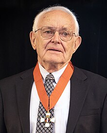 Graeme Bydder合作论文数University of California, San Diego2
 Geoffrey Burnstock合作论文数Autonomic Neuroscience Institute, School of Medicine, Royal Free Hospital2
 Pieter Dagnelie合作论文数Department of Internal Medicine, School for Cardiovascular Diseases, Maastricht University2Catherine J. Waterfield合作论文数2Agrippino Ravagna合作论文数2S. E. C. Davies合作论文数2K. L. Allen合作论文数2S.C.R. Williams合作论文数2Richard A. Iles合作论文数2Maureen S. Thorniley合作论文数2Kate J Brooks合作论文数2A. M. Giuffrida Stella合作论文数2Sandra Simpkin合作论文数2




