Patients with an activation mutation of the Lrp5 gene exhibit high bone mass (HBM). Limited information is available regarding compartment-specific changes in bone. The relationship between the phenotype and serum serotonin is not well documented. To evaluate bone, serotonin, and bone turnover markers (BTM) in Lrp5-HBM patients, we studied 19 Lrp5-HBM patients (T253I) and 19 age- and sex-matched controls. DXA and HR-pQCT were used to assess BMD and bone structure. Serum serotonin, sclerostin, dickkopf-related protein 1 (DKK1), and BTM were evaluated. Z-scores for the forearm, total hip, lumbar spine, forearm, and whole body were significantly increased (mean +/- SD) between 4.94 +/- 1.45 and 7.52 +/- 1.99 in cases versus -0.19 +/- 1.19 to 0.58 +/- 0.84 in controls. Tibial and radial cortical areas, thicknesses, and BMD were significantly higher in cases. In cases, BMD at the lumbar spine and forearm and cortical thickness were positively associated and trabecular area negatively associated with age (r=0.49, 0.57, 0.74, and -0.61, respectively, p<.05). Serotonin was lowest in cases (69.5 [29.9-110.4] ng/mL versus 119.4 [62.3-231.0] ng/mL, p<.001) and inversely associated with tibial cortical density (r=-0.49, p<.05) and directly with osteocalcin (OC), bone-specific alkaline phosphatase (B-ALP), and procollagen type 1 amino-terminal propeptide (PINP) (r=0.52-0.65, p<.05) in controls only. OC and S-CTX were lower and sclerostin higher in cases, whereas B-ALP, PINP, tartrate-resistant acid phosphatase (TRAP), and dickkopf-related protein 1 (DKK1) were similar in cases and controls. In conclusion, increased bone mass in Lrp5-HBM patients seems to be caused primarily by changes in trabecular and cortical bone mass and structure. The phenotype appeared to progress with age, but BTM did not suggest increased bone formation. (C) 2011 American Society for Bone and Mineral Research.
The Lrp5 gene is a major determinant of bone mass accrual. It has been demonstrated recently to achieve this function by hampering the synthesis of gut-derived serotonin, which is a powerful inhibitor of bone formation. In this study we analyzed plasma serotonin levels in patients with a high-bone-mass (HBM) phenotype owing to gain-of-function mutation of Lrp5 (T253I). A total of 9 HBM patients were compared with 18 sex- and age-matched controls. In HBM patients, the serotonin concentrations in platelet-poor plasma were significantly lower than in the controls (mean +/- SEM: 2.16 +/- 0.28 ng/mL versus 3.51 +/- 0.49 ng/mL, respectively, p < .05). Our data support the hypothesis that circulating serotonin levels mediate the increased bone mass resulting from gain-of-function mutations in Lrp5 in humans.
HLA class-I expression is weak in embryonic stem cells but increases rapidly during lineage progression. It is unknown whether all three classical HLA class-I antigens follow the same developmental program. In the present study, we investigated allele-specific expression of HLA-A, -B, and -C at the mRNA and protein levels on human mesenchymal stem cells from bone marrow and adipose tissue as well as striated muscle satellite cells and lymphocytes. Using multicolour flow cytometry, we found high cell surface expression of HLA-A on all stem cells and PBMC examined. Surprisingly, HLA-B was either undetectable or very weakly expressed on all stem cells protecting them from complement-dependent cytotoxicity (CDC) using relevant human anti-B and anti-Cw sera. IFN gamma stimulation for 48-72 h was required to induce full HLA-B protein expression. Quantitative real-time RT-PCR showed that IFN gamma induced a 9-42 fold increase of all six HLA-A,-B,-C gene transcripts. Interestingly, prior to stimulation, gene transcripts for all but two alleles were present in similar amounts suggesting that post-transcriptional mechanisms regulate the constitutive expression of HLA-A,-B, and -C. Locus-restricted expression of HLA-A, -B and -C challenges our current understanding of the function of these molecules as regulators of CD8(+) T-cell and NK-cell function and should lead to further inquiries into their expression on other cell types.
Mesenchymal stem cells (MSC) are capable of multilineage differentiation into cells like osteoblasts, chondrocytes or adipocytes. MSCs can be isolated from bone marrow and expanded ex vivo for up to 25-40 population doublings while maintaining genetic stability and differentiation potential. MSCs have great potential in the field of tissue engineering and regenerative medicine where cartilage and bone conditions which are non-treatable or show very slow improvement can be effectively handled. Several clinical trials have been performed using MSC and show very promising results.
Mechanisms controlling human multipotent mesenchymal (stromal) stem cell (hMSC) differentiation into osteoblasts or adipocytes are poorly understood. We have previously demonstrated that Wnt signaling in hMSC enhanced osteoblast differentiation and inhibited adipogenesis by comparing two hMSC cell lines overexpressing mutated forms of the Wnt co-receptor LRP5: T253I (hMSC-LRP5T253) and T244M (hMSC-LRP5T244) conducting high and low level of Wnt signaling, respectively. To explore the underlying molecular mechanisms, we compared gene expression profiles of hMSC-LRP5T253 and hMSC-LRP5T244 treated with Wnt3a using whole genome expression microarrays and found that TNFRSF19 is differentially up-regulated between the two cells lines. Bioinformatic analysis and dual luciferase assay of its promoter revealed that TNFRSF19 transcript 2 (TNFRSF19.2) is a target of canonical Wnt signaling. Knocking down TNFRSF19 in hMSC-LRP5T253 cells decreased Wnt3a-induced osteoblast differentiation marker alkaline phosphate activity and its overexpression in hMSC-LRP5T244 cells increased alkaline phosphate activity. In addition, TNFRSF19 was negatively regulated by adipogenic transcription factor CCAAT/enhancer-binding proteins (C/EBP). Knocking down TNFRSF19 in hMSC-LRP5T253 cells or its overexpression in hMSC-LRP5T244 cells significantly increased or decreased adipogenesis, respectively. In conclusion, we revealed a novel function of TNFRSF19 as a factor mediating differentiation signals that determine the hMSC differentiating fate into osteoblasts or adipocytes.
Genetic mutations in the LRP5 gene affect Wnt signaling and lead to changes in bone mass in humans. Our in vivo and in vitro results show that activated mutation T253I of LRP5 enhances osteogenesis and inhibits adipogenesis. Inactivating mutation T244M of LRP5 exerts opposite effects.
 Moustapha Kassem合作论文数Laboratory of the Molecular Endocrinology, Department of Endocrinology, Diabetes and Metabolism, University Hospital of Odense;Sundhedsvidenskabelige Fakultet, Københavns Universitet;Department of Clinical Research, Sundhedsvidenskabelige Fakultet, Syddansk Universitet8
 Morten Frost合作论文数Danish Ageing Research Centre, University of Southern Denmark;Department of Endocrinology and Metabolism, Odense University Hospital;University of Southern Denmark, Odense University Hospital2
 Isa Adiba合作论文数2
 Sabir Hardee J合作论文数Odense University Hospital and Clinical Institute, University of Southern Denmark2
 Jens Bollerslev合作论文数Department of Endocrinology, Morbid Obesity and Preventive Medicine, Institute of Clinical Medicine, Faculty of Medicine, University of Oslo;Oslo Diabetes Research Centre2
 Basem M Abdallah合作论文数Department of Endocrinology, Odense University Hospital2
 Torben Barington合作论文数Department of Clinical Immunology, Odense University Hospital2Kim Brixen合作论文数Department of Endocrinology, Odense University Hospital2
 Weimin Qiu合作论文数2Jan O. Nehlin合作论文数2J. Adjaye合作论文数1Katharina Drews合作论文数1
 Abbas Jafari Kermani合作论文数Department of Cellular and Molecular Medicine, Faculty of Health and Medical Sciences, University of Copenhagen1W. Qiu合作论文数1
 Yuhui Hu (胡宇慧)合作论文数Department of Pharmacology, School of Medicine, Southern University of Science and Technology1
 Na Li合作论文数1
 Gerard Karsenty合作论文数Department of Genetics and Development, Columbia University Medical Center;Department of Biomedical Engineering, The Fu Foundation School of Engineering and Applied Science, Columbia University in the City of New York1Larsen Kenneth H合作论文数1
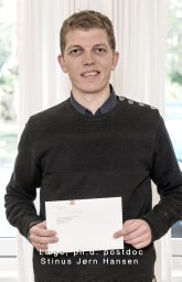 Hansen Stinus合作论文数1Gossiel Fatma合作论文数1Wim Van Hul合作论文数1
 Wei Chen (陈炜)合作论文数Department of Systems Biology, School of Life Sciences, Southern University of Science and Technology1
 Li Chen合作论文数1
 Richard Eastell合作论文数Division of Clinical Medicine, School of Medicine and Population Health, University of Sheffield;Mellanby Centre for Musculoskeletal Research, University of Sheffield1
 Susanne Mandrup合作论文数Department of Biochemistry and Molecular Biology, Faculty of Science, University of Southern Denmark;Center of Excellence in Functional Genomics and Tissue Plasticity, University of Southern Denmark1Michael Gaster合作论文数1
 Vijay Kumar Yadav合作论文数Medical Center, Columbia University1




