Background: Regional periarticular bone mineral density may influence fixation and survivorship in total knee arthroplasty, but its relationship to coronal alignment remains unclear. This study assessed the association between coronal knee alignment and osteoporosis using CT Hounsfield Unit (HU)-based thresholds. Methods: Patients aged ≥ 50 years with standing long-leg radiographs and phantomless knee-CT (2008–2025) were retrospectively identified. Exclusion criteria included >12 months between studies, incomplete CT, non-120 kV acquisition, prior fracture or surgery, or metabolic bone disease other than osteopenia or osteoporosis. Mean trabecular attenuation was measured over 15 mm of epiphyseal bone in the distal femur and proximal tibia. Osteoporosis was defined using CT-HU thresholds. Coronal alignment was measured using hip–knee–ankle angle (HKAA) and categorized as varus (<178°), neutral (178–182°), or valgus (>182°). Measurements were performed by a reviewer blinded to osteoporosis classification. Multivariable logistic regression adjusted for age, sex, body mass index, and Kellgren–Lawrence grade. Results: Among 306 patients (mean age 66.9 ± 9.0 years; 51.3% female), 99.3% underwent CT for arthroplasty planning. Osteoporosis prevalence was 34.3% of varus, 58.2% of neutral, and 68.6% of valgus knees. Increasing valgus alignment was associated with higher osteoporosis odds, whereas varus alignment showed lower odds. Female sex (OR 3.06; p < 0.001), age (OR 1.06/year; p < 0.001), and HKAA (OR 1.05/degree; p = 0.042) remained independently associated with osteoporosis, whereas Kellgren–Lawrence grade was nonsignificant. Conclusions: Coronal alignment was associated with CT-defined regional knee osteoporosis. Valgus alignment showed increased odds of osteoporosis, whereas varus alignment showed lower prevalence.
Background Treatment of stiffness following total knee arthroplasty (TKA) is complicated by a lack of a specific diagnosis for arthrofibrosis. Magnetic resonance imaging (MRI) provides a noninvasive means to generate and evaluate clinically diagnostic images of deep structures within the knee. The purpose of this study was to assess how the severity and location of arthrofibrosis observed on MRI relate to MRI synovial classifications and range of motion in primary TKA patients undergoing revision surgery. Methods There were 137 patients (140 knees) scheduled for TKA revision surgery who underwent preoperative MRI and were assigned a global synovial classification (normal, abnormal, arthrofibrosis, infection, focal scarring, polymeric) and severity of arthrofibrosis (none, mild, moderate, severe) within the individual anterior and posterior compartments. Passive joint flexion and extension measurements were recorded intraoperatively. Results The global synovial classification of arthrofibrosis had a greater prevalence of severe fibrosis within the anterior (25 of 42) and posterior (16 of 42) compartments as compared to other classifications (P < 0.0001). Reduced flexion and extension were found in patients who had a global synovial classification of arthrofibrosis (P < 0.0001). Reduced knee flexion was observed with greater anterior fibrosis severity, and reduced knee extension was observed with greater posterior fibrosis severity (P < 0.0001). Conclusions Anterior and posterior fibrosis severity differs across MRI synovial classifications. Further, the extent of joint flexion and extension differs by MRI synovial classifications and severity of compartment arthrofibrosis. The results of this study support that MRI presents an effective and noninvasive means to discern global and local synovial reactions within individuals undergoing revision TKA.
BACKGROUND:The burden of revision total joint arthroplasty (rTJA) is increasing. Revision procedures are associated with an increased risk of perioperative complications. Obese patients undergoing rTJA may have a higher risk of wound complications due to their soft-tissue envelope. Closed-incision negative pressure wound therapy (ciNPWT) has been rigorously investigated for its effect on reducing wound complications; however, the literature is limited to primary total joint arthroplasty. The purpose of the current study was to investigate the association between ciNPWT and clinical outcomes for obese patients undergoing aseptic revision total hip and knee arthroplasty. METHODS:This was a retrospective study examining aseptic rTJA procedures from January 2017 to December 2021 at a high-volume institution. Patients were included if their body mass index was > 35 and had a minimum follow-up of 2 years. The cohorts were rTJA procedures that used ciNPWT versus procedures without ciNPWT use. A total of 214 rTJA (Hip, n = 61; Knee, n = 153) patients qualified for analysis. After surgery, ciNPWT was applied on 130 (60.7%) patients (92 knees and 38 hips), and standard dressing was used on 84 (39.3%) patients (61 knees and 23 hips). There were no significant baseline differences between the groups. Outcomes evaluated were as follows: (1) revision for infection; (2) superficial wound complications; (3) overall complications; (4) all-cause revision; and (5) all-cause readmissions. The association between ciNPWT use and outcomes was evaluated using Chi-square tests. RESULTS:When controlling for all variables, ciNPWT use was associated with significantly fewer revisions for infection (0.8 versus 8.3%), overall complications (3.8 versus 11.9%), all-cause revisions (3.1 versus 13.1%), and readmissions (10.8 versus 21.7%) compared to those who did not use ciNPWT. Secondary superficial wound complication-specific outcomes did not show significant differences. CONCLUSIONS:To our knowledge, this study is the largest retrospective study to date examining the effect of ciNPWT on outcomes in obese aseptic revision rTJA patients. Our results demonstrated major benefits associated with the use of ciNPWT in obese patients undergoing aseptic rTJA in reference to infection-related revision, overall complications, all-cause revisions, and early readmission for any reason.
BACKGROUND:Understanding the sources of patients' expectations of total knee arthroplasty (TKA) can foster realistic expectations. Sources of expectations may be influenced by clinical and psychological variables, including general disposition or attitude. METHODS:Patients undergoing TKA completed a 19-item survey preoperatively that addressed the amount of improvement expected (i.e., "complete" to "a little") for symptoms and physical and psychological well-being. Patients reported the sources of these expectations, which were grouped into categories. Disposition or attitude was assessed for positive affect (e.g., openness to change) and negative affect (e.g., irritability) with use of the Positive and Negative Affect Schedule (PANAS). Clinical status was assessed using the Knee injury and Osteoarthritis Outcome Score-Joint Replacement (KOOS-JR). Expectations and sources of expectations were compared with KOOS-JR and PANAS scores and demographic and clinical characteristics (e.g., symptom duration) with use of statistical analyses. RESULTS:A total of 232 patients (mean age, 65 years; 60% women; 17% Latino ethnicity or non-White race) were enrolled 17 ± 8 days preoperatively. Seventy-two percent expected ≥15 of the survey items; complete improvement was expected for 59% of items. Expecting more items was associated with better preoperative KOOS-JR scores (p < 0.0001), more negative affect (p = 0.003), a shorter duration of symptoms (p = 0.01), and not being a college graduate (p = 0.04). Patients volunteered multiple sources of expectations, including favorable outcomes from a previous orthopaedic surgery (11% of patients), knowledge of favorable (56%) and unfavorable (7%) TKA outcomes in others, their current surgeon (15%), avoidance of further knee deterioration (10%), internet information (16%), and general optimism (18%). Citing more sources was associated with worse KOOS-JR scores (p = 0.02) and less positive affect (p = 0.009). Sources of expectations were associated with patient variables; specifically, knowledge of favorable outcomes was associated with better KOOS-JR scores (p = 0.04), knowledge of unfavorable outcomes was associated with more negative affect (p = 0.04), optimism was associated with more positive affect (p = 0.01), and information from the internet was associated with longer symptom duration (p = 0.04) and less negative affect (p = 0.02). CONCLUSIONS:Patients had high expectations of TKA and derived these expectations from diverse sources spanning personal and social network experiences. Patients' expectations and/or sources of expectations were associated with functional status and positive and negative affect. LEVEL OF EVIDENCE:Prognostic Level II . See Instructions for Authors for a complete description of levels of evidence.
Aims Periprosthetic femoral fracture (PPF) is a major complication following total hip arthroplasty (THA). Uncemented femoral components are widely preferred in primary THA, but are associated with higher PPF risk than cemented components. Collared components have reduced PPF rates following uncemented primary THA compared to collarless components, while maintaining similar prosthetic designs. The purpose of this study was to analyze PPF rate between collarless and collared component designs in a consecutive cohort of posterior approach THAs performed by two high-volume surgeons. Methods This retrospective series included 1,888 uncemented primary THAs using the posterior approach performed by two surgeons (PKS, JMV) from January 2016 to December 2022. Both surgeons switched from collarless to collared components in mid-2020, which was the only change in surgical practice. Data related to component design, PPF rate, and requirement for revision surgery were collected. A total of 1,123 patients (59.5%) received a collarless femoral component and 765 (40.5%) received a collared component. PPFs were identified using medical records and radiological imaging. Fracture rates between collared and collarless components were analyzed. Power analysis confirmed 80% power of the sample to detect a significant difference in PPF rates, and a Fisher's exact test was performed to determine an association between collared and collarless component use on PPF rates. Results Overall, 17 PPFs occurred (0.9%). There were 16 fractures out of 1,123 collarless femoral components (1.42%) and one fracture out of 765 collared components (0.13%; p = 0.002). The majority of fractures (n = 14; 82.4%) occurred within 90 days of primary THA. There were ten reoperations for PPF with collarless components (0.89%) and one reoperation with a collared component (0.13%; p = 0.034). Conclusion Collared femoral components were associated with significant decreases in PPF rate and reoperation rate for PPF compared to collarless components in uncemented primary THA. Future studies should investigate whether new-generation collared components reduce PPF rates with longer-term follow-up.
Background Modular metaphyseal engaging (MME) femoral components in total hip arthroplasty (THA) allow optimized femoral length, offset, and anteversion and are useful in patients with unusual proximal femoral anatomy. Fretting, corrosion, and stem fractures above the modular sleeve are complications associated with these implants. The purpose of this study was to identify failure mechanisms of retrieved MME femoral components at our institution, identify all broken stem cases, and evaluate how often an extended trochanteric osteotomy (ETO) was required for removal. Methods All consecutively retrieved MME femoral components from September 2002 to May 2023 were reviewed. Patient demographics, procedure information, component specifications, indications for removal, and requirements for further revision surgery were reviewed. Descriptive statistics were calculated for variables of interest. Results There were 131 retrieved MME components. The mean age at surgery was 59 years (range, 28 to 75), 49% were women, mean body mass index was 29.4 (range, 20.7 to 33.3), and mean American Society of Anesthesiologists score was 2.4 ± 0.5. There were 102 (78%) stems of 1 design (stem A), and 29 (22%) stems of a different design (stem B). Of 131 components, 10 (7.6%) failed secondary to stem fracture proximal to the modular sleeve. Four of 102 (4%) of stem A and 6 of 29 (21% of stem B) fractured. All broken stems required additional intervention for removal during revision THA, using an ETO (N = 9) or cortical window (N = 1) in which an intraoperative proximal femoral fracture occurred. Conclusions Broken MME stems present a challenge for orthopaedic surgeons during revision THA. When a stem fracture occurs above the ingrown sleeve, the distal splines may have osseous interdigitation into the clothespin. Thus, when revising a broken MME stem, an ETO should be performed, and the segment should be long enough to allow distal access.
BACKGROUND:Identification of femoral component debonding in the work-up of painful total knee arthroplasty (TKA) often poses a diagnostic challenge. The purpose of this study was to compare the sensitivity and specificity of radiographs, computed tomography (CT), and magnetic resonance imaging (MRI) for identifying femoral component loosening with debonding at the time of revision of a primary TKA with a recalled polyethylene insert. METHODS:Using an institutional database, we identified all cases of revision TKA performed for this specific implant recall following a primary TKA between 2014 and 2022. Patients who had a preoperative radiograph, CT, and MRI were included (n = 77). Sensitivity, specificity, and positive and negative likelihood ratio (LR) for predicting loosening were compared among the imaging modalities, using the intraoperative evidence of implant loosening as the gold standard. RESULTS:At the time of revision surgery, the femoral component was noted to have aseptic loosening with debonding in 46 of the 77 (60%) of the TKAs. There were no significant differences in demographics in the cohort with femoral debonding compared to those with well-fixed implants. The CT demonstrated a sensitivity of 28% and a specificity of 97%, while the MRI demonstrated a sensitivity of 37% and a specificity of 94% for detecting femoral loosening due to debonding. Both CT and MRI demonstrated poor negative LRs for femoral loosening (LR 0.7). CONCLUSIONS:In this series of revision TKAs for a specific recalled component, neither CT nor MRI accurately diagnosed femoral component debonding. For patients who have this implant, it is imperative to interrogate the implant-cement interface intraoperatively and prepare for full revision surgery as well as marked bone loss secondary to osteolysis.
BackgroundChronic extensor mechanism disruption after total knee arthroplasty (TKA) is a rare but challenging condition. There are several surgical approaches for quadriceps tendon repairs. In this report, we present a modified surgical technique for quadriceps tendon repair in chronic extensor mechanism disruption without the use of allografts or mesh augmentation.MethodsWe retrospectively reviewed twelve consecutive cases of chronic extensor mechanism with complete quadriceps tendon ruptures after TKA that underwent the advancement and imbrication technique. Patient outcomes were evaluated using the Knee Injury and Osteoarthritis Outcome Score for Joint Replacement (KOOS Jr.), the range of motion and extensor lag measurements, and standardized lateral radiographs were reviewed for Insall-Salvati-Ratio (ISR) pre-operatively and at their most recent follow-up visit.ResultsThere were twelve knees from patients who had a mean age of 72 years (range, 62 to 81) and were evaluated with a mean follow-up of 15.9 months (range, 11.4 to 50.9). The extensor lag significantly improved from 40.8 ± 31.9° (range, 10 to 90°) to 2.9 ± 6.9° (P = 0.014), the ISR significantly changed from 0.87 to 1.07 (P = 0.010), and the KOOS Jr. showed a significant difference: a raw score of 13.0 ± 5.8 versus 8.7 ± 5.6 (P = 0.002) and an interval score of 54.1 ± 14.0 versus 66.2 ± 15.6 (P = 0.001).ConclusionReconstruction of extensor mechanism in chronic quadriceps tendon ruptures after TKA with the advancement and imbrication technique showed excellent functional outcomes with no extensor lag and excellent restoration of motion. This technique can be combined with TKA revision surgery or used on its own. To ensure successful outcomes, the authors favor rigid immobilization for 12 weeks before starting mobilization.
BACKGROUND:Research on hip instability has focused on establishing "safe" ranges of combined component position in supine posture or functional placement of the acetabular component based on the hip-spine relationship. A new angle, the polar axis angle (PAA), of the total hip arthroplasty (THA) components describes the concentricity of both components and can be evaluated in functional positions that confer a greater risk of instability (ie, sitting). The goal of this study was to compare the PAA in functional positions between patients who experienced a postoperative dislocation and a matched control group who did not have a dislocation. METHODS:An institutional database was searched for patients experiencing a dislocation after primary THA. Patients who had postoperative full-length standing and lateral-seatedradiographs were included in the dislocator group. A control group of nondislocator patients was matched 2:1 by age, body mass index, sex, and hip-spine classification. Radiographic measurements of the neck angle, acetabular ante-inclination, and PAA were performed by 2 separate blinded, trained reviewers. RESULTS:The lateral-seated neck angle and lateral-seated PAA measurements were significantly lower in the dislocator groups (n = 37) than the control group (n = 74) (23 versus 33 degrees, P < .001; 74 versus 83 degrees, P = .012, respectively). Significant differences were also observed in changes in the polar axes and neck angles between standing and seated positions (P < .001 and P < .001, respectively). When comparing patients who have mobile spines versus stiff spines within the dislocator group, there were no differences in the acetabular, neck, or PAAs. The effect of neck angle on the PAA showed a linear trend across cohorts. CONCLUSIONS:Patients who experience postoperative instability have a significantly lower PAA on lateral-seated radiographs when matched for age, sex, body mass index, and hip-spine classification. In addition, the lower seated PAA is driven more strongly by decreased functional femoral anteversion, which emphasizes the role of functional femoral version on stability in THA.
The evaluation, classification, and treatment of significant bone loss after total knee arthroplasty (TKA) continue to be a complex and debated topic in revision TKA (rTKA). Despite the introduction of new evidence and innovative technologies aimed at addressing the approach and care of severe bone loss in rTKA, there is no single document that systematically incorporates these newer surgical approaches. Therefore, a comprehensive review of the treatment of severe bone loss in rTKA is necessary. The Stavros Niarchos Foundation Complex Joint Reconstruction Center Hospital for Special Surgery, dedicated to clinical care and research primarily in revision hip and knee replacement, convened a Management of Bone Loss in Revision TKA symposium on June 24, 2022. At this meeting, the 42 international invited experts were divided into groups; each group was assigned to discuss questions related to 1 of the 4 topics: (1) assessing preoperative workup and imaging, anticipated bone loss, classification system, and implant surveillance; (2) achieving durable fixation in the setting of significant bone loss in revision TKA; (3) managing patellar bone loss and the extensor mechanism in cases of severe bone loss; and (4) considering the use of complex modular replacement systems: hinges, distal femoral, and proximal tibial replacements. Each group came to consensus, when possible, based on an extensive literature review and interactive discussion on their group topic. This document reviews each these 4 areas, the consensus of each group, and directions for future research.
Background: Achieving adequate implant fixation is critical to optimize survivorship and postoperative outcomes after revision total knee arthroplasty (rTKA). Three anatomical zones (ie, epiphysis, metaphysis, and diaphysis) have been proposed to assess fixation, but are not well-defined. The purpose of the study was to develop a deep learning workflow capable of automatically delineating rTKA zones and cone placements in a standardized way on postoperative radiographs. Methods: A total of 235 patients who underwent rTKA were randomly partitioned (6:2:2 training, validation, and testing split), and a U-Net segmentation workflow was developed to delineate rTKA fixation zones and assess revision cone placement on anteroposterior radiographs. Algorithm performance for zone delineation and cone placement were compared against ground truths from a fellowship-trained arthroplasty surgeon using the dice segmentation coefficient and accuracy metrics. Results: On the testing cohort, the algorithm defined zones in 98% of images (8 seconds/image) using anatomical landmarks. The dice segmentation coefficient between the model and surgeon was 0.89 +/- 0.08 (interquartile range [IQR]:0.88-0.94) for femoral zones, 0.91 +/- 0.08 (IQR: 0.91-0.95) for tibial zones, and 0.90 +/- 0.05 (IQR:0.88-0.94) for all zones. Cone identification and zonal cone placement accuracy were 98% and 96%, respectively, for the femur and 96% and 89%, respectively, for the tibia. Conclusion: A deep learning algorithm was developed to automatically delineate revision zones and cone placements on postoperative rTKA radiographs in an objective, standardized manner. The performance of the algorithm was validated against a trained surgeon, suggesting that the algorithm demonstrated excellent predictive capabilities in accordance with relevant anatomical landmarks used by arthroplasty surgeons in practice. (c) 2023 Elsevier Inc. All rights reserved.
BACKGROUND:As the demand for total hip arthroplasty (THA) rises, a predictive model for THA risk may aid patients and clinicians in augmenting shared decision-making. We aimed to develop and validate a model predicting THA within 10 years in patients using demographic, clinical, and deep learning (DL)-automated radiographic measurements.METHODS:Patients enrolled in the osteoarthritis initiative were included. DL algorithms measuring osteoarthritis- and dysplasia-relevant parameters on baseline pelvis radiographs were developed. Demographic, clinical, and radiographic measurement variables were then used to train generalized additive models to predict THA within 10 years from baseline. A total of 4,796 patients were included [9,592 hips; 58% female; 230 THAs (2.4%)]. Model performance using 1) baseline demographic and clinical variables 2) radiographic variables, and 3) all variables was compared.RESULTS:Using 110 demographic and clinical variables, the model had a baseline area under the receiver operating curve (AUROC) of 0.68 and area under the precision recall curve (AUPRC) of 0.08. Using 26 DL-automated hip measurements, the AUROC was 0.77 and AUPRC was 0.22. Combining all variables, the model improved to an AUROC of 0.81 and AUPRC of 0.28. Three of the top five predictive features in the combined model were radiographic variables, including minimum joint space, along with hip pain and analgesic use. Partial dependency plots revealed predictive discontinuities for radiographic measurements consistent with literature thresholds of osteoarthritis progression and hip dysplasia.CONCLUSION:A machine learning model predicting 10-year THA performed more accurately with DL radiographic measurements. The model weighted predictive variables in concordance with clinical THA pathology assessments.
Periprosthetic femur fracture (PFF) is a major complication following total hip arthroplasty (THA) that carries significant morbidity, mortality, and economic burden. Currently, uncemented stems are highly preferred in primary THA, but have been associated with higher risk of PFF compared to cemented stems. The use of collared stems in uncemented primary THA has shown promise in reducing PFF rates postoperatively. This retrospective study included 2,294 uncemented primary THAs using the posterior approach performed by two attending surgeons from January 2016 to December 2022. Both surgeons switched from a collarless femoral stem design to a collared design in May 2020. Data was collected regarding stem design, frequency of PFF, and requirement for revision surgery. Periprosthetic fractures were identified and confirmed using medical records and/or radiographic imaging. Fracture rates and percentages between collared and collarless stems were then analyzed. A Fisher's Exact Test was performed to determine if there was a significant association between collared and collarless stem use on PFF rates. A total of 2,294 uncemented primary THAs performed by 2 surgeons were eligible for analysis. 903 (39.4%) patients received a collared stem, and 1,391 (60.6%) patients received a collarless stem. In total, 14 (0.6%) PFFs occurred over the study period. There was 1 fracture (0.1%) out of 903 collared stems, and 13 fractures (0.9%) out of 1,391 collarless stems (p = 0.012). Collared stems were associated with a significant decrease in PFF rate when compared to collarless stems in uncemented primary THA. Future studies are encouraged to continue to investigate PFF and other complication rates with the use of a collared stem design.
BACKGROUND:Cementless tibial baseplates in total knee arthroplasty include fixation features (eg, pegs, spikes, and keels) to ensure sufficient primary bone-implant stability. While the design of these features plays a fundamental role in biologic fixation, the effectiveness of anterior spikes in reducing bone-implant micromotion remains unclear. Therefore, we asked: Can an anterior spike reduce the bone-implant micromotion of cementless tibial implants? METHODS:We performed computational finite element analyses on 13 tibiae using the computed tomography scans of patients scheduled for primary total knee arthroplasty. The tibiae were virtually implanted with a cementless tibial baseplate with 2 designs of fixation of the baseplate: 2 pegs and 2 pegs with an anterior spike. We compared the bone-implant micromotion under the most demanding loads from stair ascent between both designs. RESULTS:Both fixation designs had peak micromotion at the anterior-lateral edge of the baseplate. The design with 2 pegs and an anterior spike had up to 15% lower peak micromotion and up to 14% more baseplate area with micromotions below the most conservative threshold for ingrowth, 20 μm, than the design with only 2 pegs. The greatest benefit of adding an anterior spike occurred for subjects who had the smallest area of tibial bone below the 20 μm threshold (ie, most at risk for failure to achieve bone ingrowth). CONCLUSIONS:An anteriorly placed spike for cementless tibial baseplates with 2 pegs can help decrease the bone-implant micromotion during stair ascent, especially for subjects with increased bone-implant micromotion and risk for bone ingrowth failure.
Many patients are satisfied following total knee arthroplasty (TKA) surgery, but post-operative challenges occur. This study evaluated synovial reactions in patients with TKA and related pre-operative MRI findings to direct evaluation of retrieved tibial polyethylene liners from revision surgery. Patients with synovial response of 'arthrofibrosis' had less damage to the tibial insert than patients with a synovial response of 'polymeric'. MRI was able to distinguish specific synovial responses directly to clinically confirmed arthrofibrosis and aseptic loosening.
Dissatisfaction occurs in nearly 20% of patients after total knee arthroplasty (TKA); however, there remains only limited understanding of the biologic mechanisms that may contribute to suboptimal postoperative outcomes requiring revision surgery. Expansion of effector T and B cells, could promote an abnormal healing response via local or peripheral immune system mechanisms and contribute to inferior outcomes necessitating revision TKA. In this pilot study, we hypothesized that patients suffering from complications of arthrofibrosis or instability may exhibit differences in adaptive immune function. Patients (n = 31) undergoing revision TKA for an indication of arthrofibrosis or instability were prospectively enrolled. Whole blood and synovial fluid (SF) from the operative knee were collected at time of surgery. Peripheral blood mononuclear cells were isolated and analyzed by flow cytometry. Serum and SF were assessed for immunoglobulin levels by Luminex and antiphospholipid antibodies by enzyme-linked immunoassay. No significant differences were observed in peripheral blood T/B cell populations or serum immunoglobulins levels between groups. SF analysis demonstrated significant differences between the two groups, with higher levels of immunoglobulin G1 (IgG1) (p = 0.0184), IgG3 (p = 0.0084) and antiphosphatidyl serine IgG (p = 0.034) in arthrofibrosis relative to instability patients. Increased levels of both IgG subclasses and antiphospholipid antibodies in the SF suggest that intra-articular T-B cell interactions, potentially triggered by exposure to apoptotic components generated during post-op healing, could be functioning as a source of immune complexes that fuel fibrous tissue growth in arthrofibrotic patients.
Background: Perioperative malnutrition is associated with increased complications and mortality following revision total joint arthroplasty (rTJA). Nutritional consultations are useful in characterizing patient nutritional status but are inconsistently utilized post rTJA. We sought: 1) to describe the prevalence of nutritional consultations post rTJA, 2) to determine if septic rTJA patients needed consultations more often, and 3) if a diagnosis of 'malnutrition' from the nutritionist conferred increased readmission rates. Methods: A retrospective study of 2,697 rTJAs performed in a single institution over a 4-year period was performed. Patient demographics, reasons for rTJA, occurrences of nutritional consultation (indicated if body mass index < 20, malnutrition screening tool score >= 2, or poor oral intake postoperatively), specific nutritional diagnosis (according to 2020 Electronic Nutrition Care Process Terminology and 90-day readmission rates were recorded and analyzed). Rates of consultations and adjusted logistic regressions were calculated. Results: There were 501 patients (18.6%) who required nutritional consultations, with 55 (11.0%) of these patients receiving a 'malnutrition' diagnosis. Septic rTJA patients required significantly more nutritional consultations (P <.01) and were significantly more likely to have 'malnutrition' (P = .49). A diagnosis of malnutrition was associated with the highest odds of all-cause readmission (odds ratio (OR) = 3.89, P = .01), which was even higher than undergoing a septic rTJA. Conclusion: Nutritional consultations occur frequently following rTJA. Patients who receive a diagnosis of 'malnutrition' through consultation are at significantly higher risk for readmission and require close follow-up. Future efforts are needed to further characterize these patients in order to identify as well as optimize them preoperatively. (c) 2023 Elsevier Inc. All rights reserved.
The purpose of this study was to assess mid-term survivorship following primary total knee arthroplasty (TKA) with Optetrak Logic components and identify the most common revision indications at a single institution. We identified a retrospective cohort of 7,941 Optetrak primary TKAs performed from January 2010 to December 2018. We reviewed the intraoperative findings of 369 TKAs that required revision TKA from January 2010 to December 2021 and the details of the revision implants used. Kaplan-Meier analysis was used to determine survivorship. Cox regression analysis was used to examine the impact of patient variables and year of implantation on survival time. The estimated survivorship free of all-cause revision was 98% (95% confidence interval (CI) 97% to 98%), 95% (95% CI 95% to 96%), and 86% (95% CI 83% to 88%) at two, five, and ten years, respectively. In 209/369 revisions there was a consistent constellation of findings with varying severity that included polyethylene wear and associated synovitis, osteolysis, and component loosening. This failure mode, which we refer to as aseptic mechanical failure, was the most common revision indication. The mean time from primary TKA to revision for aseptic mechanical failure was five years (5 months to 11 years). In this series of nearly 8,000 primary TKAs performed with a specific implant, we identified a lower-than-expected mid-term survivorship and a high number of revisions with a unique presentation. This study, along with the recent recall of the implant, confirms the need for frequent monitoring of patients with Optetrak TKAs given the incidence of polyethylene failure, osteolysis, and component loosening.
 Sculco Peter K合作论文数Hospital for Special Surgery – NYC62Eduardo A. Salvati合作论文数48
 Geoffrey H. Westrich合作论文数Medical Center, Cornell University39
 Alejandro Della Valle合作论文数24
 Tim Wright合作论文数Department of Biomechanics, Hospital for Special Surgery23
 Nigel E. Sharrock合作论文数Hospital for Special Surgery22Poultsides Lazaros合作论文数19
 Friedrich Boettner合作论文数Hospital for Special Surgery, 535 East 70th Street, New York, NY 10021, USA19
 Mathias P. Bostrom合作论文数Hospital for Special Surgery;Weill Cornell Medical College;NewYork-Presbyterian Hospital19
 De Martino Ivan合作论文数Department of Orthopaedic Surgery, Hospital for Special Surgery18Steven B. Haas合作论文数15David J Mayman合作论文数Hospital for Special Surgery14
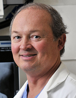 Mark P. Figgie合作论文数Orthopedic, Hospital for Special Surgery14Kethy Jules-Elysee合作论文数Hospital for Special Surgery Department of Anesthesiology 535 East 70th Street New York NY 10021 USA 535 East 70th Street New York NY 10021 USA13
 Russell E Windsor合作论文数13Allina A Nocon合作论文数12
 Stavros G. Memtsoudis合作论文数Department of Anesthesiology, Critical Care & Pain Management, Hospital for Special Surgery;Weill Cornell Medical College12
 Richard S. Laskin合作论文数Hospital for Special Surgery12Edward F. Dicarlo合作论文数Medical College, New York Hospital-Cornell University12Carol A Mancuso合作论文数Joan and Sanford I. Weill Department of Medicine, Weill Cornell Medicine, Cornell University11Pearle Andrew D合作论文数hospital for special surgery11Purdue P Edward合作论文数11PM PELLICCI合作论文数10Bryan J Nestor合作论文数10
 Yan Ma合作论文数10Malahias Michael-Alexander合作论文数2nd Orthopaedic Department, Athens University10Friedrich Bottner合作论文数10
 Edwin Su合作论文数10
 Allan E Inglis合作论文数10
 Khaled Saleh合作论文数9
 Alex Gu合作论文数8Potter H G合作论文数8
 Susan M. Goodman合作论文数Department of Rheumatology, Hospital for Special Surgery;Weill Cornell Medical College8Paul M. Pellicci合作论文数8G Go合作论文数7C S Ranawat合作论文数7Pc Harpel合作论文数CORNELL UNIV,MED CTR,COLL MED,DEPT MED,DIV HEMATOL ONCOL,NEW YORK,NY 10021, USA7
 Sakellariou Vasileios I合作论文数Athens University Medical School7Beksac Burak合作论文数Weill Medical College, Cornell University7Jerabek Seth A合作论文数7
 Andrea Baldini合作论文数7
 Panagiotis Koulouvaris合作论文数Weill Medical College, Cornell University7A Miric合作论文数7
 Lisa A. Mandl合作论文数Joan and Sanford I. Weill Department of Medicine, Weill Cornell Medicine, Cornell University6
 Mary K Crow合作论文数Joan and Sanford I. Weill Department of Medicine, Weill Cornell Medicine, Cornell University6
 Paul S Issack合作论文数6
 Triantafyllopoulos Georgios K合作论文数School of Medicine, University of Athens6
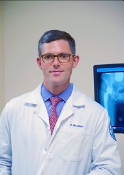 McLawhorn Alexander S合作论文数Department of Orthopedic Surgery, Hospital for Special Surgery6




