Exome (ES) or genome (GS) sequencing are recommended as first- or second-tier molecular tests for patients with developmental disorders (DD), but the clinical utility of GS continues to be debated. This prospective randomized trial involving all Belgian Human Genetics centers compared the standard of care (SoC) - combining ES and chromosomal microarray analysis or shallow GS - with GS for 567 individuals with unexplained DD. The diagnostic yield of GS was 39.8% (113/284) vs 30% for SoC (85/283) (p=0.015), mainly due to an increased detection of single nucleotide variants and indels (+8.7%). GS also enabled the detection of three non-coding (potential) pathogenic variants. Diagnostic yield was higher for females (45.5%, 97/213) compared to males (28.5%, 101/354) (p<0.001). De novo variants were found for 23.6% of patients. Analysis of inherited variants in genes associated with autosomal dominant phenotypes contributed more to the diagnostic yield (3.9%) than X-linked variants (1.9%), and to a similar extent as autosomal recessive variants (4.1%). This nationwide study indicates GS outperforms SoC for the diagnosis of patients with DD in a decentralized hospital setting and well-characterized cohort. The results also highlight the importance of evaluating autosomal dominant inherited variants in genomics analyses for DD. ### Competing Interest Statement Illumina Inc. (San Diego, CA, USA) provided the consumables for WGS of the samples used for the ring trial and the 284 probands and their parent(s) sequenced in the WGS arm, as well as access to the Emedgene platform. ### Clinical Trial NCT07051213 ### Funding Statement Illumina Inc. (San Diego, CA, USA) provided the consumables for WGS of the samples used for the ring trial and the 284 probands and their parent(s) sequenced in the WGS arm, as well as access to the Emedgene platform. This work was additionally supported by grants from the KU Leuven, C1-C14/22/125 and by FWO-TBM grant T-003819N, FWO grant G0A2622N to VJR. ### Author Declarations I confirm all relevant ethical guidelines have been followed, and any necessary IRB and/or ethics committee approvals have been obtained. Yes The details of the IRB/oversight body that provided approval or exemption for the research described are given below: The Ethical Commission of the University Hospitals of Leuven (UZ Leuven) gave ethical approval for this work (S64603). I confirm that all necessary patient/participant consent has been obtained and the appropriate institutional forms have been archived, and that any patient/participant/sample identifiers included were not known to anyone (e.g., hospital staff, patients or participants themselves) outside the research group so cannot be used to identify individuals. Yes I understand that all clinical trials and any other prospective interventional studies must be registered with an ICMJE-approved registry, such as ClinicalTrials.gov. I confirm that any such study reported in the manuscript has been registered and the trial registration ID is provided (note: if posting a prospective study registered retrospectively, please provide a statement in the trial ID field explaining why the study was not registered in advance). Yes I have followed all appropriate research reporting guidelines, such as any relevant EQUATOR Network research reporting checklist(s) and other pertinent material, if applicable. Yes Information regarding the phenotype and molecular diagnosis of study participants are shared in the supplementary files. The raw sequencing data generated during this study will be shared on EGA (EGAC00001003120).
Biallelic inactivating variants in ZNF142 underlie a clinically variable neurodevelopmental disorder. ZNF142 is a zinc-finger transcription factor with potential roles on chromatin organization, implying a possible association of ZNF142 loss of function with perturbed genome-wide DNA methylation (DNAm) pattern. We performed EPIC array-based methylation profiling of peripheral blood-derived DNA samples from 27 individuals with biallelic ZNF142 inactivating variants, together with 6 heterozygous carriers and 40 controls. A DNAm signature discovery pipeline was applied by using 440 controls for discovery and validation analyses, and a machine-learning model was trained to classify 8 individuals carrying ZNF142 variants of uncertain clinical significance. Analyses directed to explore the genome-wide DNAm landscape in affected individuals revealed 88 differentially methylated probes constituting the minimal informative set specific to ZNF142 loss of function. This reproducible pattern of DNAm changes involved regulatory regions of a small number of genes. The DNAm signature derived from peripheral blood allowed us to diagnose individuals carrying biallelic inactivating ZNF142 variants when applied to fibroblasts. Our findings provide evidence that biallelic loss-of-function ZNF142 variants result in a specific and robust DNAm signature. The identified DNAm pattern suggests occurrence of a methylation disturbance involving a small number of loci that appears to be shared by different cell lineages.
The orchestration of fetal kidney development involves the precise control of numerous genes, including HNF1A, HNF1B and PKHD1. Understanding the genetic factors influencing fetal kidney development is essential for unraveling the complexities of renal disorders. This study aimed to search for disease-causing variants in HNF1A, HNF1B, PKHD1 genes, among fetus and babies or via parental samples, using sanger sequencing, NGS technologie and MLPA. The study revealed an absence of gene deletions and disease-causing variants in the HNF1B gene. However, five previously SNPs in the HNF1A gene were identified in four patients (patients 1, 2, 3, and 4). These include c.51C > G (Exon1, p. Leu17=), c.79A > C (Exon1, p. Ile27Leu), c.1375C > T (Exon7, p. Leu459=), c.1460G > A (Exon7, p. Ser487Asn), and c.1501 + 7G > A (Intron7). Additionally, in addition to previously SNPs identified, a de novo heterozygous missense mutation (p.E508K) was detected in patient 4. Furthermore, a heterozygous mutation in exon 16 (p. Arg494*; c.1480C >T) was identified in both parents of patient 5, allowing predictions of fetal homozygosity. Bioinformatic analyses predicted the effects of the c.1522G > A mutation (p.E508K) on splicing processes, pre-mRNA structures, and protein instability and conformation. Similarly, the c.1480C > T mutation (p. Arg494*) was predicted to introduce a premature codon stop, leads to the production of a shorter protein with altered or impaired function. Identification of variants in the HNF1A and in PKHD1 genes provides valuable insights into the genetic landscape of renal abnormalities in affected patients. These findings underscore the heterogeneity of genetic variants contributing to renal disorders and emphasize the importance of genetic screening.
Background and ObjectivesBRAT1 encephalopathy is an ultra-rare autosomal recessive neonatal encephalopathy. We delineate the neonatal electroclinical phenotype at presentation and provide insights for early diagnosis.MethodsThrough a multinational collaborative, we studied a cohort of neonates with encephalopathy associated with biallelic pathogenic variants in BRAT1 for whom detailed clinical, neurophysiologic, and neuroimaging information was available from the onset of symptoms. Neuropathologic changes were also analyzed.ResultsWe included 19 neonates. Most neonates were born at term (16/19) from nonconsanguineous parents. 15/19 (79%) were admitted soon after birth to a neonatal intensive care unit, exhibiting multifocal myoclonus, both spontaneous and exacerbated by stimulation. 7/19 (37%) had arthrogryposis at birth, and all except 1 progressively developed hypertonia in the first week of life. Multifocal myoclonus, which was present in all but 1 infant, was the most prominent manifestation and did not show any EEG correlate in 16/19 (84%). Video-EEG at onset was unremarkable in 14/19 (74%) infants, and 6 (33%) had initially been misdiagnosed with hyperekplexia. Multifocal seizures were observed at a median age of 14 days (range: 1-29). During the first months of life, all infants developed progressive encephalopathy, acquired microcephaly, prolonged bouts of apnea, and bradycardia, leading to cardiac arrest and death at a median age of 3.5 months (range: 20 days to 30 months). Only 7 infants (37%) received a definite diagnosis before death, at a median age of 34 days (range: 25-126), and almost two-thirds (12/19, 63%) were diagnosed 8 days to 12 years postmortem (median: 6.5 years). Neuropathology examination, performed in 3 patients, revealed severely delayed myelination and diffuse astrogliosis, sparing the upper cortical layers.DiscussionBRAT1 encephalopathy is a neonatal-onset, rapidly progressive neurologic disorder. Neonates are often misdiagnosed as having hyperekplexia, and many die undiagnosed. The key phenotypic features are multifocal myoclonus, an organized EEG, progressive, persistent, and diffuse hypertonia, and an evolution into refractory multifocal seizures, prolonged bouts of apnea, bradycardia, and early death. Early recognition of BRAT1 encephalopathy allows for prompt workup, appropriate management, and genetic counseling.
Objectives: A growing variety of genes involved in pathologies causing severe encephalopathy with neonatal-onset epilepsy are being discovered. Recently, autosomal recessive mutations in the BRAT1 gene have been associated with clinical spectrum ranging from lethal neonatal rigidity and multifocal seizures to later onset and less-severe symptoms including developmental delay, dysautonomia, and epilepsy.
PTH resistance is characterized by elevated parathyroid hormone (PTH) levels, hypocalcemia, hyperphosphatemia and it is classically associated with GNAS locus genetic or epigenetic defects. Inactivating PTH/PTHrP signaling disorders (iPPSD) define overlapping phenotypes based on their molecular etiology. iPPSD1 is associated with PTH1R variants and variable phenotypes including ossification anomalies and primary failure of tooth eruption but no endocrine disorder. Here we report on a 10-month-old child born from consanguineous parents, who presented with mild neurodevelopmental delay, seizures, enlarged fontanelles, round face, and bilateral clinodactyly. Hand x-rays showed diffuse delayed bone age, osteopenia, short metacarpal bones and cone-shaped distal phalanges. A diagnosis of PTH resistance was made on the basis of severe hypocalcemia, hyperphosphatemia, elevated PTH and normal vitamin D levels on blood sample. The patient was treated with calcium carbonate and alfacalcidol leading to rapid bio-clinical improvement. Follow-up revealed multiple agenesis of primary teeth and delayed teeth eruption, as well as Arnold-Chiari type 1 malformation requiring a ventriculoperitoneal shunt placement. GNAS gene analysis showed no pathogenic variation, but a likely pathogenic homozygous substitution c.723C>G p.(Asp241Glu) in PTH1R gene was found by trio-based whole exome sequencing. We studied the deleterious impact of the variant on the protein conformation with bioinformatics tools. In conclusion, our study reports for the first time PTH resistance in a child with a biallelic PTH1R mutation, extending thereby the clinical spectrum of iPPSD1 phenotypes.
Biallelic variants of the gene encoding for the zinc-finger protein 142 (ZNF142) have recently been associated with intellectual disability (ID), speech impairment, seizures, and movement disorders in nine individuals from five families. In this study, we obtained phenotype and genotype information of 26 further individuals from 16 families. Among the 27 different ZNF142 variants identified in the total of 35 individuals only four were missense. Missense variants may give a milder phenotype by changing the local structure of ZF motifs as suggested by protein modeling; but this correlation should be validated in larger cohorts and pathogenicity of the missense variants should be investigated with functional studies. Clinical features of the 35 individuals suggest that biallelic ZNF142 variants lead to a syndromic neurodevelopmental disorder with mild to moderate ID, varying degrees of delay in language and gross motor development, early onset seizures, hypotonia, behavioral features, movement disorders, and facial dysmorphism. The differences in symptom frequencies observed in the unpublished individuals compared to those of published, and recognition of previously underemphasized facial features are likely to be due to the small sizes of the previous cohorts, which underlines the importance of larger cohorts for the phenotype descriptions of rare genetic disorders.
Mandibular hypoplasia, Deafness, Progeroid features, and Lipodystrophy (MDPL) syndrome is a rare autosomal dominant disorder caused by mutations in POLD1 gene and characterized by mandibular hypoplasia, deafness, progeroid features and lipodystrophy. One recurrent mutation p.(Ser605del) was reported in almost all affected patients. We report a novel de novo c.3214A>C p.(Thr1072Pro) variant in POLD1 in a 28-year-old male with MDPL syndrome. We provide a clinical description, molecular/immunohistological results, and literature review.
Purpose Despite a few recent reports of patients harboring truncating variants in NSD2, a gene considered critical for the Wolf-Hirschhorn syndrome (WHS) phenotype, the clinical spectrum associated with NSD2 pathogenic variants remains poorly understood. Methods We collected a comprehensive series of 18 unpublished patients carrying heterozygous missense, elongating, or truncating NSD2 variants; compared their clinical data to the typical WHS phenotype after pooling them with ten previously described patients; and assessed the underlying molecular mechanism by structural modeling and measuring methylation activity in vitro. Results The core NSD2-associated phenotype includes mostly mild developmental delay, prenatal-onset growth retardation, low body mass index, and characteristic facial features distinct from WHS. Patients carrying missense variants were significantly taller and had more frequent behavioral/psychological issues compared with those harboring truncating variants. Structural in silico modeling suggested interference with NSD2's folding and function for all missense variants in known structures. In vitro testing showed reduced methylation activity and failure to reconstitute H3K36me2 in NSD2 knockout cells for most missense variants. Conclusion NSD2 loss-of-function variants lead to a distinct, rather mild phenotype partially overlapping with WHS. To avoid confusion for patients, NSD2 deficiency may be named Rauch-Steindl syndrome after the delineators of this phenotype.
Background Participation in quality controls, also called external quality assessment (EQA) schemes, is required for the ISO15189 accreditation of the Medical Centers of Human Genetics. However, directives on the minimal frequency of participation in genetic quality control schemes are lacking or too heterogeneous, with a possible impact on health care quality. Objective The aim of this project is to develop Belgian guidelines on the frequency of participation in quality controls for genetic testing in the context of rare diseases. Methods A group of experts analyzed 90 EQA schemes offered by accredited providers and focused on analyses used for the diagnosis of rare diseases. On that basis, the experts developed practical recommendations about the minimal frequencies of participation of the Medical Centers of Human Genetics in quality controls and how to deal with poor performances and change management. These guidelines were submitted to the Belgian Accreditation Body and then reviewed and approved by the Belgian College of Human Genetics and Rare Diseases and by the National Institute for Health and Disability Insurance. Results The guidelines offer a decisional algorithm for the minimal frequency of participation in human genetics EQA schemes. This algorithm has been developed taking into account the scopes of the EQA schemes, the levels of experience, and the annual volumes of the Centers of Human Genetics in the performance of the tests considered. They include three key principles: (1) the recommended annual assessment of all genetic techniques and technological platforms, if possible through EQAs covering the technique, genotyping, and clinical interpretation; (2) the triennial assessment of the genotyping and interpretation of specific germline mutations and pharmacogenomics analyses; and (3) the documentation of actions undertaken in the case of poor performances and the participation to quality control the following year. The use of a Bayesian statistical model has been proposed to help the Centers of Human Genetics to determine the theoretical number of tests that should be annually performed to achieve a certain threshold of performance (eg, a maximal error rate of 1%). Besides, the guidelines insist on the role and responsibility of the national public health authorities in the follow-up of the quality of analyses performed by the Medical Centers of Human Genetics and in demonstrating the cost-effectiveness and rationalization of participation frequency in these quality controls. Conclusions These guidelines have been developed based on the analysis of a large panel of EQA schemes and data collected from the Belgian Medical Centers of Human Genetics. They are applicable to other countries and will facilitate and improve the quality management and financing systems of the Medical Centers of Human Genetics.
Purpose The variant spectrum and the phenotype of X-linked Kabuki syndrome type 2 (KS2) are poorly understood. Methods Genetic and clinical details of new and published individuals with pathogenic KDM6A variants were compiled and analyzed. Results Sixty-one distinct pathogenic KDM6A variants (50 truncating, 11 missense) from 80 patients (34 males, 46 females) were identified. Missense variants clustered in the TRP 2, 3, 7 and Jmj-C domains. Truncating variants were significantly more likely to be de novo. Thirteen individuals had maternally inherited variants and one had a paternally inherited variant. Neonatal feeding difficulties, hypoglycemia, postnatal growth retardation, poor weight gain, motor delay, intellectual disability (ID), microcephaly, congenital heart anomalies, palate defects, renal malformations, strabismus, hearing loss, recurrent infections, hyperinsulinism, seizures, joint hypermobility, and gastroesophageal reflux were frequent clinical findings. Facial features of over a third of patients were not typical for KS. Males were significantly more likely to be born prematurely, have shorter stature, and severe developmental delay/ID. Conclusion We expand the KDM6A variant spectrum and delineate the KS2 phenotype. We demonstrate that the variability of the KS2 phenotypic depends on sex and the variant type. We also highlight the overlaps and differences between the phenotypes of KS2 and KS1.
Although the prognosis of CHARGE syndrome can be highly variable from mild until severe, final diagnosis is difficult to establish in utero. The aim of our study is to compare antenatal and postnatal findings in a retrospective cohort of 10 successive patients with a positive CHD7 gene variant in order to identify the specific prenatal features for CHARGE syndrome diagnosis. Fetal ultrasound, follow-up and supplementary investigations are collected and compared to postnatal findings. Congenital heart defect (7/10), choanal atresia (7/10) and tracheoesophageal atresia (4/10) are the most frequent fetal anomalies found. Inner and external ear anomalies appear as the keystone (constant features) for prenatal diagnosis of CHARGE syndrome in fetuses with multiple anomalies and normal microarray karyotype. External ear malformations are identified in all cases by 3D ultrasound when carefully evaluated. MRI and temporal bone CT-Scan are second line useful tools to assess the diagnosis when looking for semicircular canal agenesis, arhinencephaly and/or choanal atresia. Before availability of prenatal exome sequencing in clinical routine, present findings lead to the recommendation that fetuses, with congenital heart defect (mainly septal and conotruncal), cleft lip/palate or unexplained polyhydramnios should carefully be screened for clues suggesting CHARGE syndrome using 2D and 3D ultrasound, MRI and temporal bone CT-Scan. When CHARGE syndrome is suspected with normal molecular karyotype, CHD7 gene sequencing must be offered.
Mental deficiency, epilepsy, hypogonadism, microcephaly, and obesity syndrome is a severe X-linked syndrome caused by pathogenic variants in EIF2S3. The gene encodes the γ subunit of the eukaryotic translation initiation factor-2, eIF2, essential for protein translation. A recurrent frameshift variant is described in severely affected patients while missense variants usually cause a moderate phenotype. We identified a novel missense variant (c.433A>G, p.(Met145Val)) in EIF2S3 in a mildly affected patient. Studies on zebrafish confirm the pathogenicity of this novel variant and three previously published missense variants. CRISPR/Cas9 knockout of eif2s3 in zebrafish embryos recapitulate the human microcephaly and show increased neuronal cell death. Abnormal high glucose levels were identified in mutant embryos, caused by beta cell and pancreatic progenitor deficiency, not related to apoptosis. Additional studies in patient-derived fibroblasts did not reveal apoptosis. Our results provide new insights into disease physiopathology, suggesting tissue-dependent mechanisms.
 Isabelle Maystadt合作论文数Inst Pathol & Genet, Ctr Genet Humaine, Gosselies, Belgium24
 Lederer Damien合作论文数Centre de Génétique Humaine, Institut de Pathologie et Génétique, Charleroi, Belgium. damien.lederer@ipg.be20
 Vincent Bours合作论文数Department of Oncology, University of Liège16MP Merville合作论文数9
 Stephanie Moortgat合作论文数8
 Marie Paule Merville合作论文数8X. De Leval合作论文数7Pascale Hilbert合作论文数6J Boyer合作论文数6Deprez Marie合作论文数6
 Alain Chariot合作论文数Centre Hospitalier Universitaire de Liège, University of Liège6Jean-Michel Dogné合作论文数6
 Bernard Pirotte合作论文数Laboratory of Medicinal Chemistry, University of Liège6
 Deniz Karadurmus合作论文数5Jm Meignen合作论文数4Joset Pascal合作论文数4
 Olivier Monestier合作论文数UMR1061 Unité de Génétique Moléculaire Animale, INRA4
 Anita Rauch合作论文数Institute of Medical Genetics, University of Zurich4
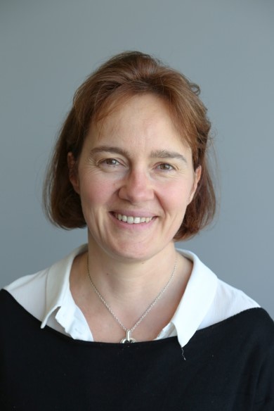 Faivre Laurence合作论文数Faculty of Medicine, University of Bourgogne;Medical Genetics Department, Hopital d'Enfants;Genetics Centre, Children's Hospital, Dijon University Hospital4
 Laurence Mercier合作论文数4
 Marie-Cécile Nassogne合作论文数Cliniques Universitaires Saint-Luc4Anne Valette合作论文数4Sandrine Mary合作论文数3Brigitte Rousseau合作论文数3K Dahan合作论文数3Anne Destrée合作论文数3
 Geert Mortier合作论文数3Cassandre Bedu-Ferrari合作论文数3Shayne F Andrew合作论文数3De Schepper Jean合作论文数3
 Rikke S Møller合作论文数Department of Regional Health Research, University of Southern Denmark;Institut for Regional Sundhedsforskning, Syddansk Universitet;Department of Epilepsy Genetics and Personalized Medicine, Danish Epilepsy Centre3Beatrice Oneda合作论文数3Alain Verloes合作论文数3
 Sarah Weckhuysen合作论文数From the Department of Neurology (F.P.), and Service of Genetic Medicine (S.E.A.),, University Hospitals of Geneva;Institute of Genetics and Genomics of Geneva (S.E.A.),, University of Geneva;Institut national de la santé et de la recherche médicale (INSERM) (V.N.;Sorbonne Universités, UPMC Univ Paris 06;CNRS (V.N., S.I., Hôpital de la Pitié-Salpêtrière;and, AP-HP Groupe hospitalier Pitié-Salpêtrière;Epilepsy, Sleep and Pediatric Neurophysiology, University Hospitals of Lyon;Hospices Civils de Lyon (D.V.),, centre de référence déficiences intellectuelles de causes rares et sclérose tubéreuse de Bourneville;Neurogenetics Group, VIB;Institute Born-Bunge, University of Antwerp;Epilepsy Centre Kempenhaeghe (S.W.),;Algemeen Stedelijk Ziekenhuis (E.F.),;Antwerp University Hospital, Antwerp University;Centre hospitalier général de Valence (M.V.R.);Department of Medical Genetics (G.L.),, Hospices Civils de Lyon;Claude Bernard Lyon I University;INSERM U1028, CRNL (G.L.),;Centre de référence épilepsies rares et Sclérose tubéreuse de Bourneville (I.A.-G., M.B.);and DNA and Cell Bank (P.C.),, Hôpital Pitié-Salpêtrière;Department of Surge;University of Geneva, University Hospitals of Geneva3Castronovo Vincenzo合作论文数3Jean-Philippe Bonjour合作论文数3
 Pasquale Striano合作论文数Center of Experimental and Translational Myology, Dipartimento Di Neuroscienze, Riabilitazione, Oftalmologia, Genetica E Scienze Materno-Infantili, Università Degli Studi Di Genova;IRCCS 'G. Gaslini'3
 Verellen-Dumoulin合作论文数Center for Human Genetics, Institut de Pathologie et de Génétique3Deborah J. Shears合作论文数3
 Rolph Pfundt合作论文数Department of Human Genetics, Radboud University Medical Centre3Biserka Relic合作论文数3
 Marwan Shinawi合作论文数Genetics and Genomic Medicine, Washington University School of Medicine3
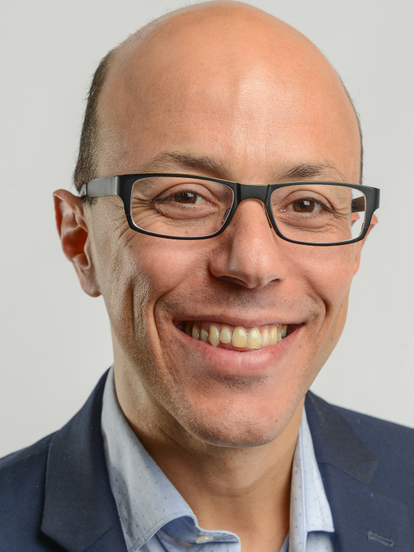 Mohamed Bentires-Alj合作论文数Department of Biomedicine, University of Basel;University Hospital Basel3Boris Keren合作论文数3
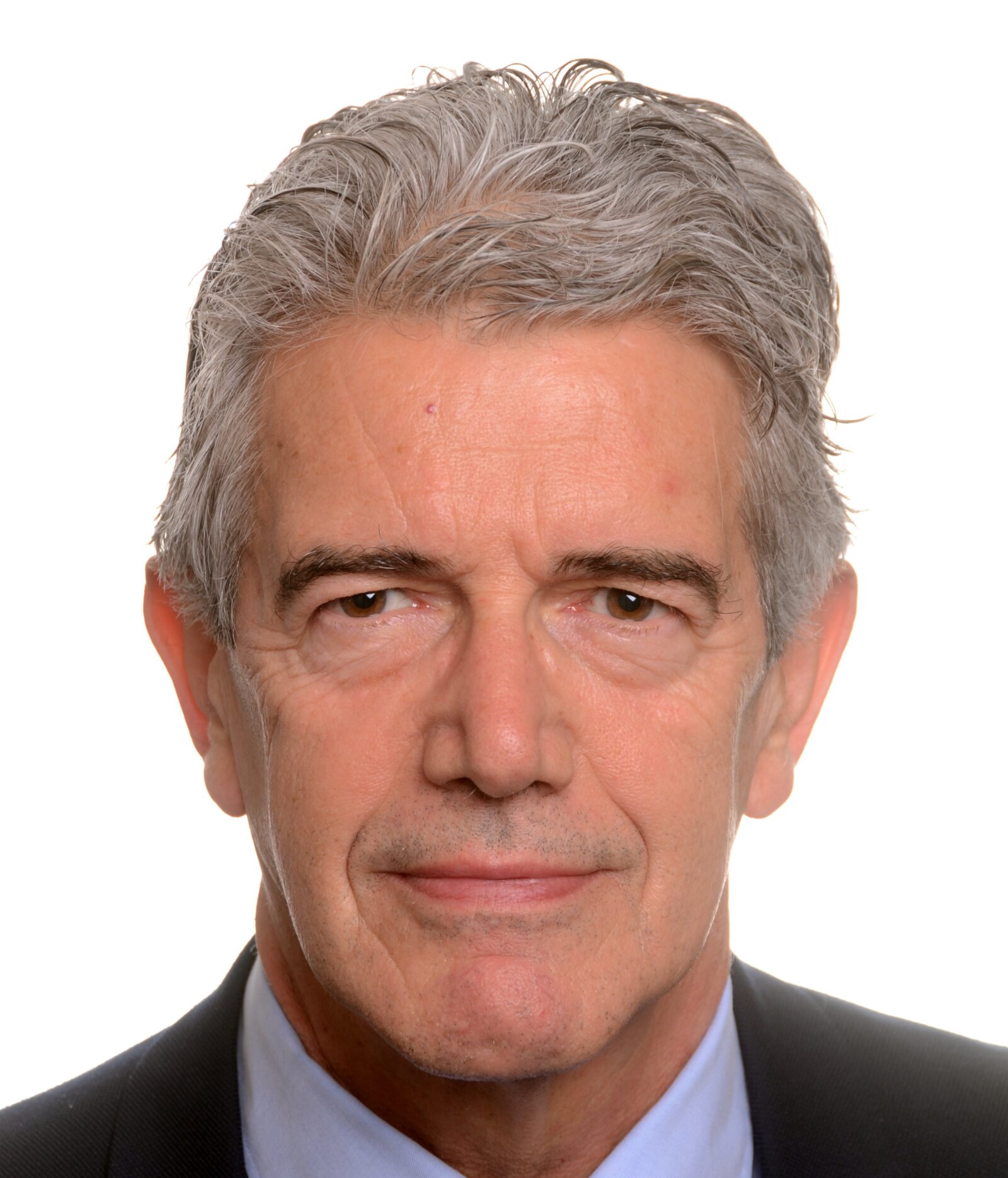 Rudy Van Coster合作论文数Ghent University Hospital3Jacques David合作论文数3Bernard Grisart合作论文数3
 Bert Callewaert合作论文数Department of Biomolecular Medicine, Ghent University3
 Berten Ceulemans合作论文数Antwerp University Hospital;Division of Pediatric Neurology, Universitair Ziekenhuis Antwerpen3




