The three-dimensional structure of the enzyme catalase from Penicillium vitale in a complex with the inhibitor aminotriazole was solved and refined by protein X-ray crystallography methods. An analysis of the three-dimensional structure of the complex showed that the inhibition of the enzyme occurs as a result of the covalent binding of aminotriazole to the amino-acid residue His64 in the active site of the enzyme. An investigation of the three-dimensional structure of the complex resulted in the amino-acid residues being more precisely identified. The binding sites of saccharide residues and calcium ions in the protein molecule were found.
A protealysin precursor (the enzyme of the peptidase family M4) was crystallized for the first time. The crystal-growth conditions were found, and single crystals of the protein with dimensions of 0.3–0.5 mm were grown. The preliminary X-ray diffraction study of the enzyme was performed. The protealysin precursor was shown to crystallize in two crystal modifications suitable for the X-ray diffraction study of the three-dimensional structure of the protein molecule at atomic resolution.
The three-dimensional structure of the complex of the enzyme SAICAR synthase with analogues of natural substrates, namely, phosphoribosylaminoimidazolecarboxamide (AICAR) and succinic acid, was determined and refined by methods of protein crystallography. Two AICAR-binding sites were revealed in the protein molecule. One of these sites is located in the active center of the enzyme in the vicinity of the ATP-binding site, which has been found in the complex of SAICAR synthase with ATP that had been studied earlier. The second AICAR-binding site is located at the periphery of the protein molecule and coincides with the additional ATP-binding site present in the complexes studied earlier. The binding site of succinic acid was revealed in the active center of the enzyme in the vicinity of the AICAR molecule. The electron density distribution for the AICAR molecule in the active center is indicative of the possible lability of the atomic group of the adenine base.
The three-dimensional structure of the complex of the enzyme SAICAR synthase with the product of the enzymatic reaction, SAICAR, was solved and refined by methods of protein crystallography. The SAICAR-binding site in the active site of the enzyme was found. The amino-acid residues providing the binding of the reaction product with the protein were revealed. These residues were compared with those involved in the substrate binding in the complex with AICAR and succinic acid studied earlier.
The crystal structures of two forms of the enzyme dimanganese catalase from Thermus Thermophilus (native and inhibited by chloride) were studied by X-ray diffraction analysis at 1.05 and 0.98 Å resolution, respectively. The atomic models of the molecules were refined to the R factors 9.8 and 10%, respectively. The three-dimensional molecular structures are characterized in detail. The analysis of electron-density distributions in the active centers of the native and inhibited enzyme forms revealed that the most flexible side chains of the amino acid residues Lys162 and Glu36 exist in two interrelated conformations. This allowed us to obtain the structural data necessary for understanding the mechanism of enzymatic activity of the dimanganese catalase.
Most of the amino acid side chains of beef liver catalase were clearly identifiable in the 2.5 R resol.utionelectron density map and are in good agreement with the sequence (W. A. Schroeder et al., Arch.Biochem.Biophys.131, 653-655, 1969).Thetertiary structure of one subunit consists of a large antiparallel S-pleated sheet domain with helica~ insertions followed by a smaller domain containing four ci:helices.
 A. I. Grebenko合作论文数8
 Vladimir V Barynin合作论文数8
 Victor Lamzin合作论文数European Molecular Biology Laboratory (EMBL),, c/o DESY6Vladimir M. Levdikov合作论文数4
 Svetlana V. Antonyuk合作论文数Molecular Biophysics Group, CCLRC Daresbury Laboratory4D.V. Urusova合作论文数3AA VAGIN合作论文数3BK VAINSHTEIN合作论文数2
 Alexander N. Popov合作论文数European Synchrotron Radiat Facil2
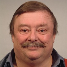 Keith S Wilson合作论文数Department of Chemistry, University of York2
 Garib N Murshudov (G. N. Murshudov)合作论文数The Medical Research Council Laboratory of Molecular Biology2T. Yu. Gromova合作论文数1Pd Hempstead合作论文数1Bk Vainshtein合作论文数1AI GREBENKO合作论文数1
 a borovik合作论文数Uzhgorod Natl Univ1
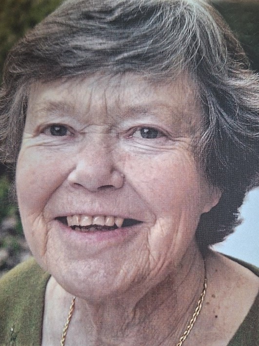 Pauline May Harrison合作论文数Department of Molecular Biology and Biotechnology, The University of Sheffield1M. E. Andrianova合作论文数1
 Ilya V Demidyuk合作论文数Institute of Molecular Genetics, Russian Academy of Sciences1Sergey V. Kostrov合作论文数1Sergei V. Khangulov合作论文数1N. I. Sosfenov合作论文数1
 Inna Petrovna Kuranova合作论文数1A. N. Popov合作论文数1Albina I Grebenko合作论文数1




