Senescence plays a key role in various physiological and pathological processes. We reported that injury-induced transient senescence correlates with heart regeneration, yet the multi-omics profile and molecular underpinnings of regenerative senescence remain obscure. Using proteomics and single-cell RNA sequencing, here we report the regenerative senescence multi-omic signature in the adult mouse heart and establish its role in neonatal heart regeneration and agrin-mediated cardiac repair in adult mice. We identified early growth response protein 1 (Egr1) as a regulator of regenerative senescence in both models. In the neonatal heart, Egr1 facilitates angiogenesis and cardiomyocyte proliferation. In adult hearts, agrin-induced senescence and repair require Egr1, activated by the integrin-FAK-ERK-Akt1 axis in cardiac fibroblasts. We also identified cathepsins as injury-induced senescence-associated secretory phenotype components that promote extracellular matrix degradation and potentially assist in reducing fibrosis. Altogether, we uncovered the molecular signature and functional benefits of regenerative senescence during heart regeneration, with Egr1 orchestrating the process. Zhang et al. show that Egr1 regulates transient senescence during neonatal heart regeneration and upon agrin-mediated cardiac repair in adult mice, acting downstream of the integrin-FAK-ERK-Akt1 axis in cardiac fibroblasts.
The p53 tumor suppressor, encoded by the TP53 gene, serves as a major barrier against malignant transformation. Patients with Li-Fraumeni syndrome (LFS) inherit a mutated TP53 allele from one parent and a wild-type TP53 allele from the other. Subsequently, the wild-type allele is lost and only the mutant TP53 allele remains. This process, which is termed loss of heterozygosity (LOH), results in only mutant p53 protein expression. We used primary dermal fibroblasts from LFS patients carrying the hotspot p53 gain-of-function pathogenic variant, R248Q to study the LOH process and characterize alterations in various pathways before and after LOH. We previously described the derivation of mutant p53 reactivating peptides, designated pCAPs (p53 Conformation Activating Peptides). In this study, we tested the effect of lead peptide pCAP-250 on LOH and on its associated cellular changes. We report that treatment of LFS fibroblasts with pCAP-250 prevents the accumulation of mutant p53 protein, inhibits LOH, and alleviates its cellular consequences. Furthermore, prolonged treatment with pCAP-250 significantly reduces DNA damage and restores long-term genomic stability. pCAPs may thus be contemplated as a potential preventive treatment to prevent or delay early onset cancer in carriers of mutant p53.
Tumor suppressor p53 (TP53) is frequently mutated in cancer, often resulting not only in loss of its tumor-suppressive function but also acquisition of dominant-negative and even oncogenic gain-of-function traits. While wild-type p53 levels are tightly regulated, mutants are typically stabilized in tumors, which is crucial for their oncogenic properties. Here, we systematically profiled the factors that regulate protein stability of wild-type and mutant p53 using marker-based genome-wide CRISPR screens. Most regulators of wild-type p53 also regulate p53 mutants, except for p53 R337H regulators, which are largely private to this mutant. Mechanistically, FBXO42 emerged as a positive regulator for a subset of p53 mutants, working with CCDC6 to control USP28-mediated mutant p53 stabilization. Additionally, C16orf72/HAPSTR1 negatively regulates both wild-type p53 and all tested mutants. C16orf72/HAPSTR1 is commonly amplified in breast cancer, and its overexpression reduces p53 levels in mouse mammary epithelium leading to accelerated breast cancer. This study offers a network perspective on p53 stability regulation, potentially guiding strategies to reinforce wild-type p53 or target mutant p53 in cancer.
Supplementary Figure Legend from hTERT-Immortalized Prostate Epithelial and Stromal-Derived Cells: an Authentic In vitro Model for Differentiation and Carcinogenesis
Supplementary Figure 2 from Transcriptional Programs following Genetic Alterations in p53, INK4A, and H-Ras Genes along Defined Stages of Malignant Transformation
Supplementary Figures 6-13 from p53 Regulates the Ras Circuit to Inhibit the Expression of a Cancer-Related Gene Signature by Various Molecular Pathways
Supplementary Figure S1 from hTERT-Immortalized Prostate Epithelial and Stromal-Derived Cells: an Authentic In vitro Model for Differentiation and Carcinogenesis
Supplementary Figure 4 from Transcriptional Programs following Genetic Alterations in <i>p53</i>, <i>INK4A</i>, and <i>H-Ras</i> Genes along Defined Stages of Malignant Transformation
Supplementary Materials & Methods from hTERT-Immortalized Prostate Epithelial and Stromal-Derived Cells: an Authentic In vitro Model for Differentiation and Carcinogenesis
Differential genes between pMSCs and their derived TLs (MSC TLs). The table contains genes that had at least 10 reads in the RNA sequencing analysis. CSC genes, ESC genes, shared target genes of Nanog, Sox2 and Oct4 or of Nanog, Sox2, Oct4, Dax1 and Nac1 and genes induced by the reprogramming process are indicated and can be filtered.
IPA analysis of the differential genes between pMSCs and their derived TLs (MSC TLs). Biological function was determined for the genes that were downregulated in p53 Mut MSC TLs compared to their parental, p53 Mut pMSC. Biological function, Pathways, upstream regulators and networks were determined for the genes that were upregulated in p53 Mut MSC TLs compared to their parental. (The threshold of significant genes; padj<0.05, -2>FC>2, max reads>10)
Supplementary Figure 1. Mutant p53 enhanced the self-renewal capacity and oncogenicity of primary bone marrow MSCs. Supplementary Figure 2. Mutant p53 MSC derived tumor lines exhibited augmented tumorigenic capacity compared with their parental cells. Supplementary Figure 3. Transcriptional differences between pMSCs and their derived MSC TLs. Supplementary Figure 4. Knocking out mutant p53, by CRISPR gene editing system, in a MSC TL led to a reduction in tumor vascularization. Supplementary Figure 5. p53 Mut MSC TL sub-clones highly expressed ESC signature derived genes and the p53 Mut dependent genes in MSC TL sub-clones are enriched for chromatin transcriptional activation marks in ESCs. Supplementary Figure 6. The MSC-TLs ESC signature derived genes are associated with poor patient survival. Supplementary Figure 7. Six hotspot p53 mutations frequency in TCGA datasets. Supplementary Figure 8. ESC gene expression signature entailed in the MSC TLs is detected in human tumors harboring p53 missense mutations. Supplementary Table 1 List of QRT PCR primers. Supplementary Table 7 Description of TCGA samples.
Li-Fraumeni syndrome (LFS) is a hereditary cancer predisposition syndrome associated with germline TP53 pathogenic variants. Here, we perform whole-genome sequence (WGS) analysis of tumors from 22 patients with TP53 germline pathogenic variants. We observe somatic mutations affecting Wnt, PI3K/AKT signaling, epigenetic modifiers and homologous recombination genes as well as mutational signatures associated with prior chemotherapy. We identify near-ubiquitous early loss of heterozygosity of TP53 , with gain of the mutant allele. This occurs earlier in these tumors compared to tumors with somatic TP53 mutations, suggesting the timing of this mark may distinguish germline from somatic TP53 mutations. Phylogenetic trees of tumor evolution, reconstructed from bulk and multi-region WGS, reveal that LFS tumors exhibit comparatively limited heterogeneity. Overall, our study delineates early copy number gains of mutant TP53 as a characteristic mutational process in LFS tumorigenesis, likely arising years prior to tumor diagnosis.
Supplementary Figure 1 from Transcriptional Programs following Genetic Alterations in p53, INK4A, and H-Ras Genes along Defined Stages of Malignant Transformation
Supplementary Figure 1 from Mutant p53 Protects Cells from 12-O-Tetradecanoylphorbol-13-Acetate–Induced Death by Attenuating Activating Transcription Factor 3 Induction
Supplementary Tables 1-4 from Mutant p53 Protects Cells from 12-O-Tetradecanoylphorbol-13-Acetate–Induced Death by Attenuating Activating Transcription Factor 3 Induction
Supplementary Figure 3 from Transcriptional Programs following Genetic Alterations in <i>p53</i>, <i>INK4A</i>, and <i>H-Ras</i> Genes along Defined Stages of Malignant Transformation
Supplementary Figure Legends from Transcriptional Programs following Genetic Alterations in p53, INK4A, and H-Ras Genes along Defined Stages of Malignant Transformation
The MSC-TLs ESC signature derived genes are associated with poor patient survival. Kaplan-Meier analyses of the indicated data sets showing significant relationship between expression levels of genes, derived from the MSC-TLs ESC signature, and patient survival. Supplementary
Supplementary Figure 2 from Transcriptional Programs following Genetic Alterations in <i>p53</i>, <i>INK4A</i>, and <i>H-Ras</i> Genes along Defined Stages of Malignant Transformation
 Naomi Goldfinger合作论文数72
 Moshe Oren合作论文数Department of Molecular Cell Biology,Weizmann Institute of Science51
 Michael Milyavsky合作论文数Department of Pathology, Sackler Faculty of Medicine, Tel Aviv University45
 Hilla Solomon合作论文数36
 Igor Shats合作论文数Department of Molecular Cell Biology, The Weizmann Institute of Science34
 Yossi Buganim (尤西·步加尼姆)合作论文数Hebrew Univesity of Jerusalem-Hadassah Medical School;Faculty of Medicine, The Hebrew University of Jerusalem33
 Ran Brosh合作论文数Department of Molecular Cell Biology;Weizmann Institute of Science;Department of Molecular Cell Biology, Weizmann Institute of Science32
 Shlomi (Shalom) Madar合作论文数Dept Mol Cell Biol, Weizmann Inst Sci31Alina Molchadsky合作论文数31
 Neta Erez合作论文数Department of Molecular Cell Biology, The Weizmann Institute of Science27
 Ido Goldstein合作论文数25Perry Stambolsky合作论文数22
 Curtis C. Harris合作论文数Laboratory of Human Carcinogenesis, Center for Cancer Research, National Cancer Institute, National Institutes of Health21
 Yuval Tabach合作论文数Department of Physics;Weizmann Institute of Science;Department of Physics, Weizmann Institute of Science20Dov Schwartz合作论文数Department of Biochemistry;Department of Molecular Cell Biology;Weizmann Institute of Science20Eyal Kalo合作论文数19Ira Kogan合作论文数18Ronit Aloni-Grinstein合作论文数17Domany, Eytan合作论文数16Nathan Trainin合作论文数16Noa Rivlin合作论文数Dept Mol Cell Biol, Weizmann Inst Sci16Gabriela Koifman合作论文数15Ira Kogan-Sakin合作论文数15Marina Kalis合作论文数15
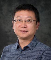 Xiaohu Tang合作论文数Michigan Technol Univ, Dept Biol Sci, Houghton, MI 49931 USA14Doron Ginsberg合作论文数14
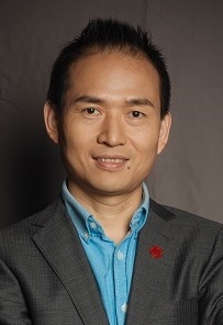 Xisong Ke (柯细松)合作论文数Institute of Interdisciplinary Integrative Medicine Research, Shanghai University of Traditional Chinese Medicine13Shetzer Yoav合作论文数13Lilach Weisz合作论文数13Anne Margrete Øyan合作论文数13
 Karl-Henning Kalland合作论文数13R Wolkowicz合作论文数12Irit Zurer合作论文数11
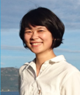 Yi Qu (屈祎)合作论文数Institute of Interdisciplinary Integrative Medicine Research, Shanghai University of Traditional Chinese Medicine10Stein Yan合作论文数10
 Irun R. Cohen合作论文数Department of Immunology and Regenerative Biology, Weizmann Institute of Science10
 Perry Tal合作论文数9Yoach Rais合作论文数The Department of Molecular Genetics, Weizmann Institute of Science9
 Gad Shaulsky合作论文数Department of Molecular and Human Genetics, Baylor College of Medicine9Johannes Herkel合作论文数9
 Amnon Peled合作论文数Goldyne Savad;Institute of Gene Therapy;Hadassah University Hospital;Institute of Gene Therapy, Hadassah University Hospital9
 J.A. Schalken合作论文数Faculty of Medical Sciences, Radboud University;Radboud University Medical Center8
 Ido Nachmany合作论文数8Yehudit Cohen合作论文数8
 David Reisman合作论文数Department of Chemistry, Bar-Ilan University8Giuseppe Lonetto合作论文数7Nava Almog合作论文数7Shay Eizenberger合作论文数7




