IntroductionThe crosstalk between immune cells through plasma extracellular vesicles (EVs) during SARS-CoV-2 infection may represent a significant determinant of clinical course in COVID-19 patients. EVs from SARS-CoV-2 virus-infected cells deliver their informational content to immune cells implicated in COVID-19 pathogenesis, thereby modulating pro-inflammatory immune responses during infection. γδ T cells are innate cells known for their pleiotropic properties spanning both innate and adaptive immunity and for their possible contribution to inflammation. This study aimed to characterize the biophysical profile and protein content of EVs derived from patients with severe and mild COVID-19, and to analyze their impact on the functional activity of Vδ2 T cells.MethodsPlasma samples from 42 COVID-19 hospitalized patients (17 severe and 25 mild) were enrolled at the National Institute for Infectious Diseases Lazzaro Spallanzani in Rome. Twenty-three healthy donors (HD) served as the control group. Plasma cytokines were quantified by an automated multiplex immunoassay. EVs were purified using nickel-based isolation (NBI) and analyzed by quantitative LC-MS proteomics. Data are available via ProteomeXchange with identifier PXD072061. Characterization of EVs was performed using multiparametric flow cytometry, as well as the Vδ2 T cell functional assays. Peripheral blood mononuclear cells from 10 HD were utilized for immunological assays.ResultsCytometric characterization revealed that EVs from severe COVID-19 patients were enriched in platelet components compared to HD and mild patients. Protein expression of EVs from severe patients clustered differently in PCA and heatmap analyses with respect to HD and mild patients. A volcano plot revealed several proteins that were differentially expressed between EVs from mild and severe patients. A significant induction of several processes, including platelet degranulation, complement, coagulation, and innate immunity, was observed in the pathway analysis. EVs from severe COVID-19 patients enhanced the responsiveness of Vδ2 T cells to phosphoantigen, increasing their activation and proinflammatory cytokine production (TNF-α).ConclusionsProteomic differential analysis reveals the expression/regulation of innate immune-related proteins in EVs from severe patients compared to mild patients/HD and supports their potential role in modulating innate immunity. Specifically, functional analysis of Vδ2 T cells suggests that EVs may contribute to the pathogenesis of severe COVID-19 by delivering molecular signals that exacerbate innate immune-driven inflammation.
Abstract Background Vδ2 T cells are promising candidates for approaches of immunotherapy due to their unique pleiotropic functions; they were recently shown to enhance antiviral protection in hematopoietic stem cell transplantation (HSCT) recipients via innate effector activity and modulation of virus-specific adaptive T-cell response. Extracellular Vesicles (EVs) are key carriers of immunomodulatory signals and Vδ2-derived EVs (Vδ2-EVs) exhibit antitumor activity but their role in viral infection remain unclear. The aim of this study was to investigate the direct and immunomodulatory antiviral functions of Vδ2-EVs in healthy subjects and HSCT patients. Methods The direct antiviral activity of Vδ2-EVs were tested in vitro using a model of Cytomegalovirus (CMV) replication. The immunomodulatory antiviral activities of Vδ2-EVs were evaluated in both healthy donors and HSCT recipients by functional immunological assays (cytokine release and proliferation capability of virus-specific T cells). Finally, their molecular cargo was characterized through miRNA sequencing. Results Our findings reveal that Vδ2-EVs efficiently inhibit CMV replication, reducing the frequency of CMV-infected fibroblast cells. Moreover, Vδ2-EVs are taken up by myeloid cells and were able to activate antigen-presenting cells, leading to an increased frequency of CMV-specific T cells, as measured by IFN-γ production. Accordingly, Vδ2-EVs enhanced the proliferation of CMV-specific T-cell clones in HSCT pediatric recipients. Finally, the analysis of miRNA content in Vδ2-EVs highlighted the enrichment of miRNAs that target genes regulating critical antiviral response processes such as SOCS1. Conclusions Altogether, this study provides new insights into the antiviral functions of Vδ2-EVs and underscores their translational therapeutic potential as modulators of antiviral immunity in immunocompromised settings.
A previously healthy 39-year-old man developed highly symptomatic post-COVID-19 condition (also known as long COVID) marked by cognitive dysfunction, disabling fatigue, and autonomic symptoms unresponsive to multiple multidisciplinary interventions. Given the presence of markedly elevated serum autoantibodies against G protein-coupled receptors, high-dose intravenous immunoglobulin therapy was initiated at 400 mg/kg per day for 5 consecutive days. After 4 weeks, a maintenance dose of 500 mg/kg was administered for 1 day, followed by two further maintenance cycles consisting of 500 mg/kg per day for 3 consecutive days, each given at 4-week intervals. In parallel, the patient underwent a cognitive stimulation intervention. Neurological symptoms were assessed with the Fatigue Assessment Scale and the WHO Disability Assessment Schedule 2.0, and the immunological profile was longitudinally analysed during intravenous immunoglobulin treatment. Fatigue scores normalised, neurocognitive performance returned to normal value, and quality of life improved after the first infusion and fully recovered within 1 year. Immunological profiling revealed the presence of an inverted CD4 to CD8 T-cell ratio that persisted during the whole follow-up. We also identified a CD8+ T cell-monocyte complex and spontaneous IFNγ release. Intravenous immunoglobulin therapy was associated with a significant reduction of these complexes, spontaneous IFNγ and TNF production, markers of endothelial inflammation, and circulating autoantibody titres. This patient provides exploratory evidence that high-dose intravenous immunoglobulin was associated with sustained clinical recovery from long COVID over 1 year of follow-up, accompanied by immunological changes consistent with modulation of post-viral immune dysregulation, including a reduction in pathogenic T cell-monocyte synapses. Although causal inference cannot be established from a single patient, these findings suggest that this cellular interaction can contribute to long COVID and that immunomodulation could represent a rational therapeutic approach to be evaluated in selected patients.
[This corrects the article DOI: 10.3389/fcimb.2025.1612198.].
IntroductionAcute COVID-19 infection causes significant alterations in the innate and adaptive immune systems. While most individuals recover naturally, some develop long COVID (LC) syndrome, marked by persistent or new symptoms weeks to months after SARS-CoV-2 infection. Despite its prevalence, there are no clinical tests to distinguish LC patients from those fully recovered. Understanding the immunological basis of LC is essential for improving diagnostic and treatment approaches.MethodsWe performed deep immunophenotyping and functional assays to examine the immunological profiles of LC patients, individuals with active COVID-19, recovered patients, and healthy donors. This analysis assessed both innate and adaptive immune features, identifying potential biomarkers for LC syndrome. A Binomial Generalized Linear Model (BGLM) was used to pinpoint immune features characterizing LC.ResultsCOVID-19 patients exhibited depletion of innate immune cell subsets, including plasmacytoid and conventional dendritic cells, classical, non-classical, and intermediate monocytes, and monocyte-derived inflammatory dendritic cells. Elevated basal inflammation was observed in COVID-19 patients compared to LC patients, whose immune profiles were closer to those of healthy donors and recovered individuals. However, LC patients displayed persistent immune alterations, including reduced T cell subsets (CD4, CD8, Tregs) and switched memory B cells, similar to COVID-19 patients. Through BGLM, a unique adaptive immune signature for LC was identified, featuring memory CD8 and gd T cells with low proliferative capacity and diminished expression of activation and homing receptors.DiscussionThe findings highlight a unique immunological signature associated with LC syndrome, characterized by persistent adaptive immune dysregulation. While LC patients displayed recovery in innate immune profiles comparable to healthy and Recovered individuals, deficits in T cell and memory B cell populations were evident, differentiating LC from full recovery. These findings provide insights into LC pathogenesis and may support the development of diagnostic tools and targeted therapies.
The impact of anti-Spike monoclonal antibody (mAbs) treatment on the immune response of COVID19-patients is poorly explored. In particular, a comparison of the immunological influence of different therapeutic regimens has not yet been performed. Aim of the study was to compare the kinetic of innate and adaptive immune response as well as the SARS-CoV-2 specific humoral and T cell response in two groups of SARS-CoV-2-infected patients treated with two different mAbs regimens: Bamlanivimab/Etesevimab (BAM/ETE) or Casirivimab/Imdevimab (CAS/IMD). SARS-CoV-2-infected patients (n = 39) with mild/moderate disease were enrolled before (T0) and after 7 days (T7) and 30 day (T30) from mAbs infusion. Patients were divided in two groups on the basis of the mAb regimen: BAM/ETE (n = 15) and CAS/IMD (n = 24). The phenotype/function of immune cell subsets was evaluated by flow-cytometry and by ELISA. The Spike-specific T cell response (IFN-γ) and anti-Nucleocapside IgG were evaluated by chemiluminescence assay. SARS CoV-2 RNA in nasal swabs was evaluated by RT-PCR. Eleven out of the thirty-nine enrolled patients tested negative at T7, among which nine (81.8 %) had been treated with CAS/IMD regimen. A comparable increase in CD4 and CD8 T cells was observed in both treatment groups. Moreover, a reduction of CD38 expression on T (CD4, CD8 and Vδ2) and on NK cells was observed in both groups, as well as a reduction overtime of the perforin expression in T (CD8, Vδ2) and in NK cells reaching significance only in CAS/IMD-treated patients. The SARS-CoV-2-specific T cells response increased at T7 in BAM/ETE-treated patients and at T30 in CAS/IND group. Of note, at T30 SARS-CoV2-specific T cells was higher in CAS/IMD than in BAM/ETE group. Furthermore, the titre of anti-N IgG increased overtime in both groups with a faster kinetic in CAS/IMD group. The spontaneous production of inflammatory cytokines by monocytes and neutrophils was similar the two mAb regimens, as well as the level of plasmatic IL-6. Finally, patients were also analysed according to sex. The male group showed a higher frequency of activated CD4 T cells, NKG2A-expressing CD8 T cells and perforin-expressing Vδ2 T cells compared to female group. Moreover, a higher specific T cell response at T30 was observed in the male compared to female group. In conclusion, these results show similar effects of both mAb regimens in restoring T and NK cell homeostasis and in reducing inflammation. In contrast, CAS/IMD allows a better humoral and cellular SARS-CoV2 specific immunization.
Splenectomy or congenital asplenia is associated with severe reduction of memory B cells and increased risk of fulminant sepsis by encapsulated bacteria. Current guidelines recommend vaccinations against these pathogens before or after splenectomy, but the longevity of immunity acquired after splenectomy has not been determined. The impact of splenectomy on innate immune cells is unknown. We analyzed frequency, differentiation stage, and function of innate and adaptive immunity in the peripheral blood of adult (n = 41) and pediatric (n = 14) patients splenectomized or born asplenic and in spleens of solid organ donors. The absence of the spleen impacts the B-cell compartment, causing a significant increase of circulating immature transitional and depletion of memory B cells. Using SARS-CoV-2 vaccination as a model, we show that 1 year after the last immunization, despite normal levels of neutralizing antibodies, memory B and T cells were significantly reduced. Analysis of post-pandemic spleens shows that spike-specific memory B and T cells homed to the spleen. We also show a previously unrecognized role of the spleen in the homeostasis of innate NK and Vδ2 T cells. These populations showed altered phenotype and impaired function in the adults, but not in children, suggesting that other tissues may support innate cell development during early life. The reduced function of innate lymphocytes must be considered as an additional immune impairment and risk factor. These findings emphasize the spleen's irreplaceable role in maintaining immune memory across all ages and suggest that its absence contributes to dysfunctions of innate and adaptive immunity in adults.
A growing number of pediatric and adult subjects worldwide suffer from impaired immune response to pathogens, due to both disease and medical treatments. Different types of immunodeficiency or immunosuppressive drugs may affect different aspects of the immune system and therefore predispose to different risks of infections (aetiology and severity) that can seriously compromise the survival of patients. In this context, the identification of immune-therapeutic strategies aimed at enhancing innate and adaptive antimicrobial immunity is desirable. Vγ9Vδ2 T cells constitute a small fraction of T cells in peripheral blood, but exhibit potent, broad, and pleiotropic antiviral activities ranging from direct cytotoxicity of infected cells to the ability to enhance both innate and adaptive immunity of virus-specific αβ T cells. These activities are not virus-specific and can potentially act against virtually any infection. For this reason, Vγ9Vδ2 T cells represent an incredible opportunity in the management of immunocompromised patients who would greatly benefit from improved antimicrobial immunity. The lack of MHC restriction and the easily ex vivo expansion protocols allow to open their possible use in allogeneic context, thus overcoming the obstacle of possible reduced immune function in immunocompromised patients, and offering an "off-the shelves" effective cell therapy. Moreover, the ability of Vγ9Vδ2 T cells to recognize and kill cells expressing stress antigens may be exploited to optimize strategies based on universal Chimeric Antigen Receptor γδ T cells and/or bispecific γδ T-cell engagers. Finally, the recent data on the use of Vγ9Vδ2-derived-vesicles as therapeutic vectors and effective delivery systems further broaden their possible applications.
Introduction:Polymorphonuclear-myeloid-derived suppressor cells (PMN-MDSC) are elevated in COVID-19 patients, playing a crucial role in suppressing the SARS-CoV-2 specific T-cell response and serving as an early marker for disease progression. In this study, we investigated the involvement of PMN-MDSC from COVID-19 patients in the formation of extracellular traps (ET). Methods:Fifty RT-PCR-confirmed severe COVID-19 patients admitted to the ICU and ten healthy donors were enrolled. PBMC were isolated from peripheral blood by density gradient centrifugation, and PMN-MDSC frequency was evaluated by flow cytometry. PMN-MDSC were isolated by immunomagnetic separation. ET extrusion was analyzed by immunofluorescence imaging. Apoptosis of pulmonary microvascular endothelial cells cultured with PMN-MDSC was measured by flow cytometry. Results:We found that platelet-rich plasma (PRP) from COVID-19 patients, unlike that from healthy donors, induced ET formation by PMN-MDSC. Furthermore, the PRP-induced ET was found to be independent of Toll-like receptor 4 (TLR4) signaling. Interestingly, the SARS-CoV-2 Spike protein itself can trigger ET formation via a TLR4-dependent pathway. Additionally, PMN-MDSC induced endothelial cell apoptosis through an ET-independent mechanism. Discussion:These findings highlight a previously unrecognized contribution of PMN-MDSCs to the thrombotic complications in severe COVID-19 cases, underscoring their detrimental impact on disease progression.
Abstract γδ T cells represent key players in immune surveillance after T-cell receptor α/β (αβ)/CD19-depleted HLA-haploidentical hematopoietic stem cell transplantation (haplo-HSCT). Although encouraging data are available on the impact of Vδ2-targeting therapy in improving HSCT clinical outcomes, their role in providing antimicrobial immunity is largely unexplored. This study aimed to investigate the antiviral protective profile of Vδ2 T cells in pediatric patients given haplo-HSCT. The characterization of γδ T cells was performed in pediatric recipients (n = 26) in the donor graft and at 30, 60, and 120 days after haplo-HSCT. The antiviral activity of Vδ2 T cells and the cytomegalovirus (CMV)-specific αβ T-cell immunity was analyzed. Early after HSCT, Vδ2 T cells was significantly higher in patients who did not experience viral reactivation (No-VR) than in patients with CMV reactivation. Interestingly, this difference was already present in the grafts. Clustering analysis identified a protective subset of Vδ2 T cells in patients with No-VR, expressing CD16, NKG2D, and CD107a, and producing Th1 cytokines. This subset directly correlated with interleukin-15 and inversely with the CMV DNA level. Stimulated Vδ2 T cells inhibit CMV replication, acquired CD86/HLA-DR molecules, induced HLA-DR on monocytes, and improved the αβ CMV–specific T-cell response. Altogether, these results identify an antiviral protective profile displayed by Vδ2 T cells early after HSCT, and define their ability to inhibit CMV replication, to induce antigen-presenting cell maturation and to improve αβ virus–specific T-cell response, opening a new application of Vδ2-targeting immunotherapy after HSCT, adding the antiviral to the antitumor potential.
Our understanding of cellular immunity in response to COVID-19 infection or vaccination is limited because of less commonly used techniques. We investigated both the cellular and humoral immune responses before and after the administration of a third dose of the SARS-CoV-2 vaccine among a group of healthcare workers. Cellular immunity was evaluated using the VIDAS interferon-gamma (IFNγ) RUO test, which enables automated measurement of IFNγ levels after stimulating peripheral blood lymphocytes.Booster doses significantly enhanced both cellular and humoral immunity. Concerning cellular response, the booster dose increased the percentage of positive IFNγ release assay (IGRA) results but no difference in IFNγ release was found. The cellular response was not associated with protection against SARS-CoV-2 infection. Interestingly, vaccinated and infected healthcare workers exhibited the highest levels of anti-spike and neutralizing antibodies.In conclusion, the IGRA is a simple method for measuring cellular immune responses after vaccination. However, its usefulness as a complement to the study of humoral responses is yet to be demonstrated in future research.
Sarcomas are rare, mesenchymal tumors, representing about 10–15
Background: During HIV infection, effective combined antiretroviral therapy suppresses viral replication and restores the number of circulating CD4+ T cells. However, 15%–30% of treated patients show a discordant response to combined antiretroviral therapy. Myeloid-derived suppressor cells (MDSC) are expanded in HIV+ patients; to better understand the role of MDSC on CD4 T-cell recovery, we evaluated the frequency of MDSC in HIV+ patients under combined antiretroviral therapy and its association with immunologic response. Methods: We enrolled 60 HIV+ patients, including complete responders (R, n = 44), virologic nonresponders (VNR, n = 5), and immunologic nonresponders (INR, n = 11). The frequency of circulating MDSC and the percentage of activated and naïve CD4 T cells were evaluated by flow cytometry. Plasmatic cytokine levels were analyzed by automated ELISA. Results: As previously observed, polymorphonuclear MDSC (PMN-MDSC) frequency was higher in HIV+ patients compared with healthy donors. Furthermore, PMN-MDSC percentage was higher in INR than R patients, and a significant association between MDSC frequency and immunologic failure was confirmed by a receiver operator characteristic analysis. Accordingly, an inverse correlation was found between the percentages of PMN-MDSC and naïve CD4 T cells. A positive correlation was observed between PMN-MDSC frequency and the percentage of human leucocyte antigen locus DR + CD4 T cells and the plasmatic level of IL-1β and IL-8. Conclusion: Our results show that a high frequency of PMN-MDSC persists in INR, possibly because of immune activation, contributing to CD4 T-cell recovery failure. These findings further highlight the detrimental role of MDSC during HIV infection, suggesting these cells as a possible new therapeutic target.
Survival and complication-free survival in patients with transfusion-dependent β-thalassemia (TDT) continue to improve in settings with adequate access to care, but several frailty traits persist and remain to be fully elucidated.1 TDT patients are characterized by an impaired innate and adaptive immune response, mainly due to chronic transfusions and iron overload.2 This favors the shift towards lymphocyte Th2 phenotype and the restriction of both T and B-cell receptor repertoires.2-5 We recently reported a rapid decline of antibody against the region-binding domain (RBD) in TDT patients exposed to anti-SARS-CoV-2 mRNA vaccine (BNT162b2) when compared to health-care workers (HCW), similarly to that reported in healthy elderly subjects.6 Taking advantage of the previously characterized cohort of TDT patients (n = 154), we evaluated whether we might identify immunomodulating factors before vaccination associated with a reduced anti-RBD antibody persistence. To this aim, we planned a nested retrospective study within the clinical trial (NCT05157256) approved by the Ethical Committee of the National Institute for Infectious Diseases “L. Spallanzani,” as National Review Committee Board for COVID-19 pandemic in Italy.6 A total of 64 TDT patients (41.5% of the whole cohort of vaccinated patients) were included in this analysis; they were selected according to the random availability of serum samples before vaccination (T0) and of data about anti-RBD response after 2 (T1) and 12 (T2) weeks from the second dose of vaccine. A group of HCW (n = 10) was also included. Demographic and clinical patients' characteristics are summarized in Table S1. No significant differences in age and gender between TDT and HCW were observed (Table S1). In TDT patients, the anti-RBD titer was lower than in HCW at T2 (TDT: 344.2 BAU/mL [IQR: 203.6–532.6] vs. HCW: 534.7 BAU/mL [IQR: 307.2–830.2], p < .0012), confirming a more rapid decline of humoral response observed in the whole TDT cohort.6 Accordingly, the fold of reduction, measured as the ratio between the anti-RBD Abs titer at T1 and at T2 (T1/T2 ratio), was higher in TDT patients than in HCW (Figure S1A, p < .0001). We assessed a possible impact of clinical variables on the extent of antibody decrease in TDT patients. No impact of age (p = .193) and splenectomy (p = .112) on T1/T2 ratio was observed in TDT patients. By contrast, a slightly faster anti-RBD decrease was observed in male than in female TDT patients, and in patients treated with deferiprone (DFP) as compared to deferasirox (DFX) (Figure S1B,C). To identify marker(s) associated with a different durability of the anti-RBD vaccine response, senescent-Associated Secretory Phenotype (SASPs), including inflammatory cytokines (IL-1β, IL-6, IL-8, TNF-α) and TIMP-1 and MMP-9, were quantified in sera of TDT and HCW at T0 by an automated ELISA assay (Biotechne). As shown in Figure S2A, before vaccination, a higher expression of SASPs factors was observed in TDT patients, with higher levels of IL1-β, TNF-α, IL-6, IL-8, MMP-9, TIMP-1 as compared to those observed in HWC (p < .001 for all comparison), this finding confirming previous reports.7-10 Noteworthy, MMP-9 and TIMP-1 play pleiotropic activities, from modulation of inflammatory network to suppression of the immune response.11 Specifically, their expression can be induced by pro-inflammatory stimuli (e.g., IL-1β and TNF-α) and, on the other hand, they can modulate the inflammatory response.11 No impact of age, gender and chelation therapy on these markers was observed in TDT patients, but higher serum levels of IL-6, IL-8 and TIMP-1 were observed in splenectomized patients. This might be possibly related to absence of the spleen, which is important in both coordination of pro-inflammatory (e.g., IL1β, TNFα) and anti-inflammatory (e.g., IL10) cytokines (Figure S2B), and in B-cell maturation.12, 13 To define a possible contribution of the basal (pre-vaccination) profile of those markers in shaping the subsequent persistence of the immune response to SARS-CoV2 vaccination, we divided the TDT patients into four groups based on the extent of anti-RBD reduction over time (four quartiles of the variable anti-RBD decrease T1/T2, Figure S3A). Specifically, TDT patients were divided in: (i) slow decrease (1st quartile, 0–25th percentile); (ii) slow/middle decrease (2nd quartile, 25th–50th percentile); (iii) middle/fast decrease (3rd quartile, 50th–75th percentile) and (iv) fast decrease (4th quartile, 75th–100th percentile). Principal component analysis (PCA) was therefore performed, in order to identify the major trends inherent to the SASP factor profile (Figure 1, left panel). PCA efficiently segregated the first (red) and the fourth (green) group of TDT patients, characterized respectively by the slowest and the fastest reduction of anti-RBD Abs overtime (Figure 1, left panel). The factors mainly responsible for this segregation were the inflammatory cytokines (IL-1β, IL-8) and MMP-9. Accordingly, before vaccination, TDT patients with a fast antibody decrease showed higher levels of IL1-β, IL-8, and MMP-9 when compared to those in the slow and slow/middle group (Figure S3B). To define possible associations among SASP factors and anti-RBD reduction, a multiple correlation analysis was performed (Figure 1, middle panel). The inflammatory markers correlated with each other (blue square), suggesting a coordinated inflammatory profile in TDT patients. Moreover, we found positive correlation between TNF-α and TIMP-1, between IL-8 and MMP-9 and between IL-1β and MMP-9 (green square). This observation supports the proposed strict relationship between inflammation and MMP-9, thus linking cell senescence to inflammaging.14, 15 Of note, this plasmatic environment impacts on the durability of anti-RBD response overtime, since a positive correlation between the anti-RBD fold decrease (T1/T2) and IL-1β, IL-8 has been reported (black square). Finally, to formally prove a predictive value of SASPs factors on the anti-RBD durability, we performed a ROC analysis (Figure 1, right panel). Results showed that IL-1β and IL-8 are the best factors able to distinguish between TDT patients with a T1/T2 ratio below (slow + slow/middle) or above (middle/fast + fast) the median (IL-1β-AUC: 0.78, p = .0002; IL-8-AUC: 0.74, p = .0009). An effective primary immune response should be characterized by a good potency in terms of specific antibody titer early after vaccination/infection and by a persistence overtime of both antibody and memory B cells. Here, we show for the first time that IL1-β, IL-8, and MMP-9 before vaccination significantly impact the kinetics of decay of humoral response to SARS-CoV2 vaccination in TDT patients. Notably, interactions between pro-inflammatory cytokines and B-cell response have been described, contributing to the downregulation of the E47 transcription factor and AID enzyme, which are required for class-switch recombination in B cells and memory cells.16 This mechanism might account for a reduced or impaired humoral response in the context of “inflammaging” or age-related immune impairment,17 as in TDT patients in view of their immunological similarities. Indeed, Russel Knode et al. have recently suggested that only specific subsets of B cells have intrinsic age-related defects in class switching.17 In our population, higher pro-inflammatory cytokines might associate with a larger subset of defective B cells, explaining the different humoral response to vaccination according to the specific cytokine profile expression. Further investigations are needed in order to define the impact of these markers on the differentiation of specific memory B- and T- cells. The faster decrease of anti-RBD antibodies observed in TDT patients after vaccination together with the lower frequency of Spike-specific memory B and T cells defines an immune frailty that could benefit of specific vaccination schedule with closer/additional booster doses, mainly in splenectomized patients or in those with more expressed SASPs. In conclusion, our data identify inflammatory and senescence-associated soluble mediators before vaccination able to predict the durability of the humoral response to SARS-CoV2 mRNA vaccination. A similar observation helps characterize the specific immune deficit in patients with TDT and highlights the key role of the plasmatic environment in modulating the effectiveness of the immune response to vaccination, opening new interesting perspectives for studies and interventions. This work was supported by the American Society of Hematology (ASH Global Research Award 2019) granted to Maddalena Casale and by the Italian Ministry of Health (Fondi Ricerca Corrente) granted to Bambino Gesù Children's Hospital, IRCCS. This project has been generated within the European Reference Network on Rare Hematological Diseases (ERN-EuroBloodNet). The authors declare no competing financial interests. All patients signed the informed written consent. The data that support the findings of this study are available on request from the corresponding author. Appendix S1: Supporting Information. Please note: The publisher is not responsible for the content or functionality of any supporting information supplied by the authors. Any queries (other than missing content) should be directed to the corresponding author for the article.
BACKGROUND:Lymphopenia, particularly when restricted to the T-cell compartment, has been described as one of the major clinical hallmarks in patients with coronavirus disease 2019 (COVID-19) and proposed as an indicator of disease severity. Although several mechanisms fostering COVID-19-related lymphopenia have been described, including cell apoptosis and tissue homing, the underlying causes of the decline in T-cell count and function are still not completely understood. OBJECTIVE:Given that viral infections can directly target thymic microenvironment and impair the process of T-cell generation, we sought to investigate the impact of severe acute respiratory syndrome coronavirus 2 (SARS-CoV-2) on thymic function. METHODS:We performed molecular quantification of T-cell receptor excision circles and κ-deleting recombination excision circles to assess, respectively, T- and B-cell neogenesis in SARS-CoV-2-infected patients. We developed a system for in vitro culture of primary human thymic epithelial cells (TECs) to mechanistically investigate the impact of SARS-CoV-2 on TEC function. RESULTS:We showed that patients with COVID-19 had reduced thymic function that was inversely associated with the severity of the disease. We found that angiotensin-converting enzyme 2, through which SARS-CoV-2 enters the host cells, was expressed by thymic epithelium, and in particular by medullary TECs. We also demonstrated that SARS-CoV-2 can target TECs and downregulate critical genes and pathways associated with epithelial cell adhesion and survival. CONCLUSIONS:Our data demonstrate that the human thymus is a target of SARS-CoV-2 and thymic function is altered following infection. These findings expand our current knowledge of the effects of SARS-CoV-2 infection on T-cell homeostasis and suggest that monitoring thymic activity may be a useful marker to predict disease severity and progression.
Several glycoconjugate-based vaccines against bacterial infections have been developed and licensed for human use. Polysaccharide (PS) analysis and characterization is therefore critical to profile the composition of polysaccharide-based vaccines. For PS content quantification, the majority of Ultra High Performance Liquid Chromatography (UHPLC) methods rely on the detection of selective monosaccharides constituting the PS repeating unit, therefore requiring chemical cleavage and tailored development: only a few methods directly quantify the intact PS. The introduction of charged aerosol detector (CAD) technology has improved the response of polysaccharide analytes, offering greater sensitivity than other detector sources (e.g., ELSD). Herein, we report the development of a universal UHPLC-CAD method (UniQS) for the quantification and quality evaluation of polysaccharide antigens (e.g., Streptococcus Pneumoniae, Neisseria meningitidis and Staphylococcus aureus). This work laid the foundation for a universal UHPLC-CAD format that could play an important role in future vaccine research and development helping to reduce time, efforts, and costs.
 Chiara Agrati合作论文数97Alessandra Sacchi合作论文数Unit of Cellular Immunology, National Institute for Infectious Diseases “Lazzaro Spallanzani” – IRCCS58
 Eleonora Cimini合作论文数National Institute for Infectious Diseases ""Lazzaro Spallanzani,""48
 Rita Casetti合作论文数Department of Experimental Research, IRCCS Rome46
 Maria R. Capobianchi合作论文数33Federico Martini合作论文数National Institute for Infectious Diseases28
 Andrea Antinori合作论文数Division of Infectious Diseases, National Institute for Infectious Diseases, National Institutes of Health26
 Claudio Ronco合作论文数Department of Nephrology, Dialysis and Transplantation International Renal Research Institute, St Bortolo Hospital;University of Padua25
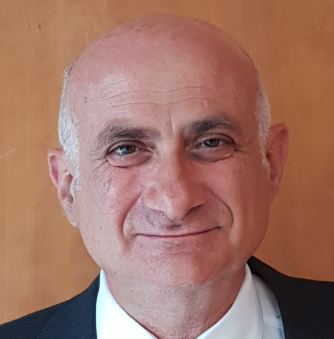 Giuseppe Ippolito合作论文数Saint Camillus International University of Health and Medical Sciences25
 Nicola Tumino合作论文数Cellular Immunology Laboratory, National Institute for Infectious Diseases 'Lazzaro Spallanzani' IRCCS21
 Germana Grassi合作论文数21Concetta Castilletti合作论文数National Institute for Infectious Diseases “L. Spallanzani”,20
 Giulia Matusali合作论文数19Stefania Notari合作论文数19V D'Intini合作论文数18
 Franco Locatelli合作论文数Department of Pediatric Hematology-Oncology, Ospedale Pediatrico Bambino Gesu, University of Pavia18
 Francesca Colavita合作论文数17M Bonello合作论文数16
 Marco Tripodi合作论文数15
 Valentina Mazzotta合作论文数Clin & Res Dept, Natl Inst Infect Dis Lazzaro Spallanzani IRCCS15Pinnetti Carmela合作论文数Department of Infectious Diseases, Catholic University15
 Davide Mariotti合作论文数13Eleonora Tartaglia合作论文数Natl Inst Infect Dis L Spallanzani IRCCS INMI Rom12
 Domenico Viola合作论文数Cellular Immunology Laboratory, National Institute for Infectious Diseases 'Lazzaro Spallanzani' IRCCS12Vergori Alessandra合作论文数12
 Gian Maria Fimia合作论文数Dipartimento di Medicina Molecolare, Università di Roma Sapienza;Laboratorio Di Biologia Cellulare E Microscopia Elettronica, INMI L. Spallanzani IRCCS12Federica Turchi合作论文数12
 Enrico Girardi合作论文数11Emanuele Nicastri合作论文数11
 Luisa Marchioni合作论文数11Adriana Ammassari合作论文数Clinic of Infectious Diseases, Catholic University11Silvia Meschi合作论文数Virology Section and Retrovirus Center, University of Pisa11Alessandra Brendolan合作论文数Department of Nephrology, Dialysis and Transplantation, St Bortolo Hospital11
 Gianpiero D'Offizi合作论文数11E. Cimini合作论文数10Eleonora Lalle合作论文数10Patrizia Lorenzini合作论文数National Center for Disease Prevention ad Heath Promotion, Italian National Institute of Health10Isabella Abbate合作论文数10
 Francesco Vaia合作论文数General Direction, National Institute for Infectious Diseases9
 Nathan W. Levin合作论文数Renal Research Institute, LLC9
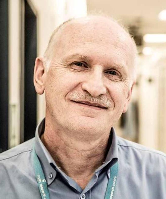 Rinaldo Bellomo合作论文数Australian and New Zealand Intensive Care Research Centre, School of Public Health and Preventative Medicine, Monash University8
 Rita Carsetti合作论文数Direzione Scientifica, IRCCS Ospedale Pediatrico Bambino Gesù8
 Mauro Piacentini合作论文数Department of Biology, University of Rome "Tor Vergata";Laboratory of Cell Biology and Electron Microscopy, National Institute for Infectious Diseases;Laboratory of Molecular Medicine, Institute of Cytology, Russian Academy of Science8Stefania Cicalini合作论文数Istituto Nazionale per le Malattie Infettive "L.Spallanzani"8
 Michele Bibas合作论文数Department of Internal Medicine;Section of Hematology;Department of Immunology;Department of Internal Medicine | Section of Hematology | Department of Immunology8G Salvatori合作论文数7
 Alessandra Amendola合作论文数7Gagliardini Roberta合作论文数Clinic of Infectious Diseases, Catholic University of Sacred Heart7
 Antonioli M合作论文数National Institute for Infectious Diseases IRCCS 'L. Spallanzani'7




