Topoisomerase III-beta (Top3b) reduces nucleic acid torsional stress and intertwining generated during RNA and DNA metabolism while protecting the genome from pathological R-loops, which otherwise result in DNA breakage and genome instability. By studying Top3b knockout mice ( Top3b -KO), we find that the loss of Top3b accelerates the development of spontaneous atypical lymphoid hyperplasia and lymphomas arising in spleens and lymph nodes, organs with prominent Top3b expression. Aging Top3b -KO mice also display splenomegaly and systemic immune alterations including neutrophilia and lymphopenia consistent with chronic inflammation. At the molecular level, Top3b deficiency causes genome-wide R-loop accumulation in splenocytes as measured by CUT&Tag sequencing. Increased R-loops are associated with genomic breaks and activation of immune signaling pathways including innate and adaptive immune cell signaling, IL-4 signaling, FAK signaling, and cGAS-STING. In addition, knocking-out Top3b promotes the rapid development of syngeneic EL4 T cell lymphomas. In conclusion, our work implies that Top3b protects from lymphoma, tumorigenesis, and immune dysregulations.
Topoisomerase III-beta (Top3b) reduces nucleic acid torsional stress and intertwining generated during RNA and DNA metabolism while protecting the genome from pathological R-loops, which otherwise result in DNA breakage and genome instability. By studying Top3b knockout mice (Top3b-KO), we find that the loss of Top3b accelerates the development of spontaneous lymphoid tumors arising in spleens and lymph nodes, the organs with prominent Top3b expression. Aging Top3b-KO mice also display splenomegaly and systemic immune alterations including neutrophilia and lymphopenia suggestive of chronic inflammation. At the molecular level, Top3b deficiency causes genome-wide R-loop accumulation in splenocytes as measured by CUT&Tag sequencing. Increased R-loops is associated with genomic DNA breaks and activation of immune signaling pathways including the IL-6 signaling, interleukin-7 signaling and cGAS-STING. Moreover, knocking-out Top3b promotes the rapid development of syngeneic EL4 T-cell lymphomas. In conclusion, our work implies that, in addition to its role in preserving the nervous system, Top3b protects from tumorigenesis and immune dysregulations.
High fat (HF) diet is a major factor in the development of metabolic dysfunction-associated steatotic liver disease (MASLD) and steatohepatis (MASH), and mitochondria have been proposed to play a role in the pathogenesis of HF diet-induced MASH. Because Mitochondrial topoisomerase I (Top1MT) is exclusively present in mitochondria and Top1MT knock-out mice are viable, we were able to assess the role of Top1MT in the development of MASH. We show that after 16 weeks of HF diet, mice lacking Top1MT are prone to the development of severe MASH characterized by liver steatosis, lobular inflammation and hepatocyte damage. Mice lacking Top1MT also show prominent mitochondrial dysfunction, ROS production and mitochondrial DNA (mtDNA) release, accompanied by hepatic inflammation and fibrosis. In summary, our study demonstrates the importance of Top1MT in sustaining hepatocyte functions and suppressing MASH.
Supplementary Figure 3: Effects of gemcitabine treatment on IL6(R) expression in vitro, impact of IL-6R and YKL-40 inhibition in BTC cell lines in vitro, expression of IL6(R) in BTC patient tumors.
Colorectal cancers (CRCs) are prevalent worldwide, yet current treatments remain inadequate. Using chemical genetic screens, we identify that co-inhibition of topoisomerase I (TOP1) and NEDD8 is synergistically cytotoxic in human CRC cells. Combination of the TOP1 inhibitor irinotecan or its bioactive metabolite SN38 with the NEDD8-activating enzyme inhibitor pevonedistat exhibits synergy in CRC patient-derived organoids and xenografts. Mechanistically, we show that pevonedistat blocks the ubiquitin/proteasome-dependent repair of TOP1 DNA-protein crosslinks (TOP1-DPCs) induced by TOP1 inhibitors and that the CUL4-RBX1 complex (CRL4) is a prominent ubiquitin ligase acting on TOP1-DPCs for proteasomal degradation upon auto-NEDD8 modification during replication. We identify DCAF13, a DDB1 and Cullin Associated Factor, as the receptor of TOP1-DPCs for CRL4. Our study not only uncovers a replication-coupled ubiquitin-proteasome pathway for the repair of TOP1-DPCs but also provides molecular and translational rationale for combining TOP1 inhibitors and pevonedistat for CRC and other types of cancers.
PDF file - 390K, Supplementary Figure 1. Histopathology of early lesions in DEN-injected Hmgn1+/+ and Hmgn1 tm1/tm1 at 25 weeks of age. Supplementary Table S1. Primers used in the study. Supplementary Table S2. List of differentially expressed genes (FC>2, p?0.01) in livers of Hmgn1+/+ and Hmgn1tm1/tm1 mice in groups A, B, and C. Supplementary Table S3. GO analysis of the differentially expressed genes
Supplementary Discussion from Regulation of α-Fetoprotein by Nuclear Factor-κB Protects Hepatocytes from Tumor Necrosis Factor-α Cytotoxicity during Fetal Liver Development and Hepatic Oncogenesis
Supplementary Tables 1-6, Figures 1-4 from Central Role of c-Myc during Malignant Conversion in Human Hepatocarcinogenesis
Supplementary Figures 1-3, Tables 1-3, Methods from Definition of Ubiquitination Modulator COP1 as a Novel Therapeutic Target in Human Hepatocellular Carcinoma
Supplementary Figure Legends 1-4 from Central Role of c-Myc during Malignant Conversion in Human Hepatocarcinogenesis
Supplementary Figure 5: Histopathology of the HuCCT-1 xenograft tumors from control group and groups treated with gemcitabine and gemcitabine in combination with tocilizumab.
PDF file - 279KB, Co-localization of CLPTM1L mutants (CLPTM1L-delta-Cterm and CLPTM1L-delta-Loop) with an ER marker, indicating a similar localization for WT and mutant CLPTM1L proteins to the endoplasmic reticulum.
Background & aims: Several types of human stem cells from embryonic (ESCs) and induced pluripotent (iPSCs) to adult tissue-specific stem cells are commonly used to generate 3D liver organoids for modeling tissue physiology and disease. We have recently established a protocol for direct conversion of primary human hepatocytes (hPHs) from healthy donor livers into bipotent progenitor cells (hCdHs). Here we extended this culture system to generate hCdH-derived liver organoids for diverse biomedical applications. Methods: To obtain hCdHs, hPHs were cultured in reprogramming medium containing A83-01 and CHIR99021 for 7 days. Liver organoids were established from hCdHs (hCdHOs) and human liver cells (hLOs) using the same donor livers for direct comparison, as well as from hiPSCs. Organoid properties were analyzed by standard in vitro assays. Molecular changes were determined by RT-qPCR and RNA-seq. Clinical relevance was evaluated by transplantation into FRG mice, modeling of alcohol-related liver disease (ARLD), and in vitro drug-toxicity tests. Results: hCdHs were clonally expanded as organoid cultures with low variability between starting hCdH lines. Similar to the hLOs, hCdHOs stably maintained stem cell phenotype based on accepted criteria. However, hCdHOs had an advantage over hLOs in terms of EpCAM expression, efficiency of organoid generation and capacity for directed hepatic differentiation as judged by molecular profiling, albumin secretion, glycogen accumulation, and CYP450 activities. Accordingly, FRG mice transplanted with hCdHOs survived longer than mice injected with hLOs. When exposed to ethanol, hCdHOs developed stronger ARLD phenotype than hLOs as evidenced by transcriptional profiling, lipid accumulation and mitochondrial dysfunction. In drug-induced injury assays in vitro, hCdHOs showed a similar or higher sensitivity response than hPHs. Conclusion: hCdHOs provide a novel patient-specific stem cell-based platform for regenerative medicine, toxi-cology testing and modeling liver diseases.
Supplementary Figure 4: Pathway over-representation analysis of differentially expressed miRNAs across cellular compartments (parent cells, EV) and treatment groups (anti-IL6R (tocilizumab), anti-YKL40) in biliary tract cancer cells in vitro.
Upregulation of c-MYC in cancer stem cell enriched populations (S1); The doxycycline (Dox)-controlled mCherry marker expression in c-MYC inducible hepatoma cell lines (S2); The doxycycline (Dox) dose-dependent effects of c-MYC induction on CSC properties (S3); The doxycycline (Dox) dose-dependent effects of c-MYC induction on self-renewal potential (S4); c-MYC switch-on and switch-off effects on self-renewal of PLC cells (S5); Effect of p53 knockdown on self-renewal of hepatoma cell lines with doxycycline (Dox) regulated c-MYC expression (S6); p53 knockdown increases the growth rate of HepG2 cells with doxycycline (Dox) regulated c-MYC expression (S7).
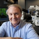 Snorri Thorgeirsson合作论文数National Cancer Institute189
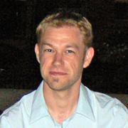 Jesper Bøje Andersen合作论文数Biotech Research and Innovation Centre, Department of Health and Medical Sciences, University of Copenhagen61
 Elizabeth A Conner合作论文数59
 Jens Marquardt合作论文数University of Lübeck;University Hospital Schleswig-Holstein48
 Seo Daekwan合作论文数Expt Carcinogenesis Lab, NIH41
 Chiara Raggi合作论文数29
 Yun-Han Lee合作论文数26
 Mitsuteru Kitade合作论文数23
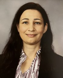 Agnes Holczbauer合作论文数21Marian E. Durkin合作论文数20
 Itzhak Avital合作论文数17EA Conner合作论文数16
 Luis E Gomez-Quiroz合作论文数Dept Ciencias Salud, Univ Autonoma Metropolitana Iztapalapa16
 Diego F Calvisi合作论文数16
 Pal Novak Kaposi合作论文数14
 Aranzazu Sanchez Munoz合作论文数Departamento de Química Orgánica, Universidad de Alcalá14Adam Judge合作论文数Tekmira Pharmaceuticals13
 Cedric Coulouarn合作论文数Université de Rennes;French Institute of Health and Medical Research12
 Ian Maclachlan合作论文数12
 Insa S Schroeder合作论文数Differentiation Group, Leibniz-Institute of Plant Genetics (IPK),11Adam Cheuk合作论文数10Jinping Jia合作论文数10Thorkell Andresson合作论文数Protein Characterization Laboratory, Cancer Research Technology Program, Frederick National Laboratory for Cancer Research, Leidos Biomedical Research, Inc., Frederick, MD 21702, USA10
 Laufey Amundadottir合作论文数NCI, Lab Translat Genom, Div Canc Epidemiol & Genet, NIH, Bethesda, MD 20892 USA10Christopher J Westlake合作论文数NCI, Lab Cell & Dev Signaling, Frederick, MD 21702 USA10
 Michael Rugaard Jensen合作论文数10
 William R Bamlet合作论文数10Thompson Abbey合作论文数10
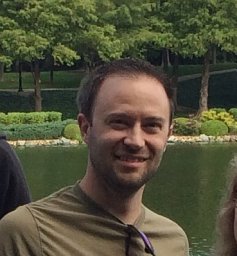 Hoskins Jason合作论文数10
 Charles Lynch合作论文数Department of Epidemiology, College of Public Health, University of Iowa;Department of Pathology, College of Public Health, University of Iowa10
 Zhen Xiao合作论文数Laboratory of Proteomics and Analytical Technologies, National Cancer Institute at Frederick10Irene Collins合作论文数10
 Hemang Parikh合作论文数Lund University, University Hospital Malmö;Laboratory of Translational Genomics, National Cancer Institute, National Institutes of Health;University Hospital Malmö, Lund University10
 Wendy Cozen合作论文数School of Medicine, University of California, Irvine;Chao Family Comprehensive Cancer Center, University of California, Irvine10Lizhi Zhang合作论文数10
 Brenda Y. Hernandez合作论文数Department of Tropical Medicine, Medical Microbiology, and Pharmacology, University of Hawaiʻi Cancer Center10Jadranka Loncarek合作论文数10Tsuyoshi Ishikawa合作论文数9
 Hirofumi Akita (秋田裕史)合作论文数Department of Gastroenterological Surgery, Graduate School of Medicine, Osaka University,9Allen D Bosley合作论文数8Marta Dzyadyk合作论文数NCI, Lab Translat Genom, Div Canc Epidemiol & Genet, NIH,Dept Hlth & Human Serv, Bethesda, MD 20892 USA8
 Gloria M. Petersen合作论文数Comprehensive Cancer Center, Mayo Clinic8Altekruse Sean F合作论文数8
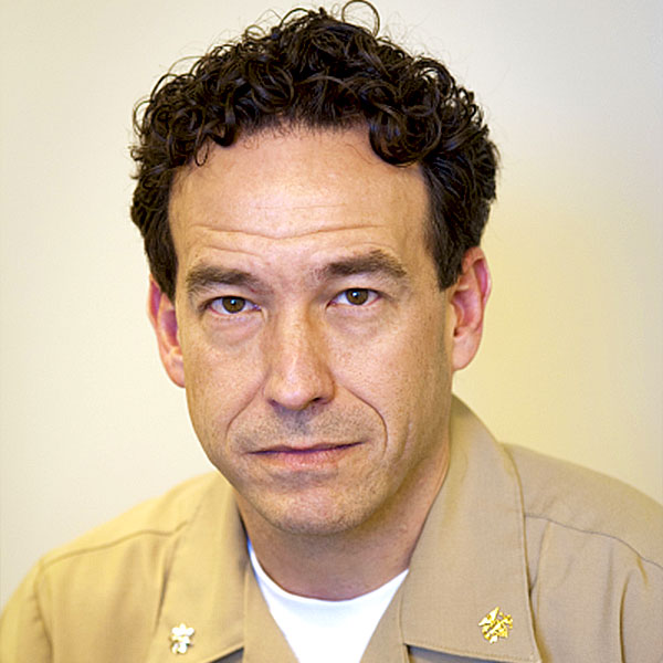 Stephen M. Hewitt合作论文数Laboratory of Pathology, Center for Cancer Research, National Cancer Institute, National Institutes of Health8
 Colm O'Rourke合作论文数8Koichi Uchida合作论文数8Sara Ladu合作论文数8
 Dongho Choi合作论文数College of Medicine, Soon Chun Hyang University8
 Eric Santoni-Rugiu合作论文数Dept. of Pathology, Rigshospitalet, University of Copenhagen;Dept. of Clinical Medicine, University of Copenhagen8




