A seizure is to the brain as a cough is to the lung the outward sign of an internal tissue irritation. An epileptic attack thus presents as a symptom that can be expressed by many different brain disorders. The most frequent of these include trauma or infection leading to scar formation in the brain, tumors, blood vessel abnormalities, developmental anomalies, metabolic errors, and genetic defects. The pattern of a seizure may range from a generalized convulsion without warning, to focal attacks with auras or initial features of onset that reflect the part of the brain where the seizure begins, such as sensory, motor, auditory, olfactory, visual, or memory systems.
We report a long-term follow-up investigation of a patient who was operated in 1954 to relieve intractable temporal lobe seizures characterized by automatism and amnesia. Neuropsychological review at 16 months after surgery showed a slight residual impairment of verbal comprehension and verbal recall and good nonverbal skills. Seizure-free since the operation except for two attacks in the early postoperative years, the patient has been off medication for 25 years and has pursued a successful career as an artist. Our investigation at 56 postoperative years focused on cognitive skills, with some emphasis on learning and memory; a clinical examination was also performed, and the anatomical extent of the resection was determined on 3-Tesla magnetic resonance imaging. Four age- and IQ-appropriate women were tested as healthy control subjects. The patient showed material-specific impairments in language and verbal memory compared with the control subjects and also compared with her own earlier performance, but her performance on other cognitive tasks did not differ from that of the control subjects. Thus, her specific deficits had worsened over time, and she was also impaired compared with healthy individuals of her age, but her deficits remained confined to the verbal sphere, consistent with her temporal lobe seizure focus and surgery.
During the 1930s, white matter tracts began to assume relevance for neurosurgery, especially after Cajal’s work. In many reviews of white matter neurobiology, the seminal contributions of Josef Klingler (1888-1963) and their neurological applications have been overlooked. In 1934 at the University of Basel under Eugen Ludwig, Klingler developed a new method of dissection based on a freezing technique for brain tissue that eloquently revealed the white matter tracts. Klingler worked with anatomists, surgeons, and other scientists, and his models and dissections of white matter tracts remain arguably the most elegant ever created. He stressed 3-dimensional anatomic relationships and laid the foundation for defining mesial temporal, limbic, insular, and thalamic fiber and functional relationships and contributed to the potential of stereotactic neurosurgery. Around 1947, Klingler was part of a Swiss-German group that independently performed the first stereotactic thalamotomies, basing their targeting and logic on Klingler’s white matter studies, describing various applications of stereotaxy and showing Klingler’s work integrated into a craniocerebral topographic system for targeting with external localization of eloquent brain structures and stimulation of deep thalamic nuclei. Klingler’s work has received renewed interest because it is applicable for correlating the results of the fiber-mapping paradigms from diffusion tensor imaging to actual anatomic evidence. Although others have described white matter tracts, none have had as much practical impact on neuroscience as Klinger’s work. More importantly, Josef Klingler was an encouraging mentor, influencing neurosurgeons, neuroscientists, and brain imaging for more than three quarters of a century.
This review focuses on some historical highlights of the surgery of epilepsy, beginning with the reports of Horsley, Krause, and Cushing to which appeared in 1909, the year that The International League Against Epilepsy (ILAE) was inaugurated. We then outline key contributions from Europe and North America, and examine particularly the evolution of our understanding of temporal lobe seizures, which have now become the most common form of epilepsy amenable to surgical cure.
In the 15th century, brain illustration began to change from a schematic system that involved scant objective rendering of the brain, to accurate depictions based on anatomical dissections that demanded significant artistic talent. Notable examples of this innovation are the drawings of Leonardo da Vinci (1498-1504), Andreas Vesalius' association with the bottega of Titian to produce the drawings of Vesalius' De humani corporis fabrica (1543), and Christopher Wren's illustrations for Thomas Willis' Cerebri Anatome (1664). These works appeared during the Renaissance and Age of Enlightenment, when advances in brain imaging, or really brain rendering, reflected not only the abilities and dedications of the artists, but also the influences of important cultural and scientific factors. Anatomy and human dissection became popular social phenomena as well as scholarly pursuits, linked with the world of the fine arts. The working philosophy of these artists involved active participation in both anatomical study and illustration, and the belief that their discoveries of the natural world could best be communicated by rendering them in objective form (that is, with realistic perspective). From their studies emerged the beginning of contemporary brain imaging. In this article, the authors examine how the brain began to be imaged in realism within a cultural and scientific milieu that witnessed the emergence of anatomical dissection, the geometry of linear perspective, and the closer confluence of art and science.
On December 14, 1883, William Osler, then pathologist at the Montreal General Hospital, presented the specimen of a brain with an almond-sized glioma beneath the right motor cortex to the Montreal Medico-Chirurgical Society. The brain specimen was from a young woman who had suffered from intermittent Jacksonian seizures for 14 years and had eventually died in status epilepticus. Aware of the pioneering removal of a tumor from the cortex reported on in 1885 by Bennett and Godlee, Osler wrote of his case, "this was an instance in which operation would have been justifiable and possibly have been the means of saving life." In 1953, a young man with Jacksonian attacks that began in his foot underwent removal of a Grade I glioma from the central fissure. The operation was performed in an awake craniotomy during which cortical mapping was used to define the motor and sensory cortices. Treatment with focal radiation followed, and afterward the patient became seizure-free, stopped taking anticonvulsant medication, and has led an active life over the past 50 years. Reference is made to the experiences of Sherrington, Cushing, and Penfield with cortical stimulation in the awake patient under regional anesthesia as an effective aid to surgery for epileptogenic lesions, tumors, and vascular malformations. Their technique allows for maximal resection with minimal neurological deficits. Over the past 20 years, this approach has been adopted effectively in many neurosurgical centers.
Wilder Penfield, a Rhodes scholar from Princeton University, New Jersey, was a student in the first course on mammalian physiology given in 1915 at Oxford University by Charles Sherrington, newly arrived from Liverpool where, as Holt Professor of Physiology for 20 years, he had become a leading authority on the physiology of the nervous system. The practical 'exercises' as well as graduate research on the Golgi apparatus and the decerebrate preparation, carried out by Penfield in Sherrington's laboratory, gave him the groundwork to develop his career as a physiological surgeon, who made fundamental observations on functional localization in the human brain during the surgical treatment of patients afflicted with epilepsy.
WILLIAM FEINDEL, oc, MDCM, DPHIL] glioma was also removed. (Today this pleased that he had no more of these It was a few weeks before the would have been detected on MRI fearful attacks and elaborate Christmas of 1951, and all through the see Neuro-Image for February 1989). hallucinations. It was nearing Neuro the surgeons were busily Before operation the young man's Christmas and he was eagerly making stirring. Dr. Penfield operated on a seizures were vivid he would describe plans to return with his father to young man (P.S.) sent from Paris by colored lights purple, violet, blue and France. Dr. Henry Hecaen because of seizures yellow and at night, green stars. In for thirteen years. Herbert Jasper had other attacks he heard bells or would But before he left hospital, a curious found epileptic spiking in the anterior hum a tune. In still another, he was and distressing episode occurred. The temporal region. At operation spikes convinced that he was dead and in Neuro then, perhaps more than at any were recorded from the temporal heaven he saw violet and blue saints time in its history, was jammed with cortex and also from the amygdala, and began to pray over and over patients. The Military Annex of some from which stimulation evoked one of again. When questioned later, he 30 beds had been demolished to the patient's typical attacks of seemed to recall this as not just a make way for the McConnell Pavilion confusion and automatic movement dream, but the real thing. and its eighty new beds, still under for which he had no recall afterwards construction. T o accommodate the (Fig. 1). During the temporal lobe ons heavy admissions, esp excision an unexpected low grade
"Soul Made Flesh—How the Discovery of the Brain Changed the World Carl Zimmer." Canadian Bulletin of Medical History, 22(1), pp. 196–197
Purpose: Controversy persists about when EEG became a fundamental tool in the preoperative investigation for epilepsy surgery. We revisit Penfield's first use of invasive EEG monitoring, emphasizing its historical importance for the evolution of epilepsy surgery.Methods: Patients' hospital charts and articles published before 1940 regarding EEG and epilepsy or EEG and cerebral lesions were reviewed to evaluate the historical context of the surgery.Results: In April 1939, Penfield performed trephination over both temporal regions and placed electrodes on the dura, intending to lateralize seizure origin in a patient with bitemporal epilepsy. The patient underwent serial EEGs with this technique. The final report of the recordings from epidural leads was "continued random delta activity in the left temporal region indicating a cortical lesion on this side." The pneumoencephalogram showed "the presence of diffuse cerebral atrophy, particularly in the left cerebral hemisphere." Based on these findings, the patient underwent surgery on April 21, revealing a meningocerebral scar in the posterior part of the left temporal lobe. Brain stimulation and electrocorticography delineated the extent of resection, while preserving the speech area. Seizures did not improve.Conclusions: We revisit the first case of epidural EEG monitoring for epilepsy surgery and show that the concept of EEG-directed surgery was already present at the Montreal Neurological Institute in the late 1930s.
Wistar rats implanted intracerebrally with AA ascites tumor were injected seven or eight days later with 11C-labelled BCNU. Radioactive compounds in samples of plasma, tumor, and contralateral brain were identified after injection at intervals by doing chloroform extraction and thin layer radiochromatography. At 60 min after injection radioactivity levels were 56% higher in the tumor than in contralateral brain. This increase was due mainly to 2-chloroethyl isocyanate, which binds to amino groups and/ or nucleic acid. The results demonstrated both a higher concentration and a faster decomposition of BCNU in the tumor.
 Y Lucas Yamamoto合作论文数66
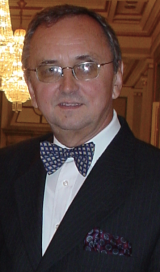 Mirko Diksic合作论文数McConnell Brain Imaging Centre, Montreal Neurological Institute, McGill University46Charles P. Hodge合作论文数19F Andermann合作论文数14Yamamoto Y L合作论文数14
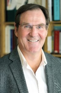 Alan C Evans合作论文数Department of Neurology and Neurosurgery, McGill University;McConnell Brain Imaging Centre, Montreal Neurological Institute-Hospital;Biospective Inc;Ludmer Centre for Neuroinformatics and Mental Health12L Yamamoto合作论文数11Ernst Meyer合作论文数11J.-G. Villemure合作论文数11R LEBLANC合作论文数10Olivier Andre合作论文数10Yves Robitaille合作论文数10Mark C. Preul合作论文数10Colin Thompson合作论文数10S FARROKHZAD合作论文数10JG VILLEMURE合作论文数8D Melanson合作论文数7
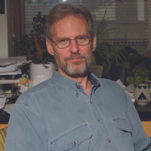 Eric Shoubridge合作论文数The Neuro Montreal Neurological Institute-Hospital;McGill University7
 Amami Kato合作论文数Department of Neurosurgery, Osaka University Medical School7K Sako合作论文数6
 Christopher J. Thompson合作论文数Department of Chemistry;University of Massachusetts6Eva Andermann合作论文数6
 Sam Berkovic合作论文数Department of Medicine, University of Melbourne;Epilepsy Research Centre, Austin Health6Jane L. Tyler合作论文数6R. Ethier合作论文数6J LITTLE合作论文数5
 Douglas Arnold合作论文数Department of Neurology and Neurosurgery, Faculty of Medicine and Health Sciences, McGill University;NeuroRx Research5R POKRUPA合作论文数4Joseph Stratford合作论文数4Jl Tyler合作论文数4J THERON合作论文数4P Gloor合作论文数4A SHERWIN合作论文数4
 Jean-Guy Villemure合作论文数Department of Neurosurgery, Centre Hospitalier Universitaire Vaudois4Hideaki Nukui合作论文数4Phanor Perot合作论文数4T STAUNTON合作论文数3MM FROJMOVIC合作论文数3zhongchuan yi (翼 中川)合作论文数3S Nagahiro合作论文数3E MEYER合作论文数3
 François Dubeau合作论文数Department of Neurology and Neurosurgery, Faculty of Medicine and Health Sciences, McGill University;Montreal Neurological Institute, McGill University;McGill University Health Centre3A HAKIM合作论文数3
 Antoine M. Hakim合作论文数Brain and Mind Research Institute, University of Ottawa;Ottawa Hospital Research Institute3Kazuhiro Sako合作论文数3
 Ruben Kuzniecky合作论文数Donald & Barbara Zucker School of Medicine, Hofstra University;School of Medicine, New York University;Northwell Health3John G. Milton合作论文数Department of Neurology, University of Chicago
Jennifer Foss Department of Psychology, University of New Orleans
John D. Hunter Department of Neurology, University of Chicago3
Ruben Kuzniecky合作论文数Donald & Barbara Zucker School of Medicine, Hofstra University;School of Medicine, New York University;Northwell Health3John G. Milton合作论文数Department of Neurology, University of Chicago
Jennifer Foss Department of Psychology, University of New Orleans
John D. Hunter Department of Neurology, University of Chicago3




