α-Synuclein aggregation is a hallmark of Parkinson's disease and related synucleinopathies. Extracellular α-synuclein fibrils enter naive cells via endocytosis, followed by transit into the cytoplasm to seed endogenous α-synuclein aggregation. Intracellular aggregates sequester numerous proteins, including subunits of the endosomal sorting complexes required for transport (ESCRT)-III system for endolysosome membrane repair, but the toxic effects of these events remain poorly understood. Using cellular models and in vitro reconstitution, we found that α-synuclein fibrils interact with a conserved α-helix in ESCRT-III proteins. This interaction sequesters ESCRT-III subunits and triggers their proteasomal destruction in a process of "collateral degradation." These twin mechanisms deplete the available ESCRT-III pool, initiating a toxic feedback loop. The ensuing loss of ESCRT function compromises endolysosome membranes, thereby facilitating escape of aggregate seeds into the cytoplasm, facilitating a "second wave" of templated aggregation and ESCRT-III sequestration. We suggest that collateral degradation and the triggering of self-perpetuating systems are general mechanisms of sequestration-induced proteotoxicity.
Localized translation broadly enables spatiotemporal control of gene expression. Here, we present LOV-domain-controlled ligase for translation localization (LOCL-TL), an optogenetic approach for monitoring translation with codon resolution at any defined subcellular location under physiological conditions. Application of LOCL-TL to mitochondrially localized translation revealed that ∼20% of human nuclear-encoded mitochondrial genes are translated on the outer mitochondrial membrane (OMM). Mitochondrially translated messages form two classes distinguished by encoded protein length, recruitment mechanism, and cellular function. An evolutionarily ancient mechanism allows nascent chains to drive cotranslational recruitment of long proteins via an unanticipated bipartite targeting signal. Conversely, mRNAs of short proteins, especially eukaryotic-origin electron transport chain (ETC) components, are specifically recruited by the OMM protein A-kinase anchoring protein 1 (AKAP1) in a translation-independent manner that depends on mRNA splicing. AKAP1 loss lowers ETC levels. LOCL-TL thus reveals a hierarchical strategy that enables preferential translation of a subset of proteins on the OMM.
Early or sorting endosomes are dynamic organelles that play key roles in proteome control by triaging plasma membrane proteins for either recycling or degradation in the lysosome1,2. These events are coordinated by numerous transiently associated regulatory complexes and integral membrane components that contribute to organelle identity during endosome maturation3. Although a subset of the several hundred protein components and cargoes known to associate with endosomes have been studied at the biochemical and/or structural level, interaction partners and higher-order molecular assemblies for many endosomal components remain unknown. Here, we combine crosslinking and native gel mass spectrometry4-7 of purified early endosomes with AlphaFold8,9 and computational analysis to create a systematic human endosomal structural interactome. We present 229 structural models for endosomal protein pairs and additional higher-order assemblies supported by experimental crosslinks from their native subcellular context, suggesting structural mechanisms for previously reported regulatory processes. Using induced neurons, we validate two candidate complexes whose interactions are supported by crosslinks and structural predictions: TMEM230 as a subunit of ATP8 and ATP11 lipid flippases10 and TMEM9 and TMEM9B as subunits of the chloride-proton antiporters CLCN3, CLCN4 and CLCN5 (ref. 11). This resource and its accompanying structural network viewer provide an experimental framework for understanding organellar structural interactomes and large-scale validation of structural predictions.
Numerous metabolic enzymes translocate from the ER membrane bilayer to the lipid droplet (LD) monolayer, where they perform essential functions. Mislocalization of certain LD-targeted membrane proteins, including HSD17B13 and PNPLA3, is implicated in metabolic dysfunction-associated steatotic liver disease (MASLD). However, the mechanisms governing the trafficking and accumulation of ER proteins on LDs remain poorly understood. Here, using MINFLUX and HILO single-molecule tracking combined with machine learning, we show that HSD17B13, GPAT4, and the model cargo LiveDrop diffuse at comparable speeds in the ER and on LDs, but become nano-confined upon reaching the LD surface. Mechanistic dissection of LiveDrop targeting revealed that this confinement, along with protein accumulation on LDs, depends on specific residues within its targeting motif. These residues mediate preferential and repeated interactions with nanoscale membrane domains, suggesting that LD-targeted proteins selectively partition into distinct lipid-protein environments that transiently retain and concentrate them at the LD surface. Single-molecule trajectories further revealed bidirectional trafficking of LiveDrop across seipin-containing ER-LD bridges, providing direct evidence for lateral protein transfer across membrane contact sites. These findings establish nanodomain-based confinement as a key mechanism driving selective protein accumulation on LDs and reveal how membrane bridges between organelles facilitate protein sorting.
Lysosomal storage diseases (LSDs) comprise ~50 monogenic disorders marked by the buildup of cellular material in lysosomes, yet systematic global molecular phenotyping of proteins and lipids is lacking. We present a nanoflow-based multiomic single-shot technology (nMOST) workflow that quantifies HeLa cell proteomes and lipidomes from over two dozen LSD mutants. Global cross-correlation analysis between lipids and proteins identified autophagy defects, notably the accumulation of ferritinophagy substrates and receptors, especially in NPC1-/- and NPC2-/- mutants, where lysosomes accumulate cholesterol. Autophagic and endocytic cargo delivery failures correlated with elevated lysophosphatidylcholine species and multilamellar structures visualized by cryo-electron tomography. Loss of mitochondrial cristae, MICOS complex components, and OXPHOS components rich in iron-sulfur cluster proteins in NPC2-/- cells was largely alleviated when iron was provided through the transferrin system. This study reveals how lysosomal dysfunction affects mitochondrial homeostasis and underscores nMOST as a valuable discovery tool for identifying molecular phenotypes across LSDs.
Metazoan cells adapt to the exhaustion of protein quality control (PQC) systems by sequestering aggregation-prone proteins in large, pericentriolar structures termed aggresomes. Defects in both aggresome formation and clearance affect proteostasis and have been linked to neurodegenerative diseases, but aggresome clearance pathways are still underexplored. Here we show that aggresomes comprising endogenous proteins are cleared via selective autophagy requiring the cargo receptor TAX1BP1. TAX1BP1 proximitomes reveal the presence of various PQC systems at aggresomes, including Hsp70 chaperones, the 26S proteasome, and the ubiquitin-selective unfoldase p97/VCP. While Hsp70 and p97/VCP with its cofactors UFD1-NPL4 and FAF1 play key roles in aggresome disassembly, the 26S proteasome is dispensable. We identify aggresomal client proteins that are degraded via different routes, in part in a p97/VCP-dependent manner via aggrephagy. Upon acute inhibition of p97/VCP, aggresomes fail to disintegrate and cannot be incorporated into autophagosomes despite the presence of factors critical for aggrephagosome formation, including p62/SQSTM1, TAX1BP1, and WIPI2. We conclude that the p97/VCP-mediated removal of ubiquitylated aggresomal clients is essential for the disintegration and subsequent piecemeal autophagy of aggresomes.
Early/sorting endosomes are dynamic organelles that play key roles in proteome control by triaging plasma membrane proteins for either recycling or degradation in the lysosome1,2,3. These events are coordinated by numerous transiently-associated regulatory complexes and integral membrane components that contribute to organelle identity during endosome maturation4. While a subset of the several hundred protein components and cargoes known to associate with endosomes have been studied at the biochemical and/or structural level, interaction partners and higher order molecular assemblies for many endosomal components remain unknown. Here, we combine cross-linking and native gel mass spectrometry5-8 of purified early endosomes with AlphaFold9,10 and computational analysis to create a systematic human endosomal structural interactome. We present dozens of structural models for endosomal protein pairs and higher order assemblies supported by experimental cross-links from their native subcellular context, suggesting structural mechanisms for previously reported regulatory processes. Using induced neurons, we validate two candidate complexes whose interactions are supported by crosslinks and structural predictions: TMEM230 as a subunit of ATP8/11 lipid flippases11 and TMEM9/9B as subunits of CLCN3/4/5 chloride-proton antiporters12. This resource and its accompanying structural network viewer provide an experimental framework for understanding organellar structural interactomes and large-scale validation of structural predictions.
Human cells consist of a complex hierarchy of components, many of which remain unexplored 1,2 . Here we construct a global map of human subcellular architecture through joint measurement of biophysical interactions and immunofluorescence images for over 5,100 proteins in U2OS osteosarcoma cells. Self-supervised multimodal data integration resolves 275 molecular assemblies spanning the range of 10 −8 to 10 −5 m, which we validate systematically using whole-cell size-exclusion chromatography and annotate using large language models 3 . We explore key applications in structural biology, yielding structures for 111 heterodimeric complexes and an expanded Rag–Ragulator assembly. The map assigns unexpected functions to 975 proteins, including roles for C18orf21 in RNA processing and DPP9 in interferon signalling, and identifies assemblies with multiple localizations or cell type specificity. It decodes paediatric cancer genomes 4 , identifying 21 recurrently mutated assemblies and implicating 102 validated new cancer proteins. The associated Cell Visualization Portal and Mapping Toolkit provide a reference platform for structural and functional cell biology.
Lysosomes, central hydrolytic organelles, are regulated by ion flow, including calcium and protons, via transporters and channels to maintain an acidified lumen for hydrolytic activity. TRPML1, a lysosomal ion channel, effluxes cations upon activation, promoting rapid conjugation of ATG8 proteins to the lysosomal membrane in a process known as conjugation of ATG8 to single membranes (CASM). However, our understanding of how TRPML1 activation reorganizes the lysosomal proteome is poorly understood. Here, we identify DMXL1 as a key regulator of lysosomal homeostasis through quantitative proteomics of lysosomes during TRPML1 activation by the agonist MLSA5. DMXL1 is recruited to lysosomes and Salmonella-containing vacuoles, both in a CASM-dependent manner. As the mammalian ortholog of yeast Rav1, DMXL1 assembles with Rav2 ortholog ROGDI and WDR7, and associates with V0 and V1 subunits of the lysosomal V-ATPase. TRPML1 activation drives V1 subunit recruitment to lysosomes in a DMXL1- and DMXL2-dependent manner. DMXL1- and DMXL2-deficient cells display reduced V1-ATPase recruitment, increased lysosomal pH and diminished hydrolytic capacity. Using AlphaFold modeling supported by cross-linking proteomics, we identify interaction interfaces within the DMXL1-ROGDI-WDR7 complex, as well as an ATP6V1A binding interface in DMXL1, whose mutation affects interaction and function. Our findings suggest CASM-dependent DMXL1 recruitment, coupled with V-ATPase assembly, is critical for maintaining lumenal pH and lysosomal function in response to TRPML1 activation.
Maintaining an optimal mitochondrial distribution is critical to ensure an adequate supply of energy and metabolites to support important cellular functions. How cells balance dynamic mitochondrial processes to achieve homeostasis is incompletely understood. Here, we show that ARMC1 partitioning between distinct mitochondrial protein complexes is a key determinant of mitochondrial distribution. In one complex, the mitochondrial trafficking adaptor MIRO recruits ARMC1, which mediates the assembly of a mitochondrial fission regulator (MTFR). MTFR stability depends on ARMC1, and MIRO-MTFR complexes specifically antagonize retrograde mitochondrial movement. In another complex, DNAJC11 facilitates ARMC1 release from mitochondria. Disrupting MIRO-MTFR assembly fails to rescue aberrant mitochondrial distributions clustered in the perinuclear area observed with ARMC1 deletion, while disrupting ARMC1 interaction with DNAJC11 leads to excessive mitochondrially localized ARMC1 and distinct mitochondrial defects. Thus, the abundance and trafficking impact of MIRO-MTFR complexes require ARMC1, whose mito-cytoplasmic shuttling balanced by DNAJC11 tunes steady-state mitochondrial distributions.
Epidermal growth factor receptor (Egfr)-driven signaling regulates fundamental homeostatic processes. Dysregulated signaling via Egfr is implicated in numerous disease pathologies and distinct Egfr-associated disease etiologies are known to be tissue-specific. The molecular basis of this tissue-specificity remains poorly understood. Most studies of Egfr signaling to date have been performed in vitro or in tissue-specific mouse models of disease, which has limited insight into Egfr signaling patterns in healthy tissues. Here, we carried out integrated phosphoproteomic, proteomic, and transcriptomic analyses of signaling changes across various mouse tissues in response to short-term stimulation with the Egfr ligand Egf. We show how both baseline and Egf-stimulated signaling dynamics differ between tissues. Moreover, we propose how baseline phosphorylation and total protein levels may be associated with clinically relevant tissue-specific Egfr-associated phenotypes. Altogether, our analyses illustrate tissue-specific effects of Egf stimulation and highlight potential links between underlying tissue biology and Egfr signaling output.
Abstract Cells regulate growth and function through a hierarchical structure of subcellular protein assemblies, in which alterations can result in cellular dysfunction leading to disease such as cancer. Much of this structure remains uncharted, resulting in recent efforts to map subcellular organization at different physical scales. Here, we report a global architectural map of human cancer cell protein assemblies spanning 10−9 to 10−5 nm, based on integration of near-proteome-wide affinity purification mass spectrometry-based protein interactions and immunofluorescent imaging in U-2 OS osteosarcoma cancer cells. The U-2 OS multi-scale integrated cell map places >5000 proteins into 270 distinct subcellular protein assemblies across biological scales, representing approximately half of expressed proteins. There are known organelles and protein complexes recovered in the map, as well as 152 putative assemblies, such as a putative interferon signaling complex consisting of a serine protease and STAT transcription factors. The map also incorporates 128 previously uncharacterized proteins into protein assemblies, such as C18orf21 association with a canonical RNAse complex. Protein subsystems in the U-2 OS map were evaluated for the potential for downstream integrative structural modeling, where available structural data (e.g. crosslinking, Protein Data Bank structures, AlphaFold predictions) are combined in a Bayesian framework to model the physical structures of protein complexes. Using this framework, we created an integrated structural model of new proteins interacting with the Rag-Ragulator complex, which regulates MAPK and mTOR signaling. In summary, the global proteome architecture map provides a resource for cellular biology discovery and the systematic determination of protein functions and structures. Citation Format: Leah V. Schaffer, Mengzhou Hu, Edward L. Huttlin, Gege Qian, Abantika Pal, Neelesh Soni, Andrew Latham, Laura Pontano Vaites, Trang Le, Yue Qin, Christopher Churas, Dexter Pratt, Ignacia Echeverria, Andrej Sali, J Wade Harper, Steven P. Gygi, Emma Lundberg, Trey Ideker. A global multiscale map of protein assemblies from integration of protein interactions and images [abstract]. In: Proceedings of the American Association for Cancer Research Annual Meeting 2024; Part 1 (Regular Abstracts); 2024 Apr 5-10; San Diego, CA. Philadelphia (PA): AACR; Cancer Res 2024;84(6_Suppl):Abstract nr 2348.
Nucleotide-binding domain and leucine-rich repeat pyrin-domain containing protein 3 (NLRP3) is an innate immune sensor that forms an inflammasome in response to various cellular stressors. Gain-of-function mutations in NLRP3 cause autoinflammatory diseases and NLRP3 signalling itself exacerbates the pathogenesis of many other human diseases. Despite considerable therapeutic interest, the primary drivers of NLRP3 activation remain controversial due to the diverse array of signals that are integrated through NLRP3. Here, we mapped subcellular proteome changes to lysosomes, mitochondrion, EEA1-positive endosomes, and Golgi caused by the NLRP3 inflammasome agonists nigericin and CL097. We identified several common disruptions to retrograde trafficking pathways, including COPI and Shiga toxin-related transport, in line with recent studies. We further characterized mouse NLRP3 trafficking throughout its activation using temporal proximity proteomics, which supports a recent model of NLRP3 recruitment to endosomes during inflammasome activation. Collectively, these findings provide additional granularity to our understanding of the molecular events driving NLRP3 activation and serve as a valuable resource for cell biological research. We have made our proteomics data accessible through an open-access Shiny browser to facilitate future research within the community, available at: https://harperlab.connect.hms.harvard.edu/inflame/. We will display anonymous peer review for this manuscript on pubpub.org (https://harperlab.pubpub.org/pub/nlrp3/) rather than a traditional journal. Moreover, we invite community feedback on the pubpub version of this manuscript, and we will address criticisms accordingly.
Neurons depend on autophagy to maintain cellular homeostasis, and defects in autophagy are pathological hallmarks of neurodegenerative disease. To probe the role of basal autophagy in the maintenance of neuronal health, we isolated autophagic vesicles from mouse brain tissue and used proteomics to identify the major cargos engulfed within autophagosomes, validating our findings in rodent primary and human iPSC-derived neurons. Mitochondrial proteins were identified as a major cargo in the absence of mitophagy adaptors such as OPTN. We found that nucleoid-associated proteins are enriched compared with other mitochondrial components. In the axon, autophagic engulfment of nucleoid-enriched mitochondrial fragments requires the mitochondrial fission machinery Drp1. We proposed that localized Drp1-dependent fission of nucleoid-enriched fragments in proximity to the sites of autophagosome biogenesis enhances their capture. The resulting efficient autophagic turnover of nucleoids may prevent accumulation of mitochondrial DNA in the neuron, thus mitigating activation of proinflammatory pathways that contribute to neurodegeneration.
Numerous proteins migrate from the bilayer membrane of the endoplasmic reticulum (ER) to the monolayer surface of lipid droplets (LD) to perform critical metabolic functions. How cargoes move between these organelles and accumulate on LDs is unclear, particularly for mammalian cells. Here, we developed a synchronized ER-to-LD protein-trafficking assay and combined it with MINFLUX- and HILO-based single-molecule tracking and machine-learning analysis. Trafficking of a model protein LiveDrop between the ER and LDs occurs by diffusion through seipin-associated ER-LD membrane bridges that allow bidirectional protein movement. Unexpectedly, LiveDrop moves with the same apparent diffusion coefficient in the ER and LDs, but becomes molecularly confined at LD surfaces through a mechanism requiring its tryptophan residues. These data show how a specific LD-targeting membrane sequence leads to confined motion and LD protein accumulation, and directly demonstrate intra-organellar protein trafficking through membrane contact sites. ### Competing Interest Statement J.M. is an employee of the company Abberior Instruments America, which commercializes super-resolution microscopy systems, including MINFLUX.
Previous studies have developed methods for the immunoisolation of lysosomes, mitochondria, EEA1-positive endosomes, Golgi, and other organelles from non-denaturing / non-detergent extracts. Here we describe our most up-to-date protocols for these approaches. Specifically, we describe methods to isolate lysosomes, mitochondria, EEA1-positive endosomes, Golgi, and the preparation of peptides for proteomics applications. We structure our protocol with a modular framework for the future addition of new cell types and organelle-IPs by anyone in the community.
The endoplasmic reticulum (ER) employs a diverse proteome landscape to orchestrate many cellular functions, ranging from protein and lipid synthesis to calcium ion flux and inter-organelle communication. A case in point concerns the process of neurogenesis, where a refined tubular ER network is assembled via ER shaping proteins into the newly formed neuronal projections to create highly polarized dendrites and axons. Previous studies have suggested a role for autophagy in ER remodelling, as autophagy-deficient neurons in vivo display axonal ER accumulation within synaptic boutons, and the membrane-embedded ER-phagy receptor FAM134B has been genetically linked with human sensory and autonomic neuropathy. However, our understanding of the mechanisms underlying selective removal of the ER and the role of individual ER-phagy receptors is limited. Here we combine a genetically tractable induced neuron (iNeuron) system for monitoring ER remodelling during in vitro differentiation with proteomic and computational tools to create a quantitative landscape of ER proteome remodelling via selective autophagy. Through analysis of single and combinatorial ER-phagy receptor mutants, we delineate the extent to which each receptor contributes to both the magnitude and selectivity of ER protein clearance. We define specific subsets of ER membrane or lumenal proteins as preferred clients for distinct receptors. Using spatial sensors and flux reporters, we demonstrate receptor-specific autophagic capture of ER in axons, and directly visualize tubular ER membranes within autophagosomes in neuronal projections by cryo-electron tomography. This molecular inventory of ER proteome remodelling and versatile genetic toolkit provide a quantitative framework for understanding the contributions of individual ER-phagy receptors for reshaping ER during cell state transitions.
 Stephen J. Elledge合作论文数Division of Genetics, Brigham and Women's Hospital;Howard Hughes Medical Institute;Department of Genetics, Harvard Medical School, Harvard University73
 Steven Gygi合作论文数Blavatnik Institute Cell Biology, Harvard Medical School;The Gygi Lab, Department of Cell Biology, Harvard Medical School55
 Joao A Paulo合作论文数53
 Alban Ordureau合作论文数Cell & Developmental Biology, Weill Cornell Graduate School of Medical Sciences, Cornell University42
 Mathew E Sowa合作论文数36
 Brenda Schulman合作论文数Max Planck Institute of Biochemistry;Department of Chemistry, Technical University of Munich;St. Jude Children's Research Hospital31
 Edward Leo Huttlin合作论文数Harvard Medical School20
 Laura Pontano Vaites合作论文数Department of Cell Biology, Harvard Medical School19
 Eric Bennett合作论文数Cell & Developmental Biology, Division of Biological Sciences, University of California, San Diego18
 Sharan Swarup合作论文数16Jin-Mi Heo合作论文数13
 Jianping Jin (金建平)合作论文数Life Sciences Institute, Zhejiang University13
 Peter M. Howley合作论文数Harvard Medical School13Ang Xiaolu L合作论文数12
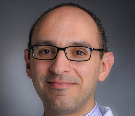 Joseph D. Mancias合作论文数Dana-Farber Cancer Institute;Harvard Medical School11Kimmelman Alec C合作论文数10John R. Lydeard合作论文数Department of Pathology, Harvard Medical School10Sebastian D. Hayes合作论文数School of Biomedical Sciences, University of Liverpool10Virginia Guarani合作论文数9Hankum Park合作论文数Department of Cell Biology, Harvard Medical School9Frances V Hundley合作论文数9James C. Powers合作论文数School of Chemistry and Biochemistry, Georgia Institute of Technology9
 Wolfram Goessling合作论文数Division of Gastroenterology, Massachusetts General Hospital;Division of Health Sciences and Technology, Harvard-MIT;Harvard Stem Cell Institute, Harvard University9Daniel C Scott合作论文数8
 Heeseon An合作论文数Tri-Institutional Training Program in Chemical Biology, Sloan Kettering Institute, Cornell University8
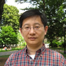 Wenyi Wei合作论文数Department of Pathology, Beth Israel Deaconess Medical Center, Harvard Medical School;Dana-Farber Harvard Cancer Center8
 Pumin Zhang (张普民)合作论文数The First Affiliated Hospital, Zhejiang University School of Medicine;Institute of Translational Medicine, Zhejiang University8Jiuchun Zhang合作论文数7X Leng合作论文数7
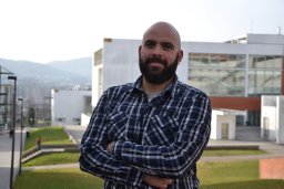 Joao A. Paulo合作论文数Department of Cell Biology, Harvard Medical School;Department of Cell Biology, Harvard Medical School7
 Felix Kraus合作论文数Department Biology II, Ludwig Maximilians University Munich7
 Xin Ye (叶鑫)合作论文数Institute of Microbiology, Chinese Academy of Sciences7
 Yang Shi (施扬)合作论文数Ludwig Institute for Cancer Research, Nuffield Department of Medicine, University of Oxford7
 Grzegorz Nalepa合作论文数AGH University of Science and Technology7
 Malavika Raman合作论文数Department of Pharmacology, University of Texas Southwestern Medical Center7Julia C Paoli合作论文数6
 Andre Van Wijnen合作论文数The Stein/Lian Laboratory, Department of Biochemistry, Larner College of Medicine, University of Vermont6Wang Xiaoxu合作论文数6
 Ian Smith合作论文数Velia Therapeutics6
 Timothy C. Thompson合作论文数Department of Genitourinary Medical Oncology, Division of Cancer Medicine, The University of Texas MD Anderson Cancer Center6
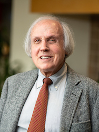 Gary S Stein合作论文数Department of Biochemistry, Larner College of Medicine, University of Vermont;Department of Surgery, Larner College of Medicine, University of Vermont;University of Vermont Cancer Center6Lisa Connell-Crowley合作论文数6Shireen A. Sarraf合作论文数6
 Mark Jedrychowski合作论文数Matchpoint Therapeutics6
 Thomas M. Wheeler合作论文数Sagis Diagnostics Laboratory, Sagis, PLLC6Takahiro Shirogane合作论文数5Meng-Kwang Marcus Tan合作论文数5Anne E White合作论文数5Tianlin Ma合作论文数5




