Purpose: Combining multi-site diffusion MRI (dMRI) data is hindered by inter-scanner variability, which confounds subsequent analysis. Previous harmonization methods require large, matched or traveling human subjects from multiple sites, which are impractical to acquire in many situations. This study aims to develop a deep learning-based dMRI harmonization framework that eliminates the reliance on multi-site in-vivo traveling human data for training. Methods: HARP employs a voxel-wise 1D neural network trained on an easily transportable diffusion phantom. The model learns relationships between spherical harmonics coefficients of different sites without memorizing spatial structures. Results: HARP reduced inter-scanner variability levels significantly in various measures. Quantitatively, it decreased inter-scanner variability as measured by standard error in FA (12
Multi-shell acquisition protocols sample diffusion signals at different b-values. While higher b-values (e.g., $$b = 2000\, \mathrm {s/mm^2}$$) offer increased sensitivity to microstructural features, their acquisition is time-intensive and suffers from reduced signal-to-noise ratio and motion sensitivity, which limits their routine clinical applicability. To address this challenge, we propose a deep learning framework for predicting high b-value ($$b = 2000\, \mathrm {s/mm^2}$$) spherical harmonic (SH) coefficients directly from low b-value ($$b = 1000\, \mathrm {s/mm^2}$$) SH coefficients. Our method is based on a simplified V-NET architecture augmented with adaptive multi-scale attention. This attention mechanism dynamically learns optimal receptive field combinations through scale-weighting networks. Unlike conventional fixed-scale approaches, our model incorporates three dilated convolutional branches with adaptive weighting, cross-scale feature fusion, and integrated spatial–channel attention. These components enable context-aware feature extraction that emphasizes the most relevant scales for diffusion pattern recognition. Experimental results demonstrate that the proposed method achieves accurate reconstruction with significantly reduced computational cost compared to modern state-of-the-art approaches.
White matter alterations are increasingly implicated in neurological diseases and their progression. Diffusion-weighted magnetic resonance imaging (DW-MRI) has been included in many international-scale studies to identify alterations in white matter microstructure and connectivity. Yet, quantitative investigation of DW-MRI data is hindered by a lack of consistency due to variations in acquisition protocols, sites, and scanners. Specifically, there is a need to harmonize the preprocessing of DW-MRI datasets to ensure that compatible and reproducible quantitative metrics are derived from each site, including (1) bundle-wise microstructure measures, (2) features of white matter fiber bundles, and (3) connectomics measures. In the MICCAI CDMRI 2023 QuantConn challenge, participants are provided raw data from the same individuals taken with two different acquisition protocols on a single 4 tesla scanner in the same scanning session and asked to preprocess the data in order to minimize acquisition differences while retaining biological variation. Here, we outline the testing framework, provide baseline pre-harmonized results, and discuss the learning implications of this challenge.
Every time a person sustains a blow to the head, they receive multiple atypical sensory inputs, often including pain. These directly stimulate the central nervous system. Yet, sensory input as a causal agent of neurophysiological dysfunction and post-concussion symptoms has never been explored. A new theory is proposed of sensory driven neurophysiological mechanisms of concussion (i) which are causally linked to the momentary blow to the head, (ii) whose time courses and other properties correspond to those observed to date for acute, sub-acute, and chronic symptoms, and (iii) which give rise to testable questions with experimentally measurable consequences. The primary assertion of the theory is that trauma induced excitation of key brain regions including the salience network (SN) and locus coeruleus (LC) can produce persistent dysfunctional alterations in the stable patterns of network excitability on which symptom-free neurological function depends. This mechanism is in play with any physical trauma, with or without a blow to the head. That is because atypical, painful, and otherwise high intensity sensory stimuli excite the SN and thence the LC, inducing plasticity widely in the brain. Many of those sensory stimuli may persist through the recovery period and while the brain is plastic, enable one or another network to learn altered and potentially dysfunctional patterns of network excitability. The secondary assertion of the theory is that with a blow to the head, convergence of high-intensity sensory stimuli within the brainstem and midbrain can cause neurophysiological coupling between brainstem nuclei which normally function independently, i.e. brainstem crosstalk (BCT). It is BCT which causes the signs and symptoms specific to head trauma, e.g., loss of consciousness, and oculomotor and vestibular dysfunction. The theory's reliance on sensory input emphasizes the importance of putative mechanisms whose initiating cause is known to have been present for every head trauma. This is in contrast to the century-long focus on mechanisms whose initiating cause, brain injury, is undetectable by clinical exam, neuroimaging, and bioassay in fully 60% of all head trauma, i.e., 70-75% of all mild TBI. As formulated and described, the theory is readily testable and falsifiable.
The purpose of this study was to assess the performance of predictive blood biomarkers for responsiveness to targeted treatments for chronic psychological issues years after traumatic brain injury (TBI). Targeted Evaluation Action and Monitoring of TBI was a prospective 6-month interventional trial of participants with chronic TBI sequelae (n = 95). Plasma biomarkers were analyzed pre-intervention: glial fibrillary acidic protein (GFAP), tau, hyperphosphorylated tau Thr231 (p-Tau), von Willebrand factor (vWF), brain lipid-binding protein (BLBP), ubiquitin C-terminal hydrolase-L1 (UCH-L1), vascular endothelial growth factor-a (VEGFa), and claudin-5 (CLDN5). Clinical outcomes included the Post-Traumatic Stress Disorder (PTSD) Checklist for DSM-5 (PCL-5) and Brief Symptom Inventory-18 (BSI-18). Regression models were built for change in PCL5/BSI-18. Biomarkers and covariates were included. Two models were built to identify responders (improved beyond the minimum clinically important difference). The model to predict change in PCL5 (R2=0.64; p < 0.001) included vWF (p = 0.032), BLBP (p = 0.001), tau (p = 0.002), VEGFa (p = 0.015), female sex (p = 0.06), and military status (p = 0.014). The model to predict change in BSI-18 (R2=0.42; p = 0.003) included vWF (p = 0.042), VEGFa (p = 0.09), BLBP (p = 0.01), CLDN5 (p < 0.001), female sex (p = 0.012), and military status (p = 0.004) as predictors. The model to differentiate participants who improved for PCL5 (R2=0.68; p < 0.001; AUC = 0.93) included vWF (p = 0.02), VEGFa (p = 0.008), and BLBP (p = 0.006). The model to differentiate participants who improved for BSI-18 (R2=0.25; p = 0.04; AUC = 0.75) included UCH-L1 (p = 0.03), GFAP (p = 0.06), and vWF (p = 0.03). Combinations of pre-intervention blood biomarkers were able to differentiate responders from nonresponders in both post-traumatic stress and overall psychological health domains.
White matter fiber reconstructions based on seeking local maxima of Orientation Distribution Functions (ODFs) typically fail to identify fibers crossing at narrow angles below 45^∘ . ODF-Fingerprinting (ODF-FP) replaces the ODF maxima localization mechanism with pattern matching, allowing the use of all information stored in ODFs. In this work, we study the ability of ODF-FP to reconstruct fibers crossing at varied angles spanning 10^∘ – 90^∘ in physical diffusion phantoms composed of textile tubes with 0.8 m diameter, approaching the anatomical scale of axons. Our results show that ODF-FP is able to correctly identify 80± 8% of the crossing fibers regardless of the crossing angle and provide the highest average reconstruction accuracy.
White matter alterations are increasingly implicated in neurological diseases and their progression. International-scale studies use diffusion-weighted magnetic resonance imaging (DW-MRI) to qualitatively identify changes in white matter microstructure and connectivity. Yet, quantitative analysis of DW-MRI data is hindered by inconsistencies stemming from varying acquisition protocols. Specifically, there is a pressing need to harmonize the preprocessing of DW-MRI datasets to ensure the derivation of robust quantitative diffusion metrics across acquisitions. In the MICCAI-CDMRI 2023 QuantConn challenge, participants were provided raw data from the same individuals collected on the same scanner but with two different acquisitions and tasked with preprocessing the DW-MRI to minimize acquisition differences while retaining biological variation. Harmonized submissions are evaluated on the reproducibility and comparability of cross-acquisition bundle-wise microstructure measures, bundle shape features, and connectomics. The key innovations of the QuantConn challenge are that (1) we assess bundles and tractography in the context of harmonization for the first time, (2) we assess connectomics in the context of harmonization for the first time, and (3) we have 10x additional subjects over prior harmonization challenge, MUSHAC and 100x over SuperMUDI. We find that bundle surface area, fractional anisotropy, connectome assortativity, betweenness centrality, edge count, modularity, nodal strength, and participation coefficient measures are most biased by acquisition and that machine learning voxel-wise correction, RISH mapping, and NeSH methods effectively reduce these biases. In addition, microstructure measures AD, MD, RD, bundle length, connectome density, efficiency, and path length are least biased by these acquisition differences. A machine learning approach that learned voxel-wise cross-acquisition relationships was the most effective at harmonizing connectomic, microstructure, and macrostructure features, but requires the same subject be scanned at each site co-registered. NeSH, a spatial and angular resampling method, was also effective and has generalizable framework not reliant co-registration. Our code is available at https://github.com/nancynewlin-masi/QuantConn/.
Purpose: To compare the performance of multi-echo (ME) and time-division multiplexing (TDM) sequences for accelerated relaxation-diffusion MRI (rdMRI) acquisition and to examine their reliability in estimating accurate rdMRI microstructure measures. Method: The ME, TDM, and the reference single-echo (SE) sequences with six TEs were implemented using Pulseq with single-band (SB) and multi-band 2 (MB2) acceleration factors. On a diffusion phantom, the image intensities of the three sequences were compared, and the differences were quantified using the normalized RMS error (NRMSE). Shinnar-Le Roux (SLR) pulses were implemented for the SB-ME and SB-SE sequences to investigate the impact of slice profiles on ME sequences. For the in-vivo brain scan, besides the image intensity comparison and T-2-estimates, different methods were used to assess sequence-related effects on microstructure estimation, including the relaxation diffusion imaging moment (REDIM) and the maximum-entropy relaxation diffusion distribution (MaxEnt-RDD). Results: TDM performance was similar to the gold standard SE acquisition, whereas ME showed greater biases (3-4x larger NRMSEs for phantom, 2x for in-vivo). T-2 values obtained from TDM closely matched SE, whereas ME sequences underestimated the T-2 relaxation time. TDM provided similar diffusion and relaxation parameters as SE using REDIM, whereas SB-ME exhibited a 60% larger bias in the map and on average 3.5x larger bias in the covariance between relaxation-diffusion coefficients. Conclusion: Our analysis demonstrates that TDM provides a more accurate estimation of relaxation-diffusion measurements while accelerating the acquisitions by a factor of 2 to 3.
In this paper, we propose a segmentation model using an anisotropic multi-well potential-based nonlinear transient PDE for colour images. A channel-wise greyscale classification approach is devised for colour image segmentation. The time evolution of the PDE model is carried out by the implicit–explicit convexity splitting approach. Further, we consider the fractional version of the time-discretised model by replacing the Laplacian with its fractional counterpart. The spatial terms are approximated by the Fourier basis under the pseudo-spectral method. The convergence and the stability of the numerical scheme are elaborated. Both models (fractional and non-fractional) are tested on some synthetic images and few real-world standard test images. The results on synthetic images are compared with those from the literature using Dice similarity index, Jaccard similarity index and BF score. Later the method is successfully applied on several medical images to classify the same.
Multi-shell HARDI scanning helps obtain a more accurate fiber orientation distribution function (fODF) than traditional DTI. However, it requires more DWI volumes, which require a longer scanning time, significantly limiting its clinical application. In this work, we have addressed this issue by proposing a GAN-based architecture to reconstruct Multi-shell Multi-tissue fODF (MSMT fODF), utilizing only a few DWI volumes obtained from single-shell scanning. The proposed GAN-based architecture leverages Residual blocks, a Sigmoid based Attention module, and a Feature Inter-dependencies based Attention module. Besides, multiple loss functions are used, including total variation, L-1 loss, and adversarial loss. Several qualitative and quantitative results justify our proposal in terms of MSMT fODF reconstruction, fiber tract estimation, and structural connectivity.
Chronic consequences of mild traumatic brain injury (mTBI) are heterogeneous, but may be treatable with targeted medical and rehabilitation interventions. A biological signature for the likelihood of response to therapy (i.e., "predictive" biomarkers) would empower personalized medicine post-mTBI. The purpose of this study was to correlate pre-intervention blood biomarker levels and the likelihood of response to targeted interventions for patients with chronic issues attributable to mTBI. Patients with chronic symptoms and/or disorders secondary to mTBI >3 months previous (104 days to 15 years; n = 74) were enrolled. Participants completed pre-intervention assessments of symptom burden, comprehensive clinical evaluation, and blood-based biomarker measurements. Multi-domain targeted interventions for specific symptoms and impairments across a 6-month treatment period were prescribed. Participants completed a follow-up testing after the treatment period. An all-possible model's backward logistic regression was built to identify predictors of improvement in relation to blood biomarker levels before intervention. The minimum clinically important difference (MCID) of the change score (post-intervention subtracted from pre-intervention) for the Post-Concussion Symptom Scale (PCSS) to identify treatment responders from non-responders was the primary outcome. The MCID for total PCSS score was 10. The model to predict change in PCSS score over the 6-month intervention was significant (R-2 = 0.09; p = 0.01) and identified ubiquitin C-terminal hydrolase L1 (odds ratio [OR] = 2.53; 95% confidence interval [CI], 1.18-5.46; p = 0.02) and hyperphosphorylated tau (p-tau; OR = 0.70; 95% CI, 0.51-0.96; p = 0.03) as significant predictors of symptom improvement beyond the PCSS MCID. In this cohort of chronic TBI subjects, blood biomarkers before rehabilitation intervention predicted the likelihood of response to targeted therapy for chronic disorders post-TBI.
BACKGROUND AND OBJECTIVE:Diffusion MRI (dMRI) has been considered one of the most popular non-invasive techniques for studying the human brain's white matter (WM). dMRI is used to delineate the brain's microstructure by approximating the WM region's fiber tracts. The achieved fiber tracts can be utilized to assess mental diseases like Multiple sclerosis, ADHD, Seizures, Intellectual disability, and others. New techniques such as high angular resolution diffusion-weighted imaging (HARDI) have been developed, providing precise fiber directions, and overcoming the limitation of traditional DTI. Unlike Single-shell, Multi-shell HARDI provides tissue fractions for white matter, gray matter, and cerebrospinal fluid, resulting in a Multi-shell Multi-tissue fiber orientation distribution function (MSMT fODF). This MSMT fODF comes up with more precise fiber directions than a Single-shell, which helps to get correct fiber tracts. In addition, various multi-compartment diffusion models, including as CHARMED and NODDI, have been developed to describe the brain tissue microstructural information. This type of model requires multi-shell data to obtain more specific tissue microstructural information. However, a major concern with multi-shell is that it takes a longer scanning time restricting its use in clinical applications. In addition, most of the existing dMRI scanners with low gradient strengths commonly acquire a single b-value (shell) upto b=1000s/mm2 due to SNR (Signal-to-noise ratio) reasons and severe imaging artifacts. METHODS:To address this issue, we propose a CNN-based ordinary differential equations solver for the reconstruction of MSMT fODF from under-sampled and fully sampled Single-shell (b=1000s/mm2) dMRI. The proposed architecture consists of CNN-based Adams-Bash-forth and Runge-Kutta modules along with two loss functions, including L1 and total variation. RESULTS:We have shown quantitative results and visualization of fODF, fiber tracts, and structural connectivity for several brain regions on the publicly available HCP dataset. In addition, the obtained angular correlation coefficients for white matter and full brain are high, showing the proposed network's utility.Finally, we have also demonstrated the effect of noise by adjusting SNR from 5 to 50 and observed the network robustness. CONCLUSION:We can conclude that our model can accurately predict MSMT fODF from under-sampled or fully sampled Single-shell dMRI volumes.
Sleep disturbances occur in up to 70% of patients with mild traumatic brain injury (mTBI). Modern mTBI management recommends targeted treatment for the patient's unique clinical manifestations (i.e., obstructive sleep apnea, insomnia). The purpose of this study was to evaluate the association of plasma biomarkers with symptom reports, overnight sleep evaluations, and response to treatment for sleep disturbances secondary to mTBI. This study is a secondary analysis of a prospective multiple interventional trial of patients with chronic issues related to mTBI. Pre- and post-intervention assessments were conducted, including overnight sleep apnea evaluation, the Pittsburgh Sleep Quality Index (PSQI), and blinded analysis of blood biomarkers. Bivariate Spearman correlations were conducted for pre-intervention plasma biomarker concentrations and 1) PSQI change scores and 2) pre-intervention sleep apnea outcomes (i.e., oxygen saturation measures). A backward logistic regression model was built to evaluate the association of pre-intervention plasma biomarkers with improvement in PSQI over the treatment period (p?<?0.05). Participants were 36.3???8.6 years old and 6.1???3.8 years from their index mTBI. Participants reported subjective improvements (PSQI?=??3.7???3.8), whereas 39.3% (n?=?11) had improved PSQI scores beyond the minimum clinically important difference (MCID). PSQI change scores correlated with von Willebrand factor (vWF; ??=??0.50; p?=?0.02) and tau (??=??0.53; p?=?0.01). Hyperphosphorylated tau correlated with average saturation (??=??0.29; p?=?0.03), lowest desaturation (??=??0.27; p?=?0.048), and baseline saturation (??=??0.31; p?=?0.02). The multi-variate model (R2?=?0.33; p?=?0.001) retained only pre-intervention vWF as a predictor (odds ratio?=?3.41; 95% confidence interval, 1.44?8.08; p?=?0.005) of improving PSQI scores beyond the MCID. vWF had good discrimination (area under the curve?=?0.83; p?=?0.01), with an overall accuracy of 77%, sensitivity of 46.2%, and specificity of 90.0%. Validation of vWF as a potential predictive biomarker of sleep improvement post-mTBI could optimize personalized management and healthcare utilization.
Diffusion MRI (dMRI) is one of the most popular techniques for studying the brain structure, mainly the white matter region. Among several sampling methods in dMRI, the high angular resolution diffusion imaging (HARDI) technique has attracted researchers due to its more accurate fiber orientation estimation. However, the current single-shell HARDI makes the intravoxel structure challenging to estimate accurately. While multi-shell acquisition can address this problem, it takes a longer scanning time, restricting its use in clinical applications. In addition, most existing dMRI scanners with low gradient-strengths often acquire single-shell up to b=1000s/mm2 because of signal-to-noise ratio issues and severe image artefacts. Hence, we propose a novel generative adversarial network, VRfRNet, for the reconstruction of multi-shell multi-tissue fiber orientation distribution function from single-shell HARDI volumes. Such a transformation learning is performed in the spherical harmonics (SH) space, as raw input HARDI volume is transformed to SH coefficients to soften gradient directions. The proposed VRfRNet consists of several modules, such as multi-context feature enrichment module, feature level attention, and softmax level attention. In addition, three loss functions have been used to optimize network learning, including L1, adversarial, and total variation. The network is trained and tested using standard qualitative and quantitative performance metrics on the publicly available HCP data-set.
HARDI-based diffusion MRI acquisition technique is a relatively recent modality of interest as it can yield more accurate fiber tracts. Besides, HARDI at higher magnetic strength is more sensitive to tissue changes and accurately estimate anatomical details in the human brain. However, a higher magnetic strength scanner is costly and not available in most clinical settings. Furthermore, due to signal-to-noise ratio issues and severe imaging artefacts, most existing 3T dMRI scanners with low gradient-strengths generally acquire single-shell up to b = 1000s/mm 2 . Hence, in this work, we consider the task of transforming the 3T single-shell HARDI signal (at b = 1000s/mm 2 ) to a 7T multi-shell HARDI signal utilizing the proposed deep learning model LF ANet. The proposed model consists of modules based on a Leapfrog method and an attention module. In addition, we have included suitable loss functions such as L 1 and total variation loss. Several quantitative and qualitative results have been presented to show the effectiveness of the proposed method.
Neuroelectric measures derived from human magnetoencephalographic (MEG) recordings hold promise as aides to diagnosis and treatment monitoring and targeting for chronic sequelae of traumatic brain injury (TBI). This study tests novel MEG-derived regional brain measures of tonic neuroelectric activation for long-term test-retest reliability and sensitivity to symptoms. Resting state MEG recordings were obtained from a normative cohort (CamCAN, baseline: n = 613; mean 16-month follow-up: n = 245) and a chronic symptomatic TBI cohort (TEAM-TBI, baseline: n = 62; mean 6-month follow-up: n = 40). The MEG-derived neuroelectric measures were corrected for the empty-room contribution using a random forest classifier. The mean 16-month correlation between baseline and 16-month follow-up CamCAN measures was 0.67; test-retest reliability was markedly improved in this study compared with previous work. The TEAM-TBI cohort was screened for depression, somatization, and anxiety with the Brief Symptom Inventory and for insomnia with the Insomnia Severity Index and was assessed via adjudication for six clinical syndromes: chronic pain, psychological health, and oculomotor, vestibular, cognitive, and sleep dysfunction. Linear classifiers constructed from the 136 regional measures from each TEAM-TBI cohort member distinguished those with and without each symptom, p < 0.0003 for each, i.e., the tonic regional neuroelectric measures of activation are sensitive to the presence/absence of these symptoms and clinical syndromes. The novel regional MEG-derived neuroelectric measures obtained and tested in this study demonstrate the necessary and sufficient properties to be clinically useful, i.e., good test-retest reliability, sensitivity to symptoms in each individual, and obtainable using automatic processing without human judgement or intervention.
Respiratory viruses are transported from an infected person to other neighboring people through respiratory droplets. These small droplets are easily advected by air currents in a room and can potentially infect others. In this work, the spread of droplets released during coughing, talking, and normal breathing is numerically analyzed in a typical conference room setting. The room space is occupied by ten people, with eight people sitting around a conference table and two people standing. Four different scenarios are considered, with the air-conditioning turned on/off and people wearing/not-wearing masks, to understand the spread of respiratory droplets inside the room. The flow in the room is simulated using a multiphase mixture model with properties computed for the inhaled and exhaled air using fundamental gas relations. The transport of respiratory droplets is analyzed using the discrete phase model with a range of droplet sizes fitted to data from previous experimental studies. The mask is modeled as porous media with the properties of a woven fabric computed using a newly developed model for multilayered homemade masks. The human inhalation and exhalation are modeled using analytical functions to mimic the biological flow patterns during breathing, coughing, and talking. Important observations about the air flow and dispersion of respiratory droplets in the conference room are presented based on the numerical analysis. Animations of all the results are included to provide insight into flow physics of the various dynamic conditions occurring in the room during an ongoing meeting. Although this study is conducted for a typical conference room, the newly developed models and techniques can be applied to other confined environments.
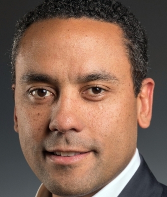 David O. Okonkwo合作论文数Neurotrauma Clinical Trials Center, Department of Neurological Surgery, School of Medicine, University of Pittsburgh40
 Sudhir Pathak合作论文数35
 Ava Puccio合作论文数Department of Neurological Surgery, School of Medicine, University of Pittsburgh;Neurotrauma Clinical Trials Center, University of Pittsburgh31
 Arthur D. Fisk合作论文数School of Psychology, Georgia Institute of Technology;Graphics, Visualization & Usability Center, College of Computing, Georgia Institute of Technology27
 Sue R Beers合作论文数21Kontos Anthony P合作论文数19Kathryn L Edelman合作论文数15
 Douglas C. Noll合作论文数Department of Biomedical Engineering, College of Engineering, University of Michigan;Department of Radiology, Medical School, University of Michigan;Michigan Alzheimer’s Disease Research Center, University of Michigan14
 Timothy Verstynen合作论文数Department of Biomedical Engineering, College of Engineering, Carnegie Mellon University13
 Ryan J. Soose合作论文数11Anthony P. Zuccolotto合作论文数Psychology Software Tools, Pittsburgh, Pennsylvania10Allison Borrasso合作论文数9
 Presson Nora合作论文数Learning Research and Development Center, University of Pittsburgh9
 Kevin Jarbo合作论文数9
 Shawn R Eagle合作论文数University of Pittsburgh Sports Medicine Concussion Program, University of Pittsburgh Medical Center8
 Christian Schunn合作论文数Department of Psychology, University of Pittsburgh8Jane Sharpless合作论文数7
 Charles Perfetti合作论文数Learning Research and Development Center, University of Pittsburgh;Department of Psychology, University of Pittsburgh;Department of Psychology, University of Michigan;Center for the Neural Basis of Cognition7
 B.V. Rathish Kumar合作论文数Department of Mathematics and Statistics, IIT Kanpur7
 Michael W. Cole合作论文数Center for Molecular and Behavioral Neuroscience, Department of Psychology, Rutgers University-Newark7
 Judith M. Shedden合作论文数7
 Aditya Nigam合作论文数Department of Computer Science and Engineering, Indian Institute of Technology Kanpur7Michael Collins合作论文数6
 Ranjeet Ranjan Jha合作论文数MANAS Lab, School of Computing and Electrical Engineering (SCEE), Indian Institute of Technology (IIT) Mandi, India. Electronic address: d16044@students.iitmandi.ac.in.6Mountz James M合作论文数6
 Michael S Worden合作论文数Department of Neuroscience, Brown University6
 Richard Shiffrin合作论文数Department of Psychological and Brain Sciences, The College of Arts & Sciences, Indiana University6
 Jason M. Chein合作论文数Department of Psychology, Temple University6
 Arnav Bhavsar合作论文数School of Computer and Electrical Engineering, Indian Institute of Technology6
 Fernando Boada合作论文数MR Research Center, University of Pittsburgh Medical Center6
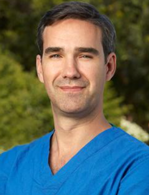 Juan Carlos Fernandez-Miranda合作论文数Department of Neurosurgery, Stanford University School of Medicine;Skull Base and Pituitary Surgery Division, Stanford University School of Medicine6Vishwesh Nath合作论文数5Donald Krieger合作论文数5
 Denes V. Agoston合作论文数Department of Primary Appointment, School of Medicine, Uniformed Services University5
 Ross C. Puffer合作论文数University of Maryland Medical Center;Walter Reed National Military Medical Center;School of Medicine, University of Maryland5
 Phillips Jeffrey S合作论文数Department of Neurology, University of Pennsylvania5
 Yue-Fang Chang合作论文数Department of Neurological Surgery, School of Medicine, University of Pittsburgh5Ray E. Eberts合作论文数5
 Elisabeth Anne Wilde合作论文数Department of Physical Medicine and Rehabilitation, Baylor College of Medicine5Richard A. Carlson合作论文数5Mark C. Detweiler合作论文数4Rebecca Hachey合作论文数4
 Savannah Tollefson合作论文数Department of Radiology, University of Pittsburgh4
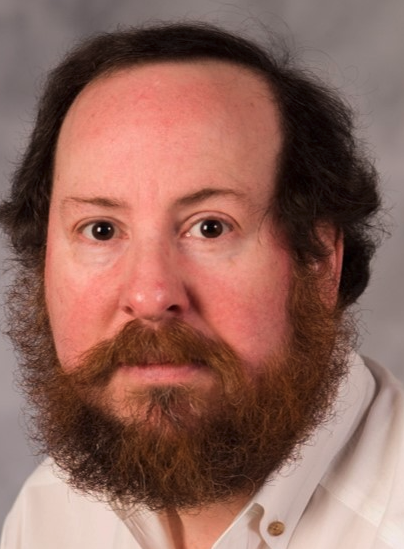 Phillip Ackerman合作论文数School of Psychology, College of Sciences, Georgia Institute of Technology4Charles M. Laymon合作论文数4
 Bird William A合作论文数4Minhas Davneet合作论文数4
 Steven Small合作论文数Department of Neuroscience, School of Behavioral and Brain Sciences, University of Texas at Dallas;The University of Texas Southwestern Medical Center;Biological Sciences Division, University of Chicago4
 Jonathan D. Cohen合作论文数Princeton Neuroscience Institute, Princeton University4




