Recurrent implantation failure (RIF) poses challenges to successful embryo implantation. In this study, we utilized isobaric tags for relative and absolute quantification (iTRAQ) to profile endometrial protein abundance in RIF patients. Through functional and pathway analyses, ECM-related proteins including fibronectin 1 (FN1), collagen type I alpha 2 chain (COL1A2), and integrin beta-1 (ITGB1) were revealed to be associated with RIF. Correlation analysis identified TGF-β1 as an upstream regulator of FN1. Knockdown experiments showed TGF-β1 downregulation could inhibit FN1 expression to inhibit decidualization markers. Our findings suggest a mechanistic link between TGF-β1/FN1 axis dysregulation and impaired decidualization observed in RIF. SIGNIFICANCE: Our study addresses the pressing issue of RIF, a significant obstacle in assisted reproductive technology. By employing isobaric tags for relative and absolute quantification (iTRAQ), we comprehensively analyzed endometrial protein abundance in RIF patients. Through functional and pathway enrichment analyses, we identified dysregulation in extracellular matrix (ECM)-related proteins, including FN1, COL1A2, and ITGB1, shedding light on their potential roles in implantation failure. Additionally, our correlation analysis revealed TGF-β1 as an upstream regulator of FN1, suggesting a novel regulatory axis involved in decidualization. Knockdown experiments further demonstrated the impact of TGF-β1 and FN1 on decidualization markers. This study contributes to a better understanding of the molecular mechanisms underlying RIF.
OBJECTIVES:This study aims to analyse the expression profiles, phenotypes, functions and cell-cell communication of various cell subpopulations in the affected aortic tissues of patients with Behçet's syndrome (BS) at the transcriptomic level. METHODS:This study recruited six participants (three with BS and three with atherosclerosis) from Beijing Anzhen Hospital between January 2023 and June 2024, collected their clinical information, and performed single-cell RNA sequencing on aortic tissue specimens using the SeekOne® MM High Flux Single Cell Transcriptome Kit V4.1. The data were analysed with Seurat and Harmony in R, including quality control, cell clustering, differential gene expression analysis, GO and KEGG enrichment analyses, subgroup analyses focusing on specific cell types, and intercellular communication analysis using CellChat v1.6.1. RESULTS:The study identified eight major cell types in aortic tissues, with significant differences in cell proportions between BS patients and controls. Compared with controls, BS patients had increased endothelial cells, fibroblasts and mesenchymal stem cells, while smooth muscle cells decreased. Subgroup analysis revealed significant differences between the BS and control groups in cell subpopulation distribution, enriched pathways and cell interactions. CONCLUSION:Our study revealed cellular and molecular changes in the aortic tissues of patients with BS, laying the foundation for elucidating the pathogenesis of BS and identifying potential therapeutic targets.
Background:Cognitive impairment is a major public health concern in aging populations, and early identification of risk factors is critical. The red blood cell distribution width to albumin ratio (RAR) has emerged as a potential biomarker reflecting inflammatory and nutritional status, but its association with cognitive impairment remains unclear. Objective:This study investigates the relationship between RAR and cognitive impairment in older adults, and explores potential mediating variables that may influence this association. Methods:A total of 2,913 participants aged ≥60 years from the National Health and Nutrition Examination Survey (NHANES) 2011-2014 cycles were analyzed, including 1,291 with cognitive impairment. Logistic regression assessed the association between RAR and cognitive impairment, adjusting for potential confounders such as age, gender, race, education, marital status, weight, height, and comorbidities. Restricted cubic spline (RCS) analysis evaluated the dose-response relationship and identified nonlinear thresholds. Subgroup analyses explored interactions between RAR and demographic/clinical factors. Causal mediation analysis, using a generalized linear model with a probit link and adjusting for age, sex, race, and education, was performed to estimate total, direct, and indirect effects via bootstrap resampling. Results:RAR was positively associated with cognitive impairment (P < 0.05). RCS analysis revealed a nonlinear threshold, with RAR ≥3.2 significantly increasing the risk of cognitive impairment (OR = 1.24, 95% CI: 1.11-1.38, P < 0.001). Subgroup analysis showed significant interactions between RAR and cardiovascular disease (CVD), hypertension, and depression (P for interaction <0.05). Stratified analysis found a stronger association between RAR and cognitive impairment in individuals without hypertension, CVD, or depression. Mediation analysis indicated that CVD (P = 0.036) and depression (P = 0.032) partially mediated the relationship, with CVD explaining 4.49% of the total effect. Hypertension had no significant mediating effect. Conclusion:RAR is significantly associated with cognitive impairment, with a stronger association when RAR ≥3.2. CVD and depression partially mediate this relationship, suggesting RAR as a potential biomarker for cognitive impairment in older adults.
Abnormalities in lipid metabolism have been proposed in Bietti’s crystalline dystrophy (BCD). We aim to characterize the lipid profiles in a case-control study. All participants were genetically confirmed by CYP4V2 gene sequencing and underwent chorioretinopathy evaluation by calculating the percentages of AF atrophy (PAFA). Fasting blood samples of BCD patients and controls were collected, and plasma was analyzed for routine lipid profiles. Targeted lipidomic evaluation includes long chain polyunsaturated fatty acids (LCPUFA) and associated eicosanoid metabolites. Routine lipids profiles showed elevated plasma levels of triglyceride (P = 0.043) and low-density lipoprotein cholesterol (P = 0.024) in BCD patients. Lipidomic analysis showed significantly decreased levels of ω-3 LCPUFA including docosahexaenoic acid (DHA, 22:6, P = 0.00068) and eicosapentaenoic acid (EPA, 20:5, P = 0.0016), as well as ω-6 LCPUFA arachidonic acid (ARA, 20:4, P < 0.0001) in BCD patients. Eicosanoid metabolites, either derived from ω-3 and/ or ω-6 LCPUFAs via cyclooxygenase (COX) or lipoxygenase (LOX) pathways, including 5-HEPE, 12-HEPE, 13-HDHA, 15-HETE, 12-HETE, 5-HETE, 6k-PGF1a, PGE2, PGJ2, and TXB2, exhibited significant differences (P < 0.0001) between BCD patients and controls. Genotypes of CYP4V2, specifically the biallelic null mutations, were observed to correlate with more remarkably reduced levels of oxylipins, involving major LOX pathway metabolites including 5-HETE, 5-HEPE, 12-HEPE and LTB4. BCD patients demonstrated significant decreases in plasma levels of ω-3 and ω-6 LCPUFA (DHA, EPA, and ARA), as well as their downstream metabolites via the COX and LOX pathways, suggesting that these might be implicated in BCD pathogenesis and could serve as biomarkers and therapeutic targets of the disease. What is known What is new
PURPOSE:To qualitatively and quantitatively characterise the genotypes and phenotypes of Bietti's crystalline dystrophy (BCD) in a cohort of patients. DESIGN:Cross-sectional and observational study. METHODS:Clinically confirmed BCD patients were recruited for genotyping and phenotyping. Multiple retinal imaging modalities were employed. Atrophy in the fovea was adopted as major consideration for staging strategy, while percentage area of autofluorescence (AF) atrophy (PAFA) in the macula was determined for quantitation. RESULTS:In 74 clinically diagnosed BCD patients, c.802-8_810del17insGC was shown the predominant variant of the CYP4V2 gene (allele frequency 55.4%). Sixty-two cases (123 eyes) with full imaging data were classified according to a modified criterion into stages 1 (n=8, 6.50%), 2A (n=9, 7.32%), 2B (n=17, 13.82%), 3A (n=30, 24.39%) and 3B (n=59, 47.97%). The eyes of the stage 2B were particularly deemed 'high risk' due to atrophy near fovea, while in stage 3A, though with remarkable foveal atrophy, preserved retinal pigment epithelium/photoreceptor islands near the fovea were found in 14 eyes. A tendency of increase in PAFA with age was found (rs=0.31, p=0.014). Significant PAFA increase was shown through stages 1 to 3B, and best-corrected visual acuity (BCVA, Logarithm of the Minimum Angle of Resolution) was shown to moderately correlate with PAFA (rs=0.56, p<0.001). CONCLUSION:The PAFA might be an efficient biomarker for BCD severities correlating with BCVA. The highly heterogeneous chorioretinopathy and BCVA of BCD cases appear to be associated with disease stages, progression types and patients' ages. Foveal involvement should be of a major concern for consideration of potential therapeutic intervention.
Background This study was to report a novel CREBBP mutation and phenotype in a child with Rubinstein–Taybi syndrome. Methods Case report of a 9-year-old boy. Results We described the patient’s clinical manifestations in detail, and found that in addition to the typical systemic manifestations of the syndrome, the outstanding manifestation of the child was severe intellectual deficiency and prominent ocular abnormalities. Whole-exome sequencing and sanger sequencing were performed on the patient and his parents, a large intragenic deletion, covering the exon 1 region and part of the intron 1 region of the TRAP1 gene, and the entire region from intron 27 to exon 30 of the CREBBP gene (chr16:3745393-3783894) was identified on the patient. This mutation affected the CREBBP histone acetyltransferase (HAT) domain. Conclusions This findings in our patient add to the spectrum of genetic variants described in Rubinstein–Taybi syndrome and present a RSTS patient with various ocular anomalies including early onset glaucoma.
Background: Spherophakia is a rare condition and compared to its occurrence with familial and systemic disorders, isolated spherophakia is even more uncommon. This rare case of isolated spherophakia will raise the alertness of physicians when dealing with a high myopia patient with shallow anterior chamber and relatively normal fundus. Case presentation: We report a case of a 17-year-old male who experienced painless decrease of vision in both eyes (OU) for 11 years, with progression of visual impairment and occasional ocular pain for one year. Examination revealed high myopia, increased intraocular pressures (IOP, 28 mmHg in the right eye (OD) and 33 mmHg in the left (OS)), shallow central anterior chambers, lenses of a spherical shape with superior subluxation, occludable angles without peripheral anterior synechiae, and healthy optic discs OU. A diagnosis of bilateral isolated spherophakia, lens subluxation, high myopia and secondary angle closure (AC) was made. Pars plana lensectomy with anterior vitrectomy and scleral suturing of an intraocular lens was performed. On postoperative follow-up at 19 days OD and 63 days OS, the visual acuity was 6/6 and the IOP was normal without any medication OU. Conclusions: A presentation with high myopia, shallow anterior chamber with a normal retina should alert the clinician to the possibility of spherophakia, as compared to high myopia caused by elongated axial length. Surgery should be considered in cases of spherophakia with AC where the IOP cannot be controlled by noninvasive means.
A number of patients with diabetes suffer from retinopathy; the pathogenesis is to be further investigated. Recent reports indicate that micro RNA (miR) plays critical roles in the development of immune inflammation. This study test a hypothesis that miR-17-92 cluster is associated with the pathogenesis of diabetes retinopathy (DR). In this study, peripheral blood samples were collected from DR patients and healthy subjects. B cells were isolated from the blood samples to be analyzed the expression of interleukin (IL)-10. The results showed that lower levels of IL-10 were detected in peripheral B cells of DR patients as compared with healthy subjects. miR-19a was increased in B cells of DR patients, which was negatively correlated with the IL-10 expression. Exposure of naive B cells to IL-17 increased the expression of miR-19a and suppression of IL-10 expression in the B cells, in which histone deacetylase 11 (HDAC 11) played a critical role. In conclusion, the IL-17 suppresses IL-10 expression in peripheral B cells via enhancing miR-19a expression and HDAC activity in DR patients. The miR-19a and HDAC 11 may be novel therapeutic targets in the treatment of DR.
Corneal epithelial barrier dysfunction is harmful to corneal health; the pathogenesis is unclear. This study aims to elucidate the mechanism by which tryptase compromises corneal epithelial barrier function. Human corneal epithelial cell line (HCE cells) was cultured into monolayers using as a study platform. Quantitative reverse transcription polymerase chain reaction and Western blotting were employed to detect the expression of matrix metalloprotenases (MMP)9. The endosome/lysosome fusion was observed by confocal microscopy. The corneal epithelial barrier function was assessed in Transwell system. The results showed that HCE cells expressed proteinase-activated receptor (PAR)2. Activation of PAR2 by tryptase induced expression of MMP9 in HCE cells, interfered with the fusion of endosome/lysosome, and compromised the epithelial barrier function, which could be prevented by pretreatment with MMP9 inhibitor. We conclude that tryptase can increase the expression of MMP9 in HCE cells and compromise the epithelial barrier function. Copyright (c) 2013 John Wiley & Sons, Ltd.
 Xiaoyan Peng (彭晓燕)合作论文数Beijing Tongren Hospital, Capital Medical University;Graduate School of Capital Medical University3
 Shengjuan Zhang合作论文数2Qian Li合作论文数Beijing Tongren Hospital(Beijing Hospital),Beijing,China2Huiling Guo合作论文数2
 Zhongjie Fu合作论文数Harvard Medical School, Harvard University
2
Zhongjie Fu合作论文数Harvard Medical School, Harvard University
2 Zibing Jin (金子兵)合作论文数Beijing Institute of Ophthalmology;Beijing Tongren Hospital, Capital Medical University2
 Xiaodong Jiao合作论文数2
 J. Fielding Hejtmancik合作论文数National Eye Institute, National Institutes of Health2Xiang Yu Shi合作论文数1Simeng Qi合作论文数1
 Dikang Pan合作论文数1
 Qian Wang合作论文数1Wang Qin合作论文数1Hui Wang合作论文数1Xiangyu Shi合作论文数1Sensen Wu合作论文数1Ye Zhang合作论文数1YanQi Li (李岩祺)合作论文数1
 Hanxue Zhao (赵汉学)合作论文数1LianRui Guo (郭连瑞)合作论文数1JianMing Guo (郭建明)合作论文数1
 Xiaokui Yang (杨晓葵)合作论文数首都医科大学附属北京妇产医院生殖医学科1
 Yingnan Zhang合作论文数Beijing Tongren Hospital, Beijing Hospital1Gao Xinxiao合作论文数Department of Ophthalmology, Osaka University Graduate School of Medicine1
 XuSheng Cao (曹绪胜)合作论文数Beijing Tongren Hospital, CMU1
 Weining Rong (容维宁)合作论文数宁夏回族自治区人民医院眼科1
 Qisheng You (游启生)合作论文数Beijing Tongren Hospital, Capital Medical University1
 Ying Jie (接英)合作论文数Beijing Tongren Hospital, Capital Medical University1
 Zhiqiang Pan (潘志强)合作论文数. Beijing Tongren Eye Bank, Beijing Ophthalmology and Visual Science Key Lab, Beijing Tongren Eye Center, Beijing Tongren Hospital, Capital Medical University, Beijing , China1
 Yongquan Gu (谷涌泉)合作论文数Xuanwu Hospital, Capital Medical University;Graduate School, Capital Medical University1Ravi Thomas合作论文数Queensland Eye Institute1
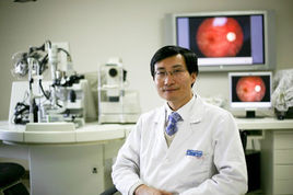 Wenbin Wei (魏文斌)合作论文数.Beijing Tongren Eye Center,Beijing Tongren Hospital,Capital Medical University,Beijing ,China1




