Although programmed cell death 1 inhibitors (PD1i) are recommended in guidelines for relapsed/refractory extranodal NK/T-cell lymphoma (ENKTL), their role in frontline therapy remains unclear. In this retrospective, multi-center study of 755 newly diagnosed ENKTL patients, 17.9% received first-line PD1i. Despite harboring more adverse risk features, the PD1i group showed significantly improved progression-free survival (hazard ratio = 0.62, p = 0.007) and overall survival (hazard ratio = 0.55, p = 0.027) after a median follow-up of 32.9 months. The benefit was also observed in key subgroups (non-upper aerodigestive tract, advanced stage, intermediate/high-risk). Conventional multivariate Cox regression before propensity score matching, robust multivariate Cox analysis after matching, and the time-varying Cox model employed in landmark analysis consistently demonstrated that PD1i therapy served as an independent favorable prognostic factor for both progression-free survival and overall survival. The random forest algorithm revealed enhanced efficacy with PD1i-asparaginase combination therapy, showing about a 50% reduction in event probability, particularly in advanced-stage and intermediate/high-risk patients. Multi-state survival modeling indicated PD1i primarily delays disease progression rather than post-progression survival. Safety data showed that adding PD1i to asparaginase-based chemotherapy resulted in an expected increase in hematologic toxicities, but with a reduced risk of severe/fatal infections and rare severe immune-related adverse events, demonstrating a favorable risk-benefit profile. Overall, in this study, frontline PD1i, particularly in combination with asparaginase, yielded significantly improved outcomes and demonstrated a manageable safety profile in intermediate-/high-risk and advanced ENKTL. This evidence supports its incorporation into first-line treatment and underscores the need for prospective randomized trials.
The co-expression of myeloid and B-cell antigens is characteristic of mixed-phenotype acute leukemia (MPAL)-B/myeloid. This finding can also be observed in AML with t(8;21)(q22;q22)/RUNX1::RUNX1T1, as well as in cases of AML with other RUNX1 rearrangements, copy number gains, or mutations. AML with plasmacytoid dendritic cell (pDC) differentiation (pDC-AML) containing clonally-related myeloblasts and neoplastic pDCs, are enriched for RUNX1 mutations and have been shown to exhibit B-cell marker expression. Here, we present two cases of pDC-AML with B-cell marker expression in which RUNX1 aberrations were not identified. Although previously we speculated on the role of RUNX1 in the aberrant expression of B-cell markers such in cases, the absence of RUNX1 lesions in the current cases supports the potential inherent ability of pDCs to express B-cell markers during maturation.
Diffuse large B-cell lymphoma (DLBCL) represents the most prevalent form of non-Hodgkin lymphoma, distinguished by its aggressive clinical presentation and poor patient outcomes. This study investigated the role of nuclear receptor subfamily 2 group F member 2 (NR2F2) and 14-3-3 epsilon (YWHAE) in DLBCL progression. NR2F2 and YWHAE were highly expressed in DLBCL cell lines and tumor tissues of patients with DLBCL. Elevated expression of both genes correlated with advanced Ann Arbor stage and higher International Prognostic Index scores in DLBCL patients. Functional assays demonstrated that knockdown of NR2F2 or YWHAE suppressed DLBCL cell proliferation, migration, and invasion, as well as tumor growth in xenograft models. Mechanistically, NR2F2 transcriptionally activated YWHAE by binding to its promoter, thereby promoting phosphoinositide 3-kinase (PI3K)/protein kinase B (AKT) signaling. Notably, treatment with a PI3K activator in YWHAE-silenced cells, or YWHAE overexpression in NR2F2-silenced cells, partially reversed the inhibitory effects on malignant behaviors of DLBCL cells. Collectively, these findings indicate that NR2F2 promotes DLBCL progression through transcriptional activation of YWHAE and subsequent activation of the PI3K/AKT pathway, suggesting a potential therapeutic target for DLBCL.
Menin inhibition leads to an antileukemic effect through hematopoietic differentiation. Treatment with the menin inhibitor revumenib results in clinical remissions in relapsed or refractory (R/R) acute myeloid leukemia (AML) with either rearrangement of lysine methyltransferase 2A (KMT2A) or mutation in nucleophosmin 1 (NPM1), leading to regulatory approval of this drug. However, determinants of response to revumenib have not been fully elucidated. We examined the immunophenotype of leukemia cells by flow cytometry, in sequential bone marrow specimens from 48 patients with R/R AML treated with revumenib. We observed dynamic changes in the immunophenotype after treatment in 16 of 31 (52%) patients, characterized by a switch from a myeloid/stem-like to a monocytic or myelomonocytic immunophenotype, or vice versa, or by substantial changes in the intensity of antigen expression or in patterns of leukemia-associated immunophenotypes. Morphologic remission with undetectable measurable residual disease (MRD) by flow cytometry following revumenib was associated with improved overall survival, with a median of 23.6 months compared with 20.8 months in patients with morphologic response and detectable MRD, and 3.2 months in non-responders. In summary, treatment monitoring of AML by flow cytometry, following menin inhibition, requires recognition of phenotypic changes associated with differentiation.
Abstract Background Renal cell carcinoma (RCC) is the most common type of kidney cancer in adults, with common metastatic sites including the lungs, bones, liver, and brain. Omental metastasis is exceedingly rare and usually occurs postoperatively. Fumarate hydratase‐deficient (FH‐deficient) RCC, a recently classified and highly aggressive subtype of RCC, is known to show early metastatic potential but remains poorly understood. We present a rare case of synchronous FH‐deficient RCC with isolated omental metastasis identified at diagnosis. Case Presentation We report the case of a 48‐year‐old man who first presented with abdominal pain, early satiety, and a 25‐pound weight loss. Imaging revealed an 11.8 cm right renal mass with a separate 15.4 cm left upper quadrant mesenteric mass with no definitive evidence of lung, bone, or liver involvement. Biopsies of both renal and mesenteric masses confirmed non‐clear‐cell FH‐deficient RCC with papillary architecture and diffuse overexpression of 2‐succinyl cysteine. Conclusion Omental metastasis of RCC is rare, especially in the absence of prior surgery. This case is distinguished by its synchronous presentation of the primary RCC and omental metastasis with a rare histologic subtype. Unlike most documented reports of RCC with omental spread, which typically involve clear cell histology and often present years after nephrectomy, this case involves a rare, aggressive subtype with atypical metastatic behavior. This case underscores the importance of considering atypical metastatic patterns in non‐clear‐cell subtypes and the need for further research to inform evidence‐based therapy.
 L. Jeffrey Medeiros合作论文数Department of Hematopathology, Division of Pathology/Lab Medicine, The University of Texas MD Anderson Cancer Center95
 Guilin Tang合作论文数61
 Sa Wang合作论文数60
 Shimin hu合作论文数Univ Texas MD Anderson Canc Ctr, Dept Hematopathol, Houston, TX 77030 USA53
 Jie Xu (许杰)合作论文数Shanghai Cancer Institute;School of Pharmaceutical Sciences, Fudan University44Hong Fang合作论文数39
 Loghavi Sanam合作论文数Dept Hematopathol, Univ Texas MD Anderson Canc Ctr35
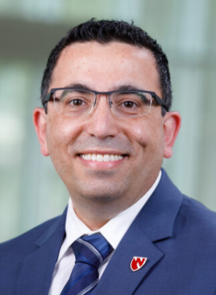 Joseph D. Khoury合作论文数Department of Pathology, Microbiology and Immunology, College of Medicine, University of Nebraska Medical Center35
 Zhenya Tang合作论文数Department of Hematopathology, The University of Texas MD Anderson Cancer Center30
 Shaoying Li合作论文数30
 Pei Lin合作论文数25
 Cheng Cameron Yin合作论文数Department of Hematopathology, Department of Hematopathology, Division of Pathology/Lab Medicine, The University of Texas MD Anderson Cancer Center;The University of Texas Graduate School of Biomedical Sciences25
 Siba El Hussein合作论文数Department of Hematopathology, The University of Texas MD Anderson Cancer Center, Houston, TX, USA.23Zhihong Hu合作论文数20Thakral Beenu合作论文数Department of Pathology and Laboratory Medicine, NorthShore University Hospital20
 M. James You合作论文数Diagnostic Bone Marrow Laboratory, Department of Hematopathology, The University of Texas MD Anderson Cancer Center;Division of Pathology/Lab Medicine, The University of Texas MD Anderson Cancer Center20
 Roberto N. Miranda合作论文数Department of Hematopathology, University of Texas MD Anderson Cancer Center;University of Missouri16
 Keyur P. Patel合作论文数Department of Hematopathology, Division of Pathology/Lab Medicine, The University of Texas MD Anderson Cancer Center;Department of Molecular Diagnostics Laboratory, Division of Pathology/Lab Medicine, The University of Texas MD Anderson Cancer Center15
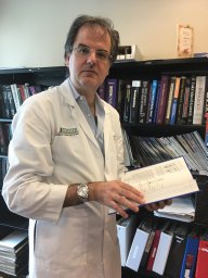 Francisco Vega合作论文数Houston Medical School, University of Texas15Fatima Zahra Jelloul合作论文数14
 Mahsa Khanlari合作论文数Department of Pathology, St. Jude Children's Research Hospital11
 Elias Jabbour合作论文数Department of Leukemia, Division of Cancer Medicine, MD Anderson Cancer Center, University of Texas11Gokce A. Toruner合作论文数Institute of Genomic Medicine, UMDNJ-New Jersey Medical School11
 Ghayas C Issa合作论文数10
 Jeffrey L. Jorgensen合作论文数Department of Hematopathology, The University of Texas M.D. Anderson Cancer Center10
 Gautam Borthakur合作论文数Department of Leukemia, Division of Cancer Medicine, The University of Texas MD Anderson Cancer Center10
 Tapan M. Kadia合作论文数Department of Leukemia, Division of Cancer Medicine, M.D. Anderson Cancer Center10
 Rashmi Kanagal Shamanna合作论文数Molecular Diagnostic Laboratory, Department of Hematopathology, Division of Pathology and Laboratory Medicine, The University of Texas MD Anderson Cancer Center;Leukemia Planning Clinic, Department of Hematopathology, Division of Pathology and Laboratory Medicine, The University of Texas MD Anderson Cancer Center9
 Naval G. Daver合作论文数Department of Leukemia, Division of Cancer Medicine, The University of Texas MD Anderson Cancer Center9
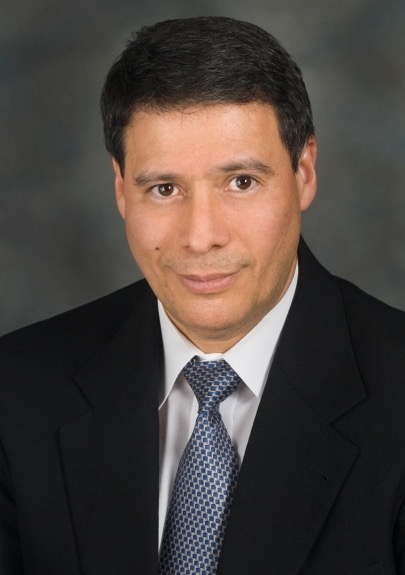 Carlos E. Bueso-Ramos合作论文数Bone Marrow Laboratory, Department of Hematopathology, The University of Texas MD Anderson Cancer Center;Division of Pathology/Lab Medicine, The University of Texas MD Anderson Cancer Center9
 Sofia Garces Narvaez合作论文数8Xiaoqiong Wang合作论文数Departments of Hematopathology, The University of Texas MD Anderson Cancer Center8
 Naveen Pemmaraju合作论文数Department of Leukemia, The University of Texas MD Anderson Cancer Center8
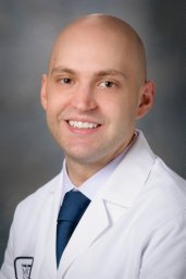 Nicholas Short合作论文数Department of Cellular and Structural Biology, University of Texas Health Science Center at San Antonio8
 Wei Xie合作论文数Wuhan University Wuhan University8Shuyu E合作论文数8Chi Young Ok合作论文数8Lianqun Qiu合作论文数7
 Koji Sasaki合作论文数6Do Hwan Kim合作论文数6
 Andres Quesada合作论文数6
 Mehrnoosh Tashakori合作论文数Department of Laboratory Medicine and Pathology, University of Minnesota6Liu Meng合作论文数6Hannah Cheung Beird合作论文数Univ Texas MD Anderson Canc Ctr, Dept Genom Med, Houston, TX 77030 USA6
 Walter Richard Burack合作论文数6
 Hagop M. Kantarjian合作论文数Department of Leukemia, Division of Cancer Medicine, The University of Texas MD Anderson Cancer Center6Yuan Ji合作论文数6
 Zimu Gong合作论文数China Medical University Shenyang, China Medical University6
 Sergej Konoplev合作论文数Memorial Sloan Kettering Cancer Center;Department of Hematopathology, The University of Texas MD Anderson Cancer Center6




