Congenital muscular dystrophy with laminin-α2 deficiency, also known as MDC1A, displays an extensive phenotypic and genetic heterogeneity. The combination of clinical, biochemical, and genetic findings must be considered to obtain the precise diagnosis and provide appropriate genetic counseling. Here we report one individual from a family presenting with clinical features including seizure attack, slight weakness of proximal leg muscles, and mild cognitive impairment with increased small angular fibers, decreased expression of α-DG and β-DG, normal expression of laminin-α2, and severe white matter changes. Targeted next-generation sequencing (NGS) revealed two homozygous missense mutations, c.2881G>A (p.Ala961Thr) and c.4406G>A (p.Cys1469Tyr), in LAMA2 in the affected member of the family. Together, these results demonstrate a role for c.2881G>A and c.4406G>A mutations in LAMA2 and show that these two mutations, especially c.4406G>A, may cause mild cognitive impairment, slight motor retardation, seizures, and severe leukoencephalopathy, which extends the clinical spectrum associated with LAMA2 mutations.
The existing data about whether acid sensing ion channels (ASICs) are proconvulsant or anticonvulsant are controversial. Particularly, acid sensing ion channel 3 (ASIC3) is the most sensitive to extracellular pH and has the characteristic ability to generate a biphasic current, but few studies have focused on the role of ASIC3 in seizure. Here we found ASIC3 expression was increased in the hippocampus of pilocarpine induced seizure rats, as well as in hippocampal neuronal cultures undergoing epileptiform discharge elicited by Mg 2+ -free media. Furthermore, ASIC3 blockade by the selective inhibitor APETx2 shortened seizure onset latency and increased seizure severity compared with the control in the pilocarpine induced seizure model. Incubation with APETx2 enhanced the excitability of primary cultured hippocampal neurons in Mg 2+ -free media. Notably, the aggravated seizure was associated with upregulation of the N -methyl- d -aspartate subtype of glutamate receptors (NMDARs), increased NMDAR mediated excitatory neurotransmission and subsequent activation of the Ca 2+ /calmodulin-dependent protein kinase II (CaMKII) and cAMP-response element binding protein (CREB) signaling pathway. Moreover, co-immunoprecipitation confirmed the interaction between ASIC3 and NMDAR subunits, and NMDARs blockade prevented the aggravated seizure caused by ASIC3 inhibition. Taken together, our findings suggest that ASIC3 inhibition aggravates seizure and potentiates seizure induced hyperexcitability at least partly by the NMDAR/CaMKII/CREB signaling pathway, which implies that ASIC3 agonists may be a promising approach for seizure treatment.
BACKGROUND/AIMS:Migraine is a disabling condition that severely impacts socioeconomic function and quality of life. The focus of this study was to develop a mouse model of trigeminal pain that mimics migraine.METHODS:After undergoing dural cannulation surgery, mice were treated with repeated dural doses of an acidic solution to induce trigeminal pain.RESULTS:The method elicited intermittent, head-directed wiping and scratching as well as the expression of both the c-FOS gene in the spinal trigeminal nucleus caudalis and calcitonin gene related peptide (CGRP) in the periaqueductal grey matter. Interestingly, the acid-induced trigeminal pain behaviour was inhibited by amiloride, an antagonist of acid-sensing ion channels (ASICs), but not by AMG-9810, an inhibitor of transient receptor potential cation channel V1(TRPV1). In addition, the relative mRNA and protein expression levels of ASIC1a and ASIC3 were increased in the acid-induced trigeminal nociceptive pathways. Furthermore, blocking CaMKII with KN-93 significantly reduced the acid-induced trigeminal pain behaviour and c-FOS gene expression.CONCLUSION:The data suggested that chronic intermittent administration of an acidic solution to mice resulted in trigeminal hypersensitivity and that dural acid-induced trigeminal pain behaviour in mice may mechanistically mimic migraine. The observations here identify an entirely novel treatment strategy for migraine.
The aim of this study was to evaluate the changes in the cognitive performance of migraine patients using a comprehensive series of cognitive/behavioral and electrophysiological tests.
Although the function of the cerebellum in neurocognition has been well-documented, the similar role of the brainstem has yet to be fully elucidated. This clinical research study aimed to combine data relating to neuropsychological assessments and P300 to explore cognitive dysfunction and affective impairment following brainstem stroke. Thirty-four patients with isolated brainstem stroke and twenty-six healthy controls were recruited; for each patient, we collated data pertaining to the P300, Addenbrooke's Cognitive Examination III (ACE-III), Montreal Cognitive Assessment Chinese version (MoCA), trail-making test (TMT), Symbol Digit Modalities Test (SDMT), Wechsler Adult Intelligence Scale-Digit Spans (DS), Stroop test, Self Rating Depression Scale (SDS), and Self Rating Anxiety Scale (SAS). Significance was analyzed using an independent T-test or the Mann-Whitney U-test. Correlation was analyzed using Pearson's correlation analysis or Spearman's correlation analysis. Collectively, data revealed that brainstem stroke caused mild cognitive impairment (MCI), and that visuospatial, attention, linguistic, and emotional disturbances may occur after isolated brainstem stroke. Cognitive decline was linked to P300 latency, ACE-III, andMoCA; P300 latency was correlated with ACE-III. Patients with right brainstem lesions were more likely to suffer memory decline. The present study provides initial data relating to the role of the brainstem in neurocognition, and will be useful for further understanding of vascular cognitive and affective impairment.
Development of a culture system that supports self-renewal and proliferation of spermatogonial stem cells (SSCs) is enormously valuable for experimental research and potential treatment for male infertility. Although several research groups had reported their successes in SSC isolation and culture, the two current accepted culture systems are different in cell enrichment methods, serum and growth factors. Previous researches also indicated SSCs from different mouse strains required different culture conditions. Here we report for the first time that SSCs from BALB/c mice could be cultured in an improved culture system for 3 months. The modified culture system consisted of an improved enzymatic procedure, the enrichment of undifferentiated spermatogonia by differential adherence selection of isolated SSCs, mouse embryonic fibroblast feeder cells, StemPro-34 SFM medium supplemented with glial cell line-derived neurotrophic factor (GDNF), basic fibroblast growth factor and GDNF-family receptor α1 (GFRα1). The improved digestion method increased the viability and enrichment efficiency of isolated testis cells. Furthermore, basal culture medium with 10% fetal bovine serum as selected medium could increase the number of germ cell colonies in the initiation stage of culture. Cultured SSCs were characterized morphologically and formed typical colonies. Immunocytochemical staining and RT-PCR showed that cultured SSCs expressed Oct-4, GFRα1, Sox2 and several other special genes resembling undifferentiated spermatogonia. Spermatogonia transplantation further confirmed that cultured SSCs were functionally normal and could restore complete spermatogenesis. The culture methods described here could serve as a paradigm to establish conditions for the culture of SSCs from other species, allowing identification of universal factors necessary for proliferation of SSCs.
 Zheman Xiao (肖哲曼)合作论文数Department of Neurology, Renmin Hospital of Wuhan University5
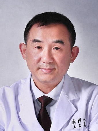 Zuneng Lu (卢祖能)合作论文数Department of Neurology, Renmin Hospital, Wuhan University4
 Lifang HUANG (黄丽芳)合作论文数3Man Ding合作论文数2XiuJuan Fu (付秀娟)合作论文数2
 Fan Zhu (朱帆)合作论文数School of Basic Medical Sciences, Wuhan University;State Key Laboratory of Virology and Biosafety, Wuhan University2
 Wenhua Zhu合作论文数1Xianjin Du合作论文数1Qian Cao合作论文数1
 Chao Weng合作论文数1Yanping Zeng合作论文数1FuJin Shen (申复进)合作论文数1HongYun Zheng (郑红云)合作论文数1HongJuan Dong (董红娟)合作论文数1WenBiao Liao (廖文彪)合作论文数1LingLong Wang (王玲珑)合作论文数1
 Sushan Luo合作论文数Huashan Hospital, Fudan University1
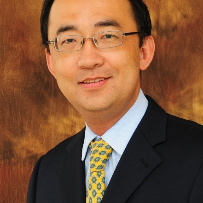 Chongbo Zhao合作论文数Department of Neurology, Huashan Hospital, Fudan University1
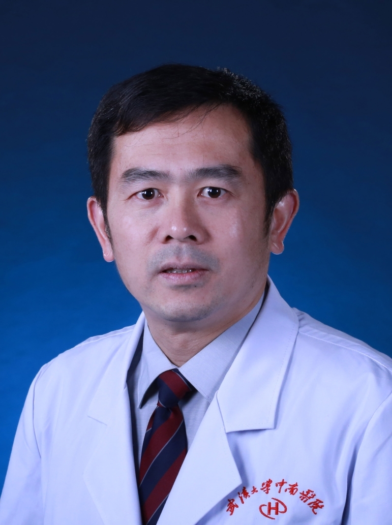 Hong Zhang (张洪)合作论文数Zhongnan Hospital of Wuhan University;Second Clinical Hospital of Wuhan University1Gao Mingshi合作论文数1Shuang Cai (蔡爽)合作论文数1
 Yan Wang (王琰)合作论文数College of Life Sciences, Wuhan University1
 Yunhe Xiong合作论文数Wuhan University, Wuhan University1
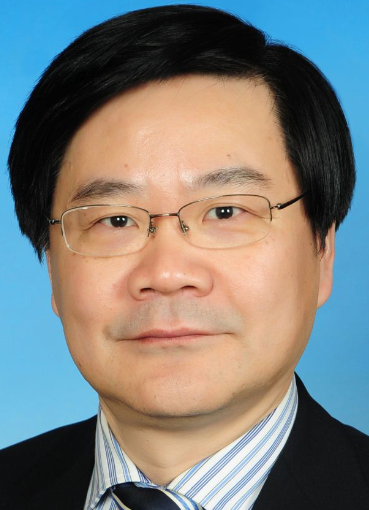 Sixing Yang (杨嗣星)合作论文数Renmin Hospital, Wuhan University1
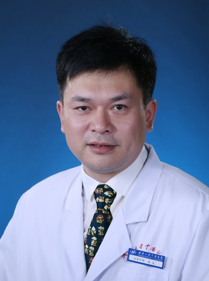 Ci Zhang (张茨)合作论文数Zhongnan Hospital of Wuhan University1




