Background Accurate identification of complete responders before neoadjuvant chemoradiotherapy (nCRT) is essential for organ-preservation strategies for rectal cancer. However, current preoperative radiological assessment methods lack sufficient accuracy. The purpose of this study is to evaluate the diagnostic value of contrast-enhanced ultrasound (CEUS) in distinguishing complete response (CR) in patients with rectal cancer after nCRT. Methods In this prospective study, 100 patients with rectal cancer treated between January 2023 and February 2025 underwent endorectal ultrasound (ERUS) and CEUS examinations before and 6–8 weeks after nCRT. Quantitative perfusion parameters were derived using time–intensity curve analysis. Surgical histopathology served as the reference standard for pathological CR, and multimodal clinical evaluation defined complete CR. Diagnostic performance was assessed using logistic regression and receiver operating characteristic curve analysis. Result A total of 100 participants (mean age, 57 ± 11 years; 77 men) were included. Pathological CR occurred in 29 (29.0%) participants, and clinical CR in 13 (13.0%), yielding an overall CR rate of 42%. Post-nCRT CEUS parameters—relative area under the time–intensity curve (rAUC*), relative enhancement intensity, and enhancement intensity difference—were significant predictors of CR (all p < 0.05). The AUCs were 0.72 (95% CI: 0.617, 0.829), .76 (95% CI: 0.665, 0.858), and 0.76 (95% CI: 0.695, 0.875), respectively. Combined models achieved AUCs of 0.80 (95% CI: 0.708, 0.887) and 0.82 (95% CI: 0.741, 0.903), respectively. Conclusion Quantitative contrast-enhanced ultrasound (CEUS) parameters, including enhancement intensity (EI), relative AUC*, and their derived ratio values (rEI, rAUC*) and difference (ΔEI), demonstrated robust discriminative capacity in complete response participants after nCRT. The CEUS-based diagnostic model also demonstrated a high level of diagnostic efficacy. The clinical diagnostic model constructed from the above parameters demonstrated higher diagnostic value.
Abstract Fluorescent nanothermometers have garnered widespread attention in the biomedical field due to their high precision, rapid temporal response, and strong tissue penetration capability. However, accurately predicting and regulating the thermometric performance is still hindered by the lack of high-precision dynamics models and data on the temperature dependence of intrinsic parameters. This leads to improved performance of nanothermometers, which heavily relies on extensive experimental exploration, characterized by a high degree of trial and error, labor-intensive efforts, and a lack of mechanistic understanding. Therefore, for fluorescence lifetime-based thermometry, this paper establishes a fluorescence dynamics model and proposes a performance prediction method for nanothermometers based on this model. First, we developed a microscopic rate equation model for core–shell nanostructures, which has enabled a precise description of luminescence for nanoparticles with various structures and doping concentrations of luminescent ions. By selecting the smallest modeling unit and simulating the random doping of luminescent ions, along with comprehensively considering physical processes such as stimulated absorption, radiative relaxation, and energy migration/transfer, we achieved accurate and computationally efficient modeling of nanoparticles with multiple shell layers. Guided by the evaluation criterion for randomness of lattice modeling, a strategy of repeated modeling and computation was proposed to mitigate the effects of such randomness. Second, within the temperature range relevant for biological tissue thermometry (25–70 °C), we experimentally measured the intrinsic parameters of the fluorescence system, including the absorption cross-section, radiative relaxation rate, and energy migration/transfer microparameters. Finally, based on the established model and measured parameters, we proposed a method for predicting the thermometric performance of nanothermometers, and the theoretical predictions of luminescence lifetime show excellent agreement with the experimentally measured values (relative deviation is only 1%). Through the simulation of the spatiotemporal distribution of excitation energy in nanoparticles, we revealed the microscopic physical mechanisms by which particle structure, luminescent ion doping concentration, and excitation conditions regulate thermometric performance. This study provides a theoretical model, fundamental data, and methodological support for the performance prediction and regulation of fluorescent nanothermometers.
Breast ultrasound (BUS) is an essential tool for diagnosing breast lesions, with millions of examinations per year. However, publicly available high-quality BUS benchmarks for AI development are limited in data scale and annotation richness. In this work, we present BUS-CoT, a BUS dataset for chain-of-thought (CoT) reasoning analysis, which contains 11,439 ultrasound images from 11,850 lesions and 4,838 patients, covering all 99 WHO-defined histopathology categories. For model training and evaluation, we provide a curated high-quality subset of 5,163 lesion-focused images annotated by experienced radiologists. To facilitate research on incentivizing CoT reasoning, we construct the reasoning processes based on observation, feature, diagnosis and pathology labels, annotated and verified by experienced experts. Moreover, by covering lesions of all histopathology types, we aim to facilitate robust AI systems in rare cases, which can be error-prone in clinical practice. The data and code are publicly available at https://doi.org/10.6084/m9.figshare.30838715.
Immunotherapy shows promise for triple-negative breast cancer (TNBC), yet its effectiveness is restricted by low response rates, poor immune cell infiltration, and systemic side effects. Here, an ultrasound-responsive cerasomal nanoplatform integrating a STING agonist (SR-717@PC-iRGD) is developed for synergistic sonodynamic-immunotherapy. The nanocarrier is self-assembled from cerasome-forming lipids (CFL), porphyrin-conjugated lipids (PL), unsaturated phospholipids (DOPC), DSPC, and DSPE-PEG2000-iRGD, with SR-717 loaded in the lipid bilayer. The resulting assembly yields nanoparticles (NPs) with high SR-717 loading and exceptional stability. The siloxane shell (cerasome) confers high stability and prevents premature drug leakage, while iRGD promotes nanoparticle binding to tumor specific integrin to facilitate accumulation and retention in the tumor. Upon ultrasound irradiation, porphyrin generates reactive oxygen species (ROS) that oxidize the lipid bilayer and disrupt the cerasome, enabling on-demand SR-717 release at tumor site. The released SR-717 activates the STING pathway, driving type-I interferon production, dendritic cell maturation, and CD8+ T-cell infiltration. This strategy integrates sonodynamic therapy (SDT) with localized immune activation, addressing challenges of instability and inefficient delivery. The platform thus offers a precise and effective approach to stimulate antitumor immunity and enhance therapeutic outcomes for TNBC where no tumor targeted therapy is currently available.
Foundation models have emerged as powerful tools for addressing various tasks in clinical settings. However, their potential development for breast ultrasound analysis remains untapped. Here we present BUSGen, the first foundation generative model designed for breast ultrasound image analysis. Pretrained on over 3.5 million breast ultrasound images, BUSGen has acquired extensive knowledge of breast structures, pathological features and clinical variations. With few-shot adaptation, BUSGen can generate repositories of realistic and informative task-specific data, facilitating the development of models for a wide range of downstream tasks. Extensive experiments highlight BUSGen's exceptional adaptability, significantly exceeding real-data-trained foundation models in breast cancer screening, diagnosis and prognosis. In breast cancer early diagnosis, our approach outperformed all board-certified radiologists (n = 9), achieving an average sensitivity improvement of 16.5% (P < 0.0001). In addition, we characterized the scaling effect of using synthetic data. Finally, BUSGen enabled de-identified data sharing, making progress forward in secure medical data utilization.
OBJECTIVE:This exploratory study aimed to develop a practical prediction model using clinical and sonographic features to identify patients with biopsy-proven ductal carcinoma in situ (DCIS) who are at low risk of pathological upstaging, with the goal of informing patient selection for DCIS active surveillance trial enrollment. METHODS:We retrospectively analyzed patients with DCIS diagnosed by core needle biopsy who underwent surgery at the National Cancer Center between February 2019 and December 2024. Clinical data and sonographic features were collected, along with selected mammographic and MRI variables for exploratory analysis. A predictive model was constructed by using multivariable logistic regression. RESULTS:We identified 224 patients diagnosed with DCIS through biopsy, including 96 pure DCIS cases (42.9%) and 128 DCIS cases with microinvasion (28.1%) or invasive carcinoma (29.0%) on final pathology. Multivariate analysis identified sonographic size (odds ratio [OR] 2.363, p = 0.02), palpable mass (OR 2.675, p = 0.02), non-parallel growth orientation on ultrasound (OR 4.449, p < 0.001), vascularity (Adler grade II-III) (OR 2.357, p = 0.014) and sonographically detected axillary lymphadenopathy (OR 5.262, p = 0.002) as independent predictors of upstaging. The predictive model constructed from these five variables achieved an area under the curve of 0.784 (95% confidence interval: 0.723-0.845) with overall accuracy of 72.3%. CONCLUSION:The proposed model based on routine clinical and sonographic features provided reasonable discrimination for upstaging risk in patients with biopsy-proven DCIS. It may serve as a useful exploratory reference for refining patient selection in active surveillance trial design.
Abstract This guideline provides a comprehensive overview of the integrative management protocols for ultrasound imaging of thyroid tumors, addressing indications, operational methods, reporting content, and advanced imaging techniques. It outlines the primary indications for thyroid ultrasound, such as evaluating thyroid enlargement or atrophy and assessing nodule characteristics and quantity. The examination method emphasizes the use of a high-frequency linear-array probe (9–16 MHz) and details patient positioning, scanning planes, and measurement standards. The content covers thyroid morphology, nodule location, echo features, lymph node status, and blood flow. Additionally, the guideline introduces the Thyroid Imaging Reporting and Data System (TI-RADS) classification criteria and management principles for stratifying the malignant risk of nodules, along with guidance on fine-needle aspiration biopsy decisions. It also includes key technical points, indications, and limitations of contrast-enhanced ultrasound and ultrasound elastography, as well as operational procedures, indications, and complication management for ultrasound-guided fine-needle aspiration biopsy. Overall, this guideline aims to enhance the standardization and precision in the diagnosis and treatment of thyroid nodules by providing integrated technical guidance for ultrasound diagnosis, risk assessment, and clinical management of thyroid tumors.
Triple-negative breast cancer (TNBC) is an aggressive malignancy characterized by poor prognosis, limited treatment options, and resistance to conventional therapies. Sonodynamic therapy (SDT) has emerged as a promising non-invasive approach that leverages ultrasound to activate sonosensitizers and generate cytotoxic reactive oxygen species (ROS). However, the therapeutic efficacy of SDT is frequently compromised by the overactivation of antioxidant pathways, notably the Keap1-Nrf2-ARE axis. In this study, we developed a multifunctional and ultrastable cerasome-based nanoplatform (ML385@PC-iRGD) that co-delivers a porphyrinbased sonosensitizer and the Nrf2 inhibitor ML385, with surface functionalization by the tumor-penetrating peptide iRGD. The cerasomes, stabilized by a siloxane surface network, exhibited excellent stability and prolonged circulation time. In vitro and in vivo studies demonstrated that ML385@PC-iRGD efficiently accumulated in tumors, enhanced cellular uptake via iRGD-mediated targeting, and triggered robust ROS production under ultrasound irradiation. Importantly, the co-delivery of ML385 suppressed Nrf2-driven antioxidant defenses, leading to amplified oxidative stress. This synergistic "ROS burst + defense blockade" strategy effectively overcome the intrinsic resistance of TNBC to oxidative therapies. Overall, our study highlights the potential of cerasome-based nanocarriers as a powerful and stable delivery system for combinatorial SDT and molecular inhibition, offering a promising therapeutic avenue for the treatment of refractory breast cancers.
Purpose:Papillary thyroid carcinoma (PTC) is a common thyroid cancer, and accurate preoperative assessment of lateral cervical lymph node metastasis is critical for surgical planning. Current methods are often subjective and prone to misdiagnosis. This study aims to improve the accuracy of metastasis evaluation using a deep learning-based segmentation method on enhanced computed tomography (CT) images. Approach:We propose a YOLOv8-based deep learning model integrated with a deformable self-attention module to enhance metastatic lymph node segmentation. The model was trained on a large dataset of pathology-confirmed CT images from PTC patients. Results:The model demonstrated diagnostic performance comparable to experienced physicians, with high precision in identifying metastatic nodes. The deformable self-attention module improved segmentation accuracy, with strong sensitivity and specificity. Conclusion:This deep learning approach improves the accuracy of preoperative assessment for lateral cervical lymph node metastasis in PTC patients, aiding surgical planning, reducing misdiagnosis, and lowering medical costs. It shows promise for enhancing patient outcomes in PTC management.
Purpose: Few studies have explored the value of radiomics signatures in predicting immunohistochemical (IHC) staining markers. This study aimed to investigate and validate radiomics models based on the Kupffer phase of Sonazoid contrast-enhanced intraoperative ultrasonography (S-CEUS) images for predicting IHC marker expression in hepatocellular carcinoma (HCC). Method: Overall, 113 consecutive patients diagnosed with HCC between November 2019 and May 2023 were retrospectively analyzed. Histopathological assessment included IHC staining for GS, CD10, GPC3, and HSP70. Radiomic features extracted from S-CEUS images were selected and analyzed. A Naïve Bayes classifier was employed to predict IHC marker expression in HCC, using selected clinical biomarkers and radiomic features. Results: For GPC3, the radiomics classifier achieved a macro-average area under the receiver operating characteristic curve (AUC) of 0.700, indicating strong performance. For GS, both radiomics and combined clinical-radiomics classifiers exhibited strong discrimination (AUCs: 0.870 and 0.882, respectively). The radiomics classifier outperformed clinical biomarkers (total and direct bilirubin) in predicting CD10, with a macro-average AUC of 0.834. However, its accuracy decreased for higher HSP70 marker expression levels (AUC: 0.694). These findings underscore the consistent effectiveness of radiomics across different IHC markers when compared to traditional clinical approaches. Conclusions: The Kupffer phase in the S-CEUS-based radiomics signature is an excellent biomarker for predicting IHC marker expression in patients with HCC.
The clinical application of artificial intelligence (AI) models based on breast ultrasound static images has been hindered in real-world workflows due to operator-dependence of standardized image acquisition and incomplete view of breast lesions on static images. To better exploit the real-time advantages of ultrasound and more conducive to clinical application, we proposed a whole-lesion-aware network based on freehand ultrasound video (WAUVE) scanning in an arbitrary direction for predicting overall breast cancer risk score. The WAUVE was developed using 2912 videos (2912 lesions) of 2771 patients retrospectively collected from May 2020 to August 2022 in two hospitals. We compared the diagnostic performance of WAUVE with static 2D-ResNet50 and dynamic TimeSformer models in the internal validation set. Subsequently, a dataset comprising 190 videos (190 lesions) from 175 patients prospectively collected from December 2022 to April 2023 in two other hospitals, was used as an independent external validation set. A reader study was conducted by four experienced radiologists on the external validation set. We compared the diagnostic performance of WAUVE with the four experienced radiologists and evaluated the auxiliary value of model for radiologists. The WAUVE demonstrated superior performance compared to the 2D-ResNet50 model, while similar to the TimeSformer model. In the external validation set, WAUVE achieved an area under the receiver operating characteristic curve (AUC) of 0.8998 (95
This study aimed to develop a multimodal imaging histological model based on computed tomography (CT) images and carcinoembryonic antigen (CEA) values to predict the efficacy of preoperative neoadjuvant chemotherapy in rectal cancer patients. Data were obtained from the Database of Colorectal Cancer of West China Hospital of Sichuan University. A total of 155 patients were enrolled and categorized into good and poor response groups based on pathological evaluation using the tumor regression grade system. Radiomics features were extracted from CT images using PyRadiomics software, and CEA data were collected and processed. Three types of models—a clinical model, a pure radiomics model, and an integrated model—were constructed using logistic regression, support vector machine, random forest (RF), and XGBoost algorithms. The results showed that the integrated model, particularly the RF and XGBoost models, demonstrated the best predictive performance. The RF model achieved an area under the curve (AUC) value of 0.96 in the testing set, with accuracy, sensitivity, and specificity of 0.88, 0.50, and 1.00, respectively. The XGBoost model had the highest AUC value of 0.97 in the testing set, with accuracy, sensitivity, and specificity of 0.91, 0.70, and 0.97, respectively. This model can be integrated into existing clinical practice to provide clinicians with additional insights for guiding treatment decisions. Future studies should recruit a larger and more diverse patient population to validate and refine the model, and prospective validation is needed to assess its real-world applicability.
Parathyroid ultrasound is widely used in clinical practice and plays a crucial role in the diagnosis and treatment of parathyroid diseases. Nevertheless, ultrasound physicians frequently encounter a number of challenges and doubts in their professional practice. For this reason, Superficial Organs and Peripheral Vessels Committee of Chinese Association of Ultrasound in Medicine and Engineering has formulated the expert consensus on certain common clinical problems of parathyroid ultrasound based on the current research progress and clinical experience, in order to guide the clinical practice. This consensus describes in detail the diagnostic and interventional common problems of parathyroid ultrasound and provides in-depth discussion on related contents.
Identifying effective predictive strategies to assess the response of immune checkpoint inhibitors (ICIs)-based combination therapy in advanced hepatocellular carcinoma (HCC) is crucial. This study presents a new longitudinal CT-based radiomics model to predict treatment response and prognosis in advanced HCC patients undergoing ICIs-based combination therapy. Longitudinal CT images were collected before and during the treatment for HCC patients across three institutions from January 2019 to April 2022. A total of 1316 radiomic features were extracted from arterial and portal venous phase abdominal CT images for each patient. A model called Longitudinal Whole-liver CT-based Radiomics (LWCTR) was developed to categorize patients into responders or non-responders using radiomic features and clinical information through support vector machine (SVM) classifiers. The area under the curve (AUC) was used as the performance metric and subsequently applied for risk stratification and prognostic assessment. The Shapley Additive explanations (SHAP) method was used to calculate the Shapley value, which explains the contribution of each feature in the SVM model to the prediction. This study included 395 eligible participants, with a median age of 57 years (IQR 51–66), comprising 344 males and 51 females. The LWCTR model performed well in predicting treatment response, achieving an AUC of 0.883 (95
Early identification of unresectable hepatocellular carcinoma (HCC) patients who may benefit from immune checkpoint inhibitors (ICIs) is crucial for optimizing outcomes. Here, we developed a multimodal fusion (MMF) system integrating CT-derived deep learning features and clinical data to predict overall survival (OS) and progression-free survival (PFS). Using retrospective multicenter data (n = 859), the MMF combining an ensemble deep learning (Ensemble-DL) model with clinical variables achieved strong external validation performance (C-index: OS = 0.74, PFS = 0.69), outperforming radiomics (29.8% OS improvement), mRECIST (27.6% OS improvement), clinical benchmarks (C-index: OS = 0.67, p = 0.0011; PFS = 0.65, p = 0.033), and Ensemble-DL (C-index: OS = 0.69, p = 0.0028; PFS = 0.66, p = 0.044). The MMF system effectively stratified patients across clinical subgroups and demonstrated interpretability through activation maps and radiomic correlations. Differential gene expression analysis revealed enrichment of the PI3K/Akt pathway in patients identified by the MMF system. The MMF system provides an interpretable, clinically applicable approach to guide personalized ICI treatment in unresectable HCC.
BACKGROUND:Trastuzumab rezetecan (also known as SHR-A1811) is a novel antibody-drug conjugate consisting of a humanised HER2-directed monoclonal antibody, cleavable tetrapeptide-based linker, and DNA topoisomerase I inhibitor. In the phase 1 portion of this phase 1/2 study, trastuzumab rezetecan showed preliminary anti-tumour activity and a favourable safety profile in patients with HER2-mutant non-small-cell lung cancer (NSCLC). We present phase 2 results from the study, which aimed to further evaluate the activity and safety of trastuzumab rezetecan at the recommended dose. METHODS:In this multicentre, single-arm, phase 2 trial, conducted in 35 hospitals in China, we recruited patients aged 18-75 years, with locally advanced or metastatic NSCLC with an activating HER2 mutation and an Eastern Cooperative Oncology Group performance status score of 0-1, who had disease progression after or were intolerant to platinum-based chemotherapy and anti-PD-1 treatment or anti-PD-L1 treatment. Trastuzumab rezetecan was administered at 4·8 mg/kg intravenously once every 3 weeks. The primary endpoint was objective response rate assessed by an independent review committee in patients who received at least one cycle of study treatment. All patients who received at least one cycle of study treatment were included in safety analyses. This study is registered with ClinicalTrials.gov, NCT04818333, and is ongoing but not recruiting. FINDINGS:Between April 14, 2023, and Dec 14, 2023, 94 patients were enrolled and treated. 42 (45%) patients were male, 52 (55%) female, 92 (98%) were Han Chinese, and two (2%) were other ethnicity Chinese. At data cutoff (June 14, 2024), the median duration of follow-up was 8·7 months (IQR 7·0-10·4). 69 (73%; 95% CI 63·3-82·0) of 94 patients had a confirmed objective response, as assessed by independent review committee. The most common grade 3-4 treatment-related adverse events were decreased neutrophil count (38 [40%] patients), decreased white blood cell count (25 [27%]), anaemia (22 [23%]), decreased platelet count (10 [11%]), and decreased lymphocyte count (seven [7%]). Treatment-related serious adverse events occurred in 22 (23%) patients, which were decreased platelet count (six [6%]), decreased neutrophil count (six [6%]), interstitial lung disease (five [5%]), decreased white blood cell count (four [4%]), anaemia (four [4%]), vomiting (three [3%]), pneumonia (three [3%]), hyponatraemia (two [2%]), and pyrexia (one [1%]), small intestinal obstruction (one [1%]), nausea (one [1%]), and chronic obstructive pulmonary disease (one [1%]). There were no treatment-related deaths. INTERPRETATION:Trastuzumab rezetecan showed clinically meaningful activity and manageable safety in patients with previously treated HER2-mutant NSCLC. Further trials are justified. FUNDING:Jiangsu Hengrui Pharmaceuticals, National Multi-disciplinary Treatment Project for Major Diseases, Collaborative Innovation Center for Clinical and Translational Science by the Ministry of Education & Shanghai. TRANSLATION:For the Chinese translation of the abstract see Supplementary Materials section.
Based on pseudo-labels, voxel-wise contrastive learning (VCL) is a prominent approach designed to learn effective feature representations for semi-supervised medical image segmentation. However, in multi-organ segmentation (MoS), the complex anatomical structures of certain organs often lead to many unreliable pseudo-labels. Directly applying VCL can introduce confirmation bias, resulting in poor segmentation performance. A common practice is to first transform these unreliable pseudo-labels into complementary ones, which represent classes that voxels are least likely to belong to, and then push voxels away from the generated complementary labels. However, we find that this approach may fail to allow voxels with unreliable pseudo-labels (unreliable voxels) to fully benefit from the advantages of VCL. In this paper, we propose DVCL, a novel distance-aware VCL method for semi-supervised MoS. DVCL is based on the observation that unreliable voxels, which may not form discriminative feature boundaries, still form clear clusters. Hence, voxels close to each other in the feature space (‘neighbors') likely belong to the same semantic class, while distant ones (‘outsiders') likely belong to different classes. In DVCL, we first identify neighbors and outsiders for all unreliable voxels, and then pull their neighbors into the same clusters while pushing outsiders away. In this way, unreliable voxels can learn more discriminative features, thereby fully enjoying the advantages of VCL. However, DVCL itself will inevitably introduce the problem of noisy neighbors and outliers. To address these challenges, we further propose a neighbor partitioning strategy and a query outlier strategy to provide more stable feature representations for DVCL. Extensive experiments demonstrate the effectiveness of our method.
Triple-negative breast cancer (TNBC), an aggressive malignancy with limited tools to predict recurrence and drug sensitivity, exhibits ferroptotic heterogeneity across subtypes. However, the tumor microenvironment (TME) mediated by ferroptosis-related genes remains poorly characterized. This study integrates single-cell and bulk RNA sequencing data from the Gene Expression Omnibus to elucidate ferroptosis-driven TME features in TNBC, employing machine learning to develop prognostic and therapeutic response prediction models. At the single-cell level, T cells were classified into three subpopulations and macrophages into two subpopulations, with their infiltration degrees significantly correlated with clinical outcomes. A risk score model constructed based on these findings demonstrated robust predictive performance, validated in external cohorts with 3-, 4-, and 5-year area under the receiver operating characteristic curves of 0.65, 0.67, and 0.71, respectively. Notably, high-risk patients exhibited enhanced sensitivity to 27 therapeutic agents. By delineating ferroptosis-associated immune heterogeneity, this work provides a risk stratification tool to enhance prognostic precision and therapeutic decision-making in TNBC, while identifying genes offer actionable targets for TNBC precision medicine.
 Chunwu Zhou (周纯武)合作论文数Cancer Hospital Chinese Academy of Medical Sciences31
 YuZhi Hao (郝玉芝)合作论文数中国医学科学院肿瘤医院影像诊断科30
 Shuangmei Zou (邹霜梅)合作论文数Cancer Hospital, Chinese Academy of Medical Sciences22
 Xuan-Tong Gong合作论文数19Cui Ning-Yi合作论文数19
 LiJuan Niu (牛丽娟)合作论文数Cancer Hospital Chinese Academy of Medical Sciences15
 Xuefeng Liu (刘雪峰)合作论文数School of Computer Science and Engineering, Beihang University14
 Jianwei Niu (牛建伟)合作论文数School of Aeronautic Science and Engineering, Beihang University13
 Dehong Luo (罗德红)合作论文数Department of Radiology, Cancer Hospital Chinese Academy of Medical Sciences, Shenzhen13
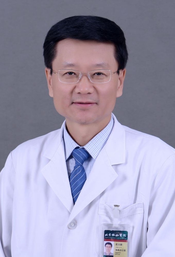 Yuxin Jiang (姜玉新)合作论文数Peking Union Medical College Hospital12
 Lin Li (李琳)合作论文数Beijing Hospital12Di Yang合作论文数10Rui Zhang (张蕊)合作论文数10Yanfeng Zhao (赵彦锋)合作论文数10
 Qingfeng Li合作论文数9Meng Lin合作论文数9Li-Shuang Gu合作论文数8Mengjia Liu合作论文数8Junying LIU (刘隽颖)合作论文数7
 Chao Sun (孙超)合作论文数北京协和医学院北京医院7
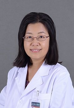 Qingli Zhu (朱庆莉)合作论文数Peking Union Medical College Hospital7
 Ni Li (李霓)合作论文数Cancer Hospital, Chinese Academy of Medical Sciences & Peking Union Medical College7Jinghui Wang (王景慧)合作论文数6dongnan fu (傅冬楠)合作论文数6SuLi Huang (黄苏里)合作论文数6YongXin Lin (林永新)合作论文数6Jian Geng (耿健)合作论文数6Huiqin Guo (郭会芹)合作论文数6
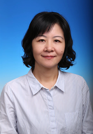 Yu Chen (陈宇)合作论文数Cancer Hospital Chinese Academy of Medical Sciences6Ming Xu (徐明)合作论文数6
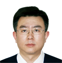 Wanqing Chen (陈万青)合作论文数Cancer Hospital, Chinese Academy of Medical Sciences6Bo Wang合作论文数5Hao Qin合作论文数5Hui Meng合作论文数5
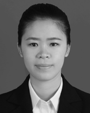 Xiaozheng Xie合作论文数5Han Jie合作论文数5
 Min Dai (代敏)合作论文数National Cancer Center;Cancer Hospital Chinese Academy of Medical Sciences5Bing Zhang (张兵)合作论文数5
 Xinming Zhao (赵心明)合作论文数中国医学科学院肿瘤医院5
 Wei Huang (黄蔚)合作论文数School of Medicine, Southeast University;Zhongda Hospital, Southeast University4Xu Teng合作论文数4MinJie Wang (王慜杰)合作论文数4Wei Tang (唐威)合作论文数4
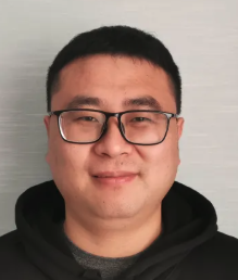 Jingyao Zhang (张靖垚)合作论文数Department of Hepatobiliary Surgery, First Affiliated Hospital of Xi’an Jiaotong University4
 Jie He (赫捷)合作论文数Chinese Academy of Medical Sciences & Peking Union Medical College;Cancer Hospital, Chinese Academy of Medical Sciences4Jiansong Ren (任建松)合作论文数Cancer Hospital Chinese Academy of Medical Sciences4Huan Zhao合作论文数4




