In this novel large-scale multiplexed immunofluorescence study we comprehensively characterized and compared layer-specific proteomic features within regions of interest of the widely divergent dorsolateral prefrontal cortex (A46) and primary visual cortex (A17) of adult rhesus monkeys. Twenty-eight markers were imaged in rounds of sequential staining, and their spatial distribution precisely quantified within gray matter layers and superficial white matter. Cells were classified as neurons, astrocytes, oligodendrocytes, microglia, or endothelial cells. The distribution of fibers and blood vessels were assessed by quantification of staining intensity across regions of interest. This method revealed multivariate similarities and differences between layers and areas. Protein expression in neurons was the strongest determinant of both laminar and regional differences, whereas protein expression in glia was more important for intra-areal laminar distinctions. Among specific results, we observed a lower glia-to-neuron ratio in A17 than in A46 and the pan-neuronal markers HuD and NeuN were differentially distributed in both brain areas with a lower intensity of NeuN in layers 4 and 5 of A17 compared to A46 and other A17 layers. Astrocytes and oligodendrocytes exhibited distinct marker-specific laminar distributions that differed between regions; notably, there was a high proportion of ALDH1L1-expressing astrocytes and of oligodendrocyte markers in layer 4 of A17. The many nuanced differences in protein expression between layers and regions observed here highlight the need for direct assessment of proteins, in addition to RNA expression, and set the stage for future protein-focused studies of these and other brain regions in normal and pathological conditions.
The laminar cellular and circuit mechanisms by which the anterior cingulate cortex (ACC) exerts flexible control of motor and affective information for goal-directed behavior have not been elucidated. Using multimodal tract-tracing, in vitro patch-clamp recording and computational approaches in rhesus monkeys (M. mulatta), we provide evidence that specialized motor and affective network dynamics can be conferred by layer-specific biophysical and structural properties of ACC pyramidal neurons targeting two key downstream structures —the dorsal premotor cortex (PMd) and the amygdala (AMY). AMY-targeting neurons exhibited significant laminar differences, with L5 more excitable (higher input resistance and action potential firing rates) than L3 neurons. Between-pathway differences were found within L5, with AMY-targeting neurons exhibiting greater excitability, apical dendritic complexity, spine densities, and diversity of inhibitory inputs than PMd-targeting neurons. Simulations using a pyramidal-interneuron network model predict that these layer- and pathway-specific single-cell differences contribute to distinct network oscillatory dynamics. L5 AMY-targeting networks are more tuned to slow oscillations well-suited for affective and contextual processing timescales, while PMd-targeting networks showed strong beta/gamma synchrony implicated in rapid sensorimotor processing. These findings are fundamental to our broad understanding of how layer-specific cellular and circuit properties can drive diverse laminar activity found in flexible behavior.
Behavioral studies have shown spatial working memory impairment with aging in several animal species, including humans. Persistent activity of layer 3 pyramidal dorsolateral prefrontal cortex (dlPFC) neurons during delay periods of working memory tasks is important for encoding memory of the stimulus. In vitro studies have shown that these neurons undergo significant age-related structural and functional changes, but the extent to which these changes affect neural mechanisms underlying spatial working memory is not understood fully. Here we confirm previous studies showing impairment on the Delayed Recognition Span Task in the spatial condition (DRSTsp), and increased in vitro action potential firing rates (hyperexcitability), across the adult life span of the rhesus monkey. We use a bump attractor model to predict how empirically observed changes in the aging dlPFC affect performance on the Delayed Response Task (DRT), and introduce a model of memory retention in the DRSTsp. Persistent activity—and, in turn, cognitive performance—in both models was affected much more by hyperexcitability of pyramidal neurons than by a loss of synapses. Our DRT simulations predict that additional changes to the network, such as increased firing of inhibitory interneurons, are needed to account for lower firing rates during the DRT with aging reported in vivo . Synaptic facilitation was an essential feature of the DRSTsp model, but it did not compensate fully for the effects of the other age-related changes on DRT performance. Modeling pyramidal neuron hyperexcitability and synapse loss simultaneously led to a partial recovery of function in both tasks, with the simulated level of DRSTsp impairment similar to that observed in aging monkeys. This modeling work integrates empirical data across multiple scales, from synapse counts to cognitive testing, to further our understanding of aging in non-human primates.
Functional recovery after cortical injury, such as stroke, is associated with neural circuit reorganization, but the underlying mechanisms and efficacy of therapeutic interventions promoting neural plasticity in primates are not well understood. Bone marrow mesenchymal stem cell-derived extracellular vesicles (MSC-EVs), which mediate cell-to-cell inflammatory and trophic signaling, are thought be viable therapeutic targets. We recently showed, in aged female rhesus monkeys, that systemic administration of MSC-EVs enhances recovery of function after injury of the primary motor cortex, likely through enhancing plasticity in perilesional motor and premotor cortices. Here, using in vitro whole-cell patch-clamp recording and intracellular filling in acute slices of ventral premotor cortex (vPMC) from rhesus monkeys (Macaca mulatta) of either sex, we demonstrate that MSC-EVs reduce injury-related physiological and morphologic changes in perilesional layer 3 pyramidal neurons. At 14-16 weeks after injury, vPMC neurons from both vehicle- and EV-treated lesioned monkeys exhibited significant hyperexcitability and predominance of inhibitory synaptic currents, compared with neurons from nonlesioned control brains. However, compared with vehicle-treated monkeys, neurons from EV-treated monkeys showed lower firing rates, greater spike frequency adaptation, and excitatory:inhibitory ratio. Further, EV treatment was associated with greater apical dendritic branching complexity, spine density, and inhibition, indicative of enhanced dendritic plasticity and filtering of signals integrated at the soma. Importantly, the degree of EV-mediated reduction of injury-related pathology in vPMC was significantly correlated with measures of behavioral recovery. These data show that EV treatment dampens injury-related hyperexcitability and restores excitatory:inhibitory balance in vPMC, thereby normalizing activity within cortical networks for motor function.SIGNIFICANCE STATEMENT Neuronal plasticity can facilitate recovery of function after cortical injury, but the underlying mechanisms and efficacy of therapeutic interventions promoting this plasticity in primates are not well understood. Our recent work has shown that intravenous infusions of mesenchymal-derived extracellular vesicles (EVs) that are involved in cell-to-cell inflammatory and trophic signaling can enhance recovery of motor function after injury in monkey primary motor cortex. This study shows that this EV-mediated enhancement of recovery is associated with amelioration of injury-related hyperexcitability and restoration of excitatory-inhibitory balance in perilesional ventral premotor cortex. These findings demonstrate the efficacy of mesenchymal EVs as a therapeutic to reduce injury-related pathologic changes in the physiology and structure of premotor pyramidal neurons and support recovery of function.
In the BACHD mouse model of Huntington's disease (HD), deletion of the N17 domain of the Huntingtin gene (BACHDΔN17, Q97) has been reported to lead to nuclear accumulation of mHTT and exacerbation of motor deficits, neuroinflammation and striatal atrophy (Gu et al., 2015). Here we characterized the effect of N17 deletion on dorsolateral striatal medium spiny neurons (MSNs) in BACHDΔN17 (Q97) and BACWTΔN17 (Q31) mice by comparing them to MSNs in wildtype (WT) mice. Mice were characterized on a series of motor tasks and subsequently whole cell patch clamp recordings with simultaneous biocytin filling of MSNs in in vitro striatal slices from these mice were used to comprehensively assess their physiological and morphological features. Key findings include that: Q97 mice exhibit impaired gait and righting reflexes but normal tail suspension reflexes and normal coats while Q31 mice do not differ from WT; intrinsic membrane and action potential properties are altered -but differentially so- in MSNs from Q97 and from Q31 mice; excitatory and inhibitory synaptic currents exhibit higher amplitudes in Q31 but not Q97 MSNs, while excitatory synaptic currents occur at lower frequency in Q97 than in WT and Q31 MSNs; there is a reduced total dendritic length in Q31 -but not Q97- MSNs compared to WT, while spine density and number did not differ in MSNs in the three groups. The findings that Q31 MSNs differed from Q97 and WT neurons with regard to some physiological features and structurally suggest a novel role of the N17 domain in the function of WT Htt. The motor phenotype seen in Q97 mice was less robust than that reported in an earlier study (Gu et al., 2015), and the alterations to MSN physiological properties were largely consistent with changes reported previously in a number of other mouse models of HD. Together this study indicates that N17 plays a role in the modulation of the properties of MSNs in both mHtt and WT-Htt mice, but does not markedly exacerbate HD-like pathogenesis in the BACHD model.
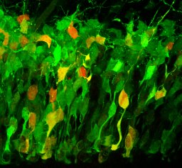
How the variety of neurons that organize into neocortical layers and functional areas arises is a central question in the study of cortical development. While both intrinsic and extrinsic cues are known to influence this process, whether distinct neuronal progenitor groups contribute to neuron diversity and allocation is poorly understood. Using in vivo genetic fate-mapping combined with whole-cell patch clamp recording, we show that the firing pattern and apical dendritic morphology of excitatory neurons in layer 4 of the barrel cortex are specified in part by their neural precursor lineage. Further, we show that separate precursors contribute to unique features of barrel cortex topography including the intralaminar position and thalamic innervation of the neurons they generate. Importantly, many of these lineage-specified characteristics are different from those previously measured for pyramidal neurons in layers 2-3 of the frontal cortex. Collectively, our data elucidate a dynamic temporal program in neuronal precursors that fine-tunes the properties of their progeny according to the lamina of destination.
Huntington's Disease (HD) is an autosomal dominant, progressive neurodegenerative disorder caused by deleterious expansion of CAG repeats in the Huntingtin gene and production of neurotoxic mutant Huntingtin protein (mHTT). The key pathological feature of HD is a profound degeneration of the striatum and a loss of cortical volume. The initial loss of indirect pathway (D2) medium spiny neuron (MSN) projections in early stages of HD, followed by a loss of direct pathway (D1) projections in advanced stages has important implications for the trajectory of motor and cognitive dysfunction in HD, but is not yet understood. Mouse models of HD have yielded important information on the effects and mechanisms of mHTT toxicity; however, whether these models recapitulate differential vulnerability of D1 vs. D2 MSNs is unknown. Here, we employed 12-month-old Q175+/- x D2-eGFP mice to examine the detailed structural and functional properties of D1 vs. D2 MSNs. While both D1 and D2 MSNs exhibited increased input resistance, depolarized resting membrane potentials and action potential threshold, only D1 MSNs showed reduced rheobase, action potential amplitude and frequency of spontaneous excitatory postsynaptic currents. Furthermore, D1 but not D2 MSNs showed marked proliferative changes to their dendritic arbors and reductions in spine density. Immunohistochemical assessment showed no loss of glutamatergic afferent inputs from cortical and subcortical sources onto identified D1 and D2 MSNs. Computational models constrained by empirical data predict that the increased dendritic complexity in Q175+/- D1 MSNs likely leads to greater dendritic filtering and attenuation of signals propagating to the soma from the dendrites. Together these findings reveal that, by twelve months, D1 and D2 MSNs exhibit distinctive responses to the presence of mHTT in this important mouse model of HD. This further highlights the need to incorporate findings from D1 and D2 MSNs independently in the context of HD models.
 Jennifer I. Luebke合作论文数Department of Anatomy, Boston University School of Medicine6
 Maria Medalla合作论文数Department of Health Sciences;Boston University;Department of Health Sciences, Boston University5Joseph W. Goodliffe合作论文数3
 Christina M. Weaver合作论文数Department of Applied Mathematics and Statistics, State University of New York at Stony Brook3Anastasia Al Rubakovic合作论文数2Teresa Guillamon-Vivancos合作论文数2
 s ibanez合作论文数Dept Anat & Neurobiol, Boston Univ2
 Douglas L. Rosene合作论文数Department of Anatomy & Neurobiology, School of Medicine, Boston University2
 Dhruba Pathak合作论文数Institute for Pathological physiology, Medical faculty Belgrade2
 Tara Moore合作论文数Department of Anatomy & Neurobiology, School of Medicine, Boston University;Department of Neurology, School of Medicine, Boston University2William A. Tyler合作论文数1Diego De Alba合作论文数1Danel Draguljic合作论文数1Anastasia Kapitonava合作论文数1
 Lisa Lowery合作论文数1Alexandra Tsolias合作论文数1
 Silas E. Busch合作论文数Dept Neurobiol, Univ Chicago1Benjamin Buller合作论文数1
 Veronica Go合作论文数1
 Samantha M Calderazzo合作论文数1
 Haydar Tarik F合作论文数Childrens Natl Med Ctr, Neurosci Res Ctr1Elizabeth McDonough合作论文数1Okamoto Mayumi合作论文数1
 Patrick R Hof合作论文数Nash Family Department of Neuroscience, Icahn School of Medicine at Mount Sinai1
 Song Hanbing合作论文数Department of Applied Science, The College of William and Mary1Kathleen S. Rockland合作论文数1Dan E. Meyer合作论文数1
 Merina Varghese合作论文数Division of Clinical and Experimental Neuroscience, Indian Institute of Chemical Biology1Monica A. Pessina合作论文数Department of Anatomy and Neurobiology, Boston University School of Medicine1




