Adult neural progenitor cells (aNPCs) are a potential source for cell based therapy for neurodegenerative diseases and traumatic brain injuries. These cells have been traditionally isolated from hippocampus, subventricular zone and white matter. However, there is still a need for an easily accessible source with better yield to counter the limitations of small surgical samples of previously characterized aNPCs. Here we show that ultrasonic aspirate (UA) samples currently considered as ` biological waste after surgery,' offer a good source for aNPCs. Furthermore, we show that culture conditions dictated the phenotype of cells across patients. The neurosphere-enriched cells were more similar to freshly isolated brain cells, while cells expanded adherently in serum conditions were similar to mesenchymal stem cells. However, cells expanded in these adherent conditions expressed some NPC and glial markers in addition to active canonical Wnt signaling. This suggests a mesenchymal-neuroectodermal hybrid nature of these cells. Finally, we show that UA-NPCs are comparable to those from neurogenic regions. Our findings suggest that UA samples can be used as a source for fresh and in vitro propagated aNPCs that could have various clinical applications.
In our lab we study neurogenesis and the development of brain tumors. We work towards treatment strategies for glioblastoma and towards using autologous neural stem cells for tissue regeneration strategies for brain damage and neurodegenerative disorders. It has been our policy to try to establish living cell cultures from all human biopsy material that we obtain. We hypothesized that small pieces of brain tissue could be cryopreserved and that live neural stem cells could be recovered at a later time. DMSO has been shown to possess a remarkable ability to diffuse through cell membranes and pass into cell interiors. Its chemical properties prevent the formation of damaging ice crystals thus allowing cell storage at or below −180 C. We report here a protocol for successful freezing of small pieces of tissue derived from human brain and human brain tumours. Virtually all specimens could be successfully revived. Assays of phenotype and behaviour show that the cell cultures derived were equivalent to those cultures previously derived from fresh tissue.
Glioma stem cells (GSCs) are thought to be the source of tumor growth and therapy resistance. To understand the biology of GSCs, and target these tumors therapeutically, we need robust strategies for in vitro expansion of primary GSCs. To date, tumor core biopsies have been the main established source of GSCs. Since these samples are used for diagnostic purposes, the available tissue for cell culture and therapeutic targeting can be limited. In addition, a core biopsy is usually taken from one part of the tumor, thus would be unlikely to represent intra-tumor heterogeneity. To overcome these problems, tissue fragments from all over the tumor can be collected using an ultrasonic aspirator during surgery, thus assembling a "global tumor biopsy". Usually, this ultrasonic aspirate (UA) sample is considered as biological waste after operations. Here, we show that UA samples offer a large and reliable source of live cells. Similar to core biopsies, UA samples enriched for GSCs that differentiated into neural lineages, showed inter-individual variation of GSC markers, and induced tumors. Molecular profiling showed that UA samples cover tumor heterogeneity better than core biopsies. These results suggest that UA samples can be used to establish large scale cultures for therapeutic applications.
BACKGROUND:Glioblastomas are invasive therapy resistant brain tumors with extremely poor prognosis. The Glioma initiating cell (GIC) population contributes to therapeutic resistance and tumor recurrence. Targeting GIC-associated gene candidates could significantly impact GBM tumorigenicity. Here, we investigate a protein kinase, PBK/TOPK as a candidate for regulating growth, survival and in vivo tumorigenicity of GICs.METHODS:PBK is highly upregulated in GICs and GBM tissues as shown by RNA and protein analyses. We knocked down PBK using shRNA vectors and inhibited the function of PBK protein with a pharmacological PBK inhibitor, HITOPK-032. We assessed viability, tumorsphere formation and apoptosis in three patient derived GIC cultures.RESULTS:Gene knockdown of PBK led to decreased viability and sphere formation and in one culture an increase in apoptosis. Treatment of cells with inhibitor HITOPK-032 (5 μM and 10 μM) almost completely abolished growth and elicited a large increase in apoptosis in all three cultures. HI-TOPK-032 treatment (5 mg/kg and 10 mg/kg bodyweight) in vivo resulted in diminished growth of experimentally induced subcutaneous GBM tumors in mice. We also carried out multi-culture assays of cell survival to investigate the relative effects on GICs compared with the normal neural stem cells (NSCs) and their differentiated counterparts. Normal NSCs seemed to withstand treatment slightly better than the GICs.CONCLUSION:Our study of identification and functional validation of PBK suggests that this candidate can be a promising molecular target for GBM treatment.
Glioblastoma (GBM) is both the most common and the most lethal primary brain tumor. It is thought that GBM stem cells (GSCs) are critically important in resistance to therapy. Therefore, there is a strong rationale to target these cells in order to develop new molecular therapies.To identify molecular targets in GSCs, we compared gene expression in GSCs to that in neural stem cells (NSCs) from the adult human brain, using microarrays. Bioinformatic filtering identified 20 genes (PBK/TOPK, CENPA, KIF15, DEPDC1, CDC6, DLG7/DLGAP5/HURP, KIF18A, EZH2, HMMR/RHAMM/CD168, NOL4, MPP6, MDM1, RAPGEF4, RHBDD1, FNDC3B, FILIP1L, MCC, ATXN7L4/ATXN7L1, P2RY5/LPAR6 and FAM118A) that were consistently expressed in GSC cultures and consistently not expressed in NSC cultures. The expression of these genes was confirmed in clinical samples (TCGA and REMBRANDT). The first nine genes were highly co-expressed in all GBM subtypes and were part of the same protein-protein interaction network. Furthermore, their combined up-regulation correlated negatively with patient survival in the mesenchymal GBM subtype. Using targeted proteomics and the COGNOSCENTE database we linked these genes to GBM signalling pathways.Nine genes: PBK, CENPA, KIF15, DEPDC1, CDC6, DLG7, KIF18A, EZH2 and HMMR should be further explored as targets for treatment of GBM.
BACKGROUND:Glioblastoma (GBM) is the most common primary brain malignancy and confers a dismal prognosis. GBMs harbor glioblastoma-initiating cells (GICs) that drive tumorigenesis and contribute to therapeutic resistance and tumor recurrence. Consequently, there is a strong rationale to target this cell population in order to develop new molecular therapies against GBM. Accumulating evidence indicates that Nα-terminal acetyltransferases (NATs), that are dysregulated in numerous human cancers, can serve as therapeutic targets.METHODS:Microarrays were used to study the expression of several NATs including NAT12/NAA30 in clinical samples and stem cell cultures. The expression of NAT12/NAA30 was analyzed using qPCR, immunolabeling and western blot. We conducted shRNA-mediated knockdown of NAT12/NAA30 gene in GICs and studied the effects on cell viability, sphere-formation and hypoxia sensitivity. Intracranial transplantation to SCID mice enabled us to investigate the effects of NAT12/NAA30 depletion in vivo. Using microarrays we identified genes and biochemical pathways whose expression was altered upon NAT12/NAA30 down-regulation.RESULTS:While decreased expression of the distal 3'UTR of NAT12/NAA30 was generally observed in GICs and GBMs, this gene was strongly up-regulated at the protein level in GBM and GICs. The increased protein levels were not caused by increased levels of the steady state mRNA but rather by other mechanisms. Also, shorter 3'UTR of NAT12/NAA30 correlated with poor survival in glioma patients. As well, we observed previously not described nuclear localization of this typically cytoplasmic protein. When compared to non-silencing controls, cells featuring NAT12/NAA30 knockdown exhibited reduced cell viability, sphere-forming ability, and mitochondrial hypoxia tolerance. Intracranial transplantation showed that knockdown of NAT12/NAA30 resulted in prolonged animal survival. Microarray analysis of the knockdown cultures showed reduced levels of HIF1α and altered expression of several other genes involved in the hypoxia response. Furthermore, NAT12/NAA30 knockdown correlated with expressional dysregulation of genes involved in the p53 pathway, ribosomal assembly and cell proliferation. Western blot analysis revealed reduction of HIF1α, phospho-MTOR(Ser2448) and higher levels of p53 and GFAP in these cultures.CONCLUSION:NAT12/NAA30 plays an important role in growth and survival of GICs possibly by regulating hypoxia response (HIF1α), levels of p-MTOR (Ser2448) and the p53 pathway.
The discovery of stem cells in the adult human brain has revealed new possible scenarios for treatment of the sick or injured brain. Both clinical use of and preclinical research on human adult neural stem cells have, however, been seriously hampered by the fact that it has been impossible to passage these cells more than a very few times and with little expansion of cell numbers. Having explored a number of alternative culturing conditions we here present an efficient method for the establishment and propagation of human brain stem cells from whatever brain tissue samples we have tried. We describe virtually unlimited expansion of an authentic stem cell phenotype. Pluripotency proteins Sox2 and Oct4 are expressed without artificial induction. For the first time multipotency of adult human brain-derived stem cells is demonstrated beyond tissue boundaries. We characterize these cells in detail in vitro including microarray and proteomic approaches. Whilst clarification of these cells' behavior is ongoing, results so far portend well for the future repair of tissues by transplantation of an adult patient's own-derived stem cells.
Glioblastoma is the most common brain tumor. Median survival in unselected patients is <10 months. The tumor harbors stem-like cells that self-renew and propagate upon serial transplantation in mice, although the clinical relevance of these cells has not been well documented. We have performed the first genome-wide analysis that directly relates the gene expression profile of nine enriched populations of glioblastoma stem cells (GSCs) to five identically isolated and cultivated populations of stem cells from the normal adult human brain. Although the two cell types share common stem- and lineage-related markers, GSCs show a more heterogeneous gene expression. We identified a number of pathways that are dysregulated in GSCs. A subset of these pathways has previously been identified in leukemic stem cells, suggesting that cancer stem cells of different origin may have common features. Genes upregulated in GSCs were also highly expressed in embryonic and induced pluripotent stem cells. We found that canonical Wnt-signaling plays an important role in GSCs, but not in adult human neural stem cells. As well we identified a 30-gene signature highly overexpressed in GSCs. The expression of these signature genes correlates with clinical outcome and demonstrates the clinical relevance of GSCs.
Earlier findings from our laboratory implicated RhoA in heart developmental processes. To investigate factors that potentially regulate RhoA expression, RhoA gene organisation and promoter activity were analysed. Comparative analysis indicated strict conservation of both gene organisation and coding sequence of the chick, mouse, and human RhoA genes. Bioinformatics analysis of the derived promoter region of mouse RhoA identified putative consensus sequence binding sites for several transcription factors involved in heart formation and organogenesis generally. Using luciferase reporter assays, RhoA promoter activity was shown to increase in mouse-derived P19CL6 cells that were induced to differentiate into cardiomyocytes. Overexpression of a dominant negative mutant of mouse RhoA (mRhoAN19) blocked this cardiomyocyte differentiation of P19CL6 cells and led to the accumulation of the cardiac transcription factors SRF and GATA4 and the early cardiac marker cardiac α -actin. Taken together, these findings indicate a fundamental role for RhoA in the differentiation of cardiomyocytes.
BACKGROUND:Evidence from genetic association studies implicate genes involved in neural migration associated with schizophrenia risk. Neural stem/progenitor cell cultures (neurosphere-derived cells) from olfactory mucosa of schizophrenia patients have significantly dysregulated expression of genes in focal adhesion kinase (FAK) signaling, a key pathway regulating cell adhesion and migration. The aim of this study was to investigate whether olfactory neurosphere-derived cells from schizophrenia patients have altered cell adhesion, cell motility, and focal adhesion dynamics.METHODS:Olfactory neurosphere-derived cells from nine male schizophrenia patients and nine male healthy control subjects were used. Cells were assayed for cell adhesion and cell motility and analyzed for integrins and FAK proteins. Focal adhesions were counted and measured in fixed cells, and time-lapse imaging was used to assess cell motility and focal adhesion dynamics.RESULTS:Patient-derived cells were less adhesive and more motile than cells derived from healthy control subjects, and their motility was reduced to control cell levels by integrin-blocking antibodies and by inhibition of FAK. Vinculin-stained focal adhesion complexes were significantly smaller and fewer in patient cells. Time-lapse imaging of cells expressing FAK tagged with green fluorescent protein revealed that the disassembly of focal adhesions was significantly faster in patient cells.CONCLUSIONS:The evidence for altered motility and focal adhesion dynamics in patient-derived cells is consistent with dysregulated gene expression in the FAK signaling pathway in these cells. Alterations in cell adhesion dynamics and cell motility could bias the trajectory of brain development in schizophrenia.
Several studies have implicated the aquaporins (aqp) 1, 4, and 9 in the pathogenesis of malignant brain tumours, suggesting that they contribute to motility, invasiveness, and oedema formation and facilitate metabolism in tumour cells under hypoxic conditions. We have studied the expression of aqp1, 4, and 9 in biopsies from glioblastomas, isolated tumour stem cells grown in a tumoursphere assay and analyzed the progenitor and differentiated cells from these cultures. We have compared these to the situation in normal rat brain, its stem cells, and differentiated cells derived thereof. In short, qPCR in tumour tissue showed presence of aqp1, 4, and 9. In the tumour progenitor population, aqp9 was markedly more highly expressed, whilst in tumour-derived differentiated cells, aqp4 was downregulated. However, immunostaining did not reveal increased protein expression of aqp9 in the tumourspheres containing progenitor cells; in contrast, its expression (both mRNA and protein) was high in differentiated cultures. We, therefore, propose that aquaporin 9 may have a central role in the tumorigenesis of glioblastoma.
There is worldwide enthusiasm for the prospect of some kind of cellular transplant therapy for repair of failing organs. The olfactory mucosa of a patient's nose is easily biopsied to provide a ready source of multipotent cells. In this article we address practical issues pertinent to using olfactory neural stem cells for tissue repair. These cells are emerging as potentially most significant candidates for human tissue repair strategies. Previously we have shown that stem cells from olfactory mucosa are multipotent. As well, we have recently published three potential clinical applications. Their expression of dopaminergic markers in vitro and in a Parkinson's rat transplant model has been demonstrated. Their conversion to chondrogenic phenotype in vitro and in vivo has also been described, as has their transplant into a rat model of cardiac infarction. Here we examine in detail the biology of the olfactory neural stem cell using the rat as our animal model cell source. We establish its presence by examining self-renewal capacity and for phenotypic acquisition in inductive circumstances. We determine its frequency within the cell population and show that our culture system selects for this putative stem cell. Our studies demonstrate that adult olfactory stem cells, when transplanted into an environmental niche different from that of their origin, are able to demonstrate multipotency by acquiring the phenotype of the resident cells. We investigate how immediate the instruction need be. We test the hypothesis that olfactory neurospheres contain stem cells whose capacity for differentiation is triggered by signals of the immediate environmental niche. Significantly, of importance to any tissue regeneration endeavor, stem cell numbers were shown to be enriched by our culture methods. This was confirmed whether measured by sphere-forming capacity or differentiation response rate.
Glioblastoma Multiforme (GBM) is the most common primary brain malignancy and despite multimodal therapy including surgery, irradiation and chemotherapy median survival remains 12–15 months. Increasing body of evidence from our and other groups is confirming the existence of a stem-like cell population within these tumors believed to confer tumor growth, invasiveness, and therapy resistance. Adult human neural stem cells (aHNSCs) are suggested to be the cells of origin for the glioblastoma stem-like cells (GSCs). To identify new molecular targets we performed a micro-array analysis on the transcriptome of 33000 genes identifying differentially expressed genes in 5 aHNSC and 9 GSC cultures. Amongst the genes highly up-regulated in GBM, several were involved in cell division and the regulation of cell cycle. For further analysis, we chose the Chromatid Segreration Mediator (CSM), a centromere specific gene important for appropriate chromatid segregation during mitosis. This candidate gene was strongly expressed in GSCs but was almost undetectable in aHNSCs. Abundance of the CSM protein may lead to aneupleudy, which is a common feature of GBMs. The high levels of CSM mRNA were confirmed by analyzing 16 GSC cultures with micro-array and qPCR. CSM was also over-expressed on protein level, and was found to be highly immunopositive in glioblastoma biopsies compared to the normal human brain. In silico analysis, using Rembrandt, identifies CSM-gene over-expression as a negative prognostic indicator in glioma patients. In order to study the functional role of this gene in GBM, we used lentiviral-based knock-down (KD) technology to obtain specific down regulation of the gene. We transduced different shRNA constructs in 2 GBM cultures and achieved KD-efficieny of 55–65%. Growth of tumors was measured by proliferation assay and the relative number of sphere-forming cells (widely known as GSCs) was quantified with a sphere-formation assay. In our KD-cultures we observed a moderate reduction of proliferation (25%). Interestingly, the sphere forming capacity was dramatically reduced by 60–70%, indicating a significant decrease in number of GSCs. Cell-cycle analysis revealed that it was 30–40% less cells in G2/M-phase and 15–20% more cells in G0/G1-phase in the KD compared to non-silencing control. We find our results very promising and suggest that Chromatid Segregation Mediator gene might have potential as a novel therapeutic target for GBM management. Citation Format: {Authors}. {Abstract title} [abstract]. In: Proceedings of the AACR-NCI-EORTC International Conference: Molecular Targets and Cancer Therapeutics; 2011 Nov 12-16; San Francisco, CA. Philadelphia (PA): AACR; Mol Cancer Ther 2011;10(11 Suppl):Abstract nr B186.
Sphere forming assays have been useful to enrich for stem like cells in a range of tumors. The robustness of this system contrasts the difficulties in defining a stem cell population based on cell surface markers. We have undertaken a study to describe the cellular and organizational composition of tumorspheres, directly comparing these to neurospheres derived from the adult human subventricular zone (SVZ). Primary cell cultures from brain tumors were found to contain variable fractions of cells positive for tumor stem cell markers (CD133 (2–93%)/SSEA1 (3–15%)/CXCR4 (1–72%)). All cultures produced tumors upon xenografting. Tumorspheres contained a heterogeneous population of cells, but were structurally organized with stem cell markers present at the core of spheres, with markers of more mature glial progenitors and astrocytes at more peripheral location. Ultrastructural studies showed that tumorspheres contained a higher fraction of electron dense cells in the core than the periphery (36% and 19%, respectively). Neurospheres also contained a heterogeneous cell population, but did not have an organization similar to tumorspheres. Although tumorspheres clearly display irregular and neoplastic cells, they establish an organized structure with an outward gradient of differentiation. We suggest that this organization is central in maintaining the tumor stem cell pool.
Abstract Glioblastoma multiformae (GBM) is the most common primary brain tumor and one of the most malignant human cancers (median survival 14 months from diagnosis despite combined surgery/irradiation/chemotherapy). Evidence from our and other groups indicates that glioblastoma stem-like cells (GSCs) contained within these tumors are critically important in GBM initiation, progression, and resistance to therapy. To identify new targets for glioblastoma treatment, we have systematically compared gene expression in nine primary GBM cell cultures to that in five primary neural stem cell cultures from the adult human brain (ahNSCs), using microarrays. Rigorous bioinformatic filtering identified 20 genes whose RNA expression levels were very high in primary GSC cultures but were undetectable in ahNSCs. The identified genes are involved in cell-cycle/division, epigenetic regulation, signaling and down-regulation of tumor-suppressors. Several of the candidate genes are implicated in cancer (breast, ovary and colon), while others have no known associations to cancer or have unknown functions. A total of nine new GBM primary cultures were further analyzed by real-time polymerase chain reaction (qPCR), Western blot and Immunohistochemistry on tissue sections and cell cultures. qPCR confirmed the increased expression of 17 of the candidate genes. These results were in good accordance with the information provided by public data bases (Rembrant, TCGA). Western blot performed on 17 candidates showed increased protein levels in 9 cases thus demarcating these as best candidates for molecular targeting. The rest of the proteins featured aberrant isoforms, probably due to alternate splicing, modifications, cleavage or degradation. Further bioinformatics analysis identified a subset of 5 candidates that are particularly highly up-regulated in GBM when compared to low-grade gliomas. To explore the functional importance of the potential target genes we established lentiviral-based shRNA delivery and started to test the potential of gene knock-downs (KDs) to inhibit growth of tumor cells. Our preliminary results look very promising. Three different lentiviral vectors, designed to silence a candidate gene encoding a tumor-suppressor binding protein kinase, were tested in three different primary GBM cultures and achieved a KD efficiency of 60–70%. We used proliferation essays to describe the growth speed of GBMs. To estimate relative numbers of sphere-forming cells (widely referred to as cancer stem cells) we performed sphere-forming assays (SFAs). The KD of the protein kinase gene resulted both in dramatic reduction of growth speed (50–70%) and in the number of sphere-forming cells (50–70%). In addition, the volume of the spheres was reduced to 50%. Cell cycle analysis revealed that there was doubling of the number of KD cells arrested in G2/M phase compared to non-silencing controls. There was also an increase (20–65%) in the proportion of apoptotic cells in 2 out of 3 GBM KD cultures. Citation Format: {Authors}. {Abstract title} [abstract]. In: Proceedings of the AACR-NCI-EORTC International Conference: Molecular Targets and Cancer Therapeutics; 2011 Nov 12-16; San Francisco, CA. Philadelphia (PA): AACR; Mol Cancer Ther 2011;10(11 Suppl):Abstract nr B202.
BACKGROUND:The adult human brain contains neural stem/progenitor cells (AHNPCs) that can survive transplantation into the adult rat brain, migrate toward a lesion, and display limited neuronal differentiation in vivo. OBJECTIVE:To investigate the effect of manipulating AHNPCs before grafting by predifferentiation, ie, initiating neuronal differentiation before transplantation, and to determine whether this cell priming would affect their ability to migrate in vivo. METHODS:AHNPCs were prepared from temporal lobe resections for epilepsy. Seven days after global brain ischemia, predifferentiated AHNPCs (exposed to basic fibroblast growth factor, heparin, and laminin) were transplanted to the left hippocampus. Four and 10 weeks after transplantation, brain sections were analyzed by immunohistochemistry. RESULTS:Transplanted primed cells expressed committed neuronal markers at a much earlier stage compared with nonprimed AHNPCs and were found colabeled with human markers within the damaged CA1 region 4 weeks after grafting. Furthermore, predifferentiated AHNPCs migrated preferentially into an ischemic lesion, similar to their undifferentiated counterparts. The chemoattractant effect from the expression of stromal cell–derived factor-1α (SDF-1α) in ischemic CA1 on AHNPCs expressing CXC chemokine receptor 4 (CXCR4) may explain this preference in migration in vivo. CONCLUSION:The plasticity of neural progenitors derived from the adult human brain may be greater than previously assumed in that manipulation before grafting may influence the phenotypes seen in vivo. The SDF-1α–CXCR4 axis is involved in the targeted migration toward an ischemic lesion in the adult rat brain, similar to previous reports on endogenous progenitors in rats and grafted fetal human neural progenitors.
There is a pressing need for patient-derived cell models of brain diseases that are relevant and robust enough to produce the large quantities of cells required for molecular and functional analyses. We describe here a new cell model based on patient-derived cells from the human olfactory mucosa, the organ of smell, which regenerates throughout life from neural stem cells. Olfactory mucosa biopsies were obtained from healthy controls and patients with either schizophrenia, a neurodevelopmental psychiatric disorder, or Parkinson's disease, a neurodegenerative disease. Biopsies were dissociated and grown as neurospheres in defined medium. Neurosphere-derived cell lines were grown in serum-containing medium as adherent monolayers and stored frozen. By comparing 42 patient and control cell lines we demonstrated significant disease-specific alterations in gene expression, protein expression and cell function, including dysregulated neurodevelopmental pathways in schizophrenia and dysregulated mitochondrial function, oxidative stress and xenobiotic metabolism in Parkinson's disease. The study has identified new candidate genes and cell pathways for future investigation. Fibroblasts from schizophrenia patients did not show these differences. Olfactory neurosphere-derived cells have many advantages over embryonic stem cells and induced pluripotent stem cells as models for brain diseases. They do not require genetic reprogramming and they can be obtained from adults with complex genetic diseases. They will be useful for understanding disease aetiology, for diagnostics and for drug discovery.
This study examines the hypothesis that multipotent olfactory mucosal stem cells could provide a basis for the development of autologous cell transplant therapy for the treatment of heart attack. In humans, these cells are easily obtained by simple biopsy. Neural stem cells from the olfactory mucosa are multipotent, with the capacity to differentiate into developmental fates other than neurons and glia, with evidence of cardiomyocyte differentiation in vitro and after transplantation into the chick embryo. Olfactory stem cells were grown from rat olfactory mucosa. These cells are propagated as neurosphere cultures, similar to other neural stem cells. Olfactory neurospheres were grown in vitro, dissociated into single cell suspensions, and transplanted into the infarcted hearts of congeneic rats. Transplanted cells were genetically engineered to express green fluorescent protein (GFP) in order to allow them to be identified after transplantation. Functional assessment was attempted using echocardiography in three groups of rats: control, unoperated; infarct only; infarcted and transplanted. Transplantation of neurosphere-derived cells from adult rat olfactory mucosa appeared to restore heart rate with other trends towards improvement in other measures of ventricular function indicated. Importantly, donor-derived cells engrafted in the transplanted cardiac ventricle and expressed cardiac contractile proteins.
De novo mutations are a cause of sporadic disease, but little is known about the developmental timing of such mutations. We studied concordant and discordant monozygous twins with de novo mutations in the sodium channel α1 subunit gene (SCN1A) causing Dravet's syndrome, a severe epileptic encephalopathy. On the basis of our findings and the literature on mosaic cases, we conclude that de novo mutations in SCN1A may occur at any time, from the premorula stage of the embryo (causing disease in the subject) to adulthood (with mutations in the germ-line cells of parents causing disease in offspring).
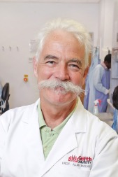 Alan Mackay-Sim合作论文数Griffith University22
 Langmoen Iver A合作论文数Institute for Surgical Research, University of Oslo11
 Biljana Stangeland合作论文数Plant Molecular Biology Laboratory;Department of Chemistry and Biotechnology;Agricultural University;Plant Molecular Biology Laboratory, Agricultural University11Andrew Wetzig合作论文数10
 François Féron合作论文数Interactions Cellulaires et Neurophysiopathologie;Faculté de Médecine;Université de la Méditerranée;Faculté de Médecine, Université de la Méditerranée10Cecilie Sandberg合作论文数Norwegian Institute for Water Research (NIVA), P.O. Box 173, Kjelsaas, Oslo, Norway9Mrinal Joel合作论文数8
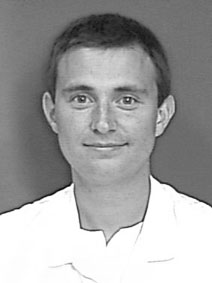 Einar Osland Vik-Mo合作论文数The Vilhelm Magnus Laboratory, Department of Neurosurgery, Oslo University Hospital;Institute of Clinical Medicine, Faculty of Medicine, University of Oslo8
 John Bianco合作论文数Princess Maxima Ctr Pediat Oncol7Zanina Grieg合作论文数7
 Denis I. Crane合作论文数School of Biomolecular and Biomedical Science, Griffith University7Awais A Mughal合作论文数6Mercy Varghese合作论文数5Ingunn Ramsnes合作论文数4Havard Olstorn合作论文数4
 Peter Silburn合作论文数Asia-Pacific Centre for Neuromodulation, Queensland Brain Institute, The University of Queensland;St Andrews War Memorial Hospital4C Masters合作论文数3Sheryl Palmero合作论文数3Chris Perry合作论文数3
 Jinan Behnan合作论文数Institute for Surgical Research and Department of Neurosurgery, Oslo University Hospital3Bellette Bernadette合作论文数Eskitis Institute for Drug Discovery, Griffith University3
 Mari Kaarbø合作论文数3G Bushell合作论文数3Hege Brincker Fjerdingstad合作论文数3Michael Donnellan合作论文数3Iver A. Langmoen合作论文数3Einar O. Vik-Mo合作论文数2Gaetano Finocchiaro合作论文数2
 Tiziana Langella合作论文数2c perry合作论文数2Yongjun Fan合作论文数2
 Thomas Burne合作论文数Queensland Brain Institute, The University of Queensland2
 Torstein R. Meling合作论文数University of Thessaly;Rigshospitalet;École polytechnique fédérale de Lausanne;Swiss Meningioma Network2
 Nygård Ståle合作论文数Bioinformat Core Facil, Oslo Univ Hosp2Abrahamsen Greger合作论文数2Palmero Emily合作论文数2Yasuhiro Watanabe合作论文数2
 John McGrath合作论文数Greater Brisbane Clinical School, Faculty of Health, Medicine and Behavioural Sciences, University of Queensland;Queensland Brain Institute, Faculty of Health, Medicine and Behavioural Sciences, University of Queensland2Nicholas Cameron合作论文数2Gabriel Lee合作论文数2
 Morten Moe合作论文数2Joel Glover合作论文数2Ratneswary Sutharsan合作论文数Eskitis Institute for Cell and Molecular Therapies, Griffith University2Havard Kjesbu Skjellegrind合作论文数2Anthony L. Cook合作论文数1Colin Masters合作论文数1Tom Burne合作论文数1Staale Nygaard合作论文数1




