Repair of damaged DNA is a complex process, particularly when it is compacted into nucleosomes. There are a number of genetic disorders with deficiencies in DNA repair. Knowledge of the genes and proteins involved in these repair deficiencies is critical in developing an understanding of the molecular mechanisms utilized by proteins in the DNA repair pathways. One of these genetic disorders is xeroderma pigmentosum (XP), which is defective in nucleotide excision repair (NER). Patients in XP complementation group A (XP-A) are among the most severely affected with the lowest levels of DNA repair. The XPA protein, which is defective in these patients, plays a number of roles in the DNA repair process. One particularly important role proposed is acting as a processivity factor enabling endonucleases (XPF and XPG) and the XPB/TFIIH translocase to localize to damage sites using a processive mechanism of action. Another proposed role is in interacting with chromatin-remodeling proteins so as to enhance accessibility of lesions in nucleosomal DNA to endonucleolytic incision and other DNA repair activities. In XP-A cells, the XPA protein is proposed to be defective in ability to act as a processivity factor; endonucleases localize damage sites by a distributive mechanism and are also defective in incision of damaged nucleosomal DNA. This defect is corrected by recombinant normal human XPA. Mutations in exons 3 and 5 in the DNA binding domain of the XPA gene lead to loss of ability of XPA to act as a processivity factor. The mutation in exon 5 was found in two XP-A patients with severe XP. These studies emphasize the importance of correlating specific mutations in an XP gene and the resulting defect in a particular repair protein with the clinical severity of XP and could lead to development of novel therapeutic approaches for this disorder.
Carcinogenesis, while traditionally attributed to the accumulation of driver mutations in genes regulating cell proliferation and apoptosis, may also be explored as a consequence of fundamental metabolic reprogramming, an idea catalyzed by the Warburg effect, where cancer cells exhibit a paradoxical preference for glycolysis over the far more efficient oxidative phosphorylation. This implies that metabolic dysregulation may be a primary instigator of neoplastic transformation. Our hypothesis proposes that the abrupt loss of cellular energy may stimulate an atavistic response, wherein rapid proliferation and migration are triggered to enhance survival in fluctuating environments. These responses lead to pathological angiogenesis and unchecked cell growth, thereby bridging the gap between genetic and metabolic pathways of carcinogenesis.
In the dermatological spectrum of oncologic manifestations, cutaneous metastases from endometrial carcinoma stand as a rarity, given the tumour’s predilection for neighbouring uterine regions. We present an exceptional case of a patient in her mid-50s, whereby an endometrial carcinoma, defying conventional pathways, manifested on the skin and nail of her distal fourth finger, an unusual site for cutaneous metastases, with a specific histology of the primary cancer.
Just as fire and electricity can be, and in many ways are, of great benefit to humanity, and as the contributions elsewhere in this issue of Clinics in Dermatology have shown, artificial intelligence (AI) can be used for the ill and help in medicine. We offer several suggestions to counter some of the more egregious and obvious ones: AI-generated material that purports to be caused by humans and AI-generated material that purports to show actual people doing things that these people would not normally do. Both suggestions rely on methods already in existence to ensure public safety.
We describe a novel assay and artificial intelligence-driven histopathologic approach identifying dermatophytes in human skin tissue sections (ie, B -DNA dermatophyte assay) and demonstrate, for the first time, the presence of dermatophytes in tissue using immunohistochemistry to detect canonical right-handed double -stranded (ds) B -DNA. Immunohistochemistry was performed using anti-ds-BDNA monoclonal antibodies with formalin-fixed paraffin -embedded tissues to determine the presence of dermatophytes. The B -DNA assay resulted in a more accurate identification of dermatophytes, nuclear morphology, dimensions, and gene expression of dermatophytes (ie, optical density values) than periodic acid-Schiff (PAS), Grocott methenamine silver (GMS), or hematoxylin and eosin (H&E) stains. The novel assay guided by artificial intelligence allowed for efficient identification of different types of dermatophytes (eg, hyphae, microconidia, macroconidia, and arthroconidia). Using the B -DNA dermatophyte assay as a clinical tool for diagnosing dermatophytes is an alternative to PAS, GMS, and H&E as a fast and inexpensive way to accurately detect dermatophytosis and reduce the number of false negatives. Our assay resulted in superior identification, sensitivity, life cycle stages, and morphology compared to H&E, PAS, and GMS stains. This method detects a specific structural marker (ie, ds-B-DNA), which can assist with diagnosis of dermatophytes. It represents a significant advantage over methods currently in use. (c) 2024 Elsevier Inc. All rights reserved.
The development of the computer and what is now known as artificial intelligence (AI) has evolved over more than two centuries in a long series of steps. The date of the invention of the first computer is estimated at 1822, when Charles Babbage (1791-1871) developed his first design of a working computer on paper, based mainly on a Jacquard loom. He worked on his project together with Augusta Ada King, Countess Lovelace (née Byron) (Ada Lovelace) (1815-1852), whom he called the "Sorceress of Numbers." This work will present the profile and achievements of Charles Babbage, Augusta Ada King, Countess Lovelace, and Alan Mathison Turing (1912 - 1954), who is considered the father of computer science and artificial intelligence, and then provide an outline of the tumultuous events affecting AI up to the present.
Artificial Intelligence (AI) is a very powerful new tool that is destined to markedly advance many areas of dermatology, including cosmetic dermatology, oculoplastics, cancer detection and treatment, dermatopathlogy, and identification of pathogens. Along with these are some special new risks and concerns, however, including ethical considerations, data analysis, interpretation of scientific studies, and recognizing systematic failures and fraud, particularly in generative AI. Each of these issues is reviewed collectively and in turn in this special of Clinics in Dermatology.
Judging whether an editor is good at the job is essential; however, this task may be difficult or even impossible. Several factors are involved, many of which are beyond the control of an editor. We examined some of such situations, which are as follows: (1) Reviewer's abuse of privileged information, in which a reviewer or an associate, who is likely to be a competitor, directs members of their laboratory to rapidly replicate the data and submit the resulting paper in the same or another journal while delaying publication of the submitted paper; (2) defective micromanagement by a stakeholder or owner, such as failure to order paper for the publication of a journal; (3) penny-wise dollar-foolish mismanagement by the owner, such as limiting the figures allowed to an absurdly low number in a dermatology journal (we have a visual specialty); (4) factional abuse, such as when members of a society use a gimmick to exercise outsized influence to effect a change in journal's content, and (5) "sto tavo (who is in charge)?," in which changes in the governance of an ownership society or publisher affect quality of the journal.
This study explored the integration and impact of artificial intelligence (AI) in diagnostic pathology, particularly dermatopathology, assessing its challenges and potential solutions for global health care enhancement. A comprehensive literature search in PubMed and Google Scholar, conducted on March 30, 2023, and using terms related to AI, pathology, and machine learning, yielded 44 relevant publications. These were analyzed under themes including the evolution of deep learning in pathology, AI's role in replacing pathologists, development challenges of diagnostic algorithms, clinical implementation hurdles, strategies for practical application in dermatopathology, and future prospects of AI in this field. The findings highlight AI's transformative potential in pathology, underscore the need for ongoing research, collaboration, and regulatory dialogue, and emphasize the importance of addressing the ethical and practical challenges in AI implementation for improved global health care outcomes.
Artificial intelligence (AI) can be a powerful tool for data analysis, but it can also mislead investigators, due in part to a fundamental difference between classic data analysis and data analysis using AI. A more or less limited data set is analyzed in classic data analysis, and a hypothesis is generated. That hypothesis is then tested using a separate data set, and the data are examined again. The premise is either accepted or rejected with a value p, indicating that any difference observed is due merely to chance. By contrast, a new hypothesis is generated in AI as each datum is added to the data set. We explore this discrepancy and suggest means to overcome it.
Treating atopic dermatitis (AD) with dupilumab, a monoclonal antibody that inhibits interleukin-4 (IL-4) and interleukin-13 (IL-13), may be associated with the progression of mycosis fungoides (MF). This study aims to examine the associations between the length of dupilumab treatment, age and sex, and the onset of MF. An institutional data registry and literature search were used for a retrospective cross-sectional study. Only patients with a diagnosis of MF on dupilumab for the treatment of AD and eczematous dermatitis were included. The primary outcome was the length of dupilumab exposure, age, sex, and the onset of MF. Linear correlations (Pearson) and Cox regression analysis were used to assess the correlation and the risk. A total of 25 patients were included in this study. Five eligible patients were identified at our institution. In addition, a PubMed review identified an additional 20 patients. At the time of MF diagnosis, the median age was 58, with 42% female. Disease history was significant for adult-onset AD in most patients (n = 17, 65.4%) or recent flare of AD previously in remission (n = 3, 11.5%). All patients were diagnosed with MF, and one patient progressed to Sézary syndrome while on dupilumab, with an average duration of 13.5 months of therapy prior to diagnosis. Tumor stage at diagnosis of MF was described in 19 of the cases and ranged from an early-stage disease (IA) to advanced disease (IV). Treatment strategies included narrow-band UVB therapy, topical corticosteroids, brentuximab, pralatrexate, and acitretin. Male gender, advanced-stage disease, and older age correlated significantly with the hazard of MF onset and a shorter time to onset during dupilumab treatment. Our results suggest a correlation between the duration of dupilumab treatment and the diagnosis of MF, the higher MF stage at diagnosis, and the shorter the duration of using dupilumab to MF onset. Furthermore, elderly male patients appeared to be more at risk as both male gender and older age correlated with a hazard of MF diagnosis. The results raise the question as to whether the patients had MF misdiagnosed as AD that was unmasked by dupilumab or if MF truly is an adverse effect of treatment with dupilumab. Close monitoring of these patients and further investigation of the relationship between dupilumab and MF can shed more light on this question .
Xeroderma pigmentosum (XP) is a rare autosomal recessive disease; relatively mild XP patients are sometimes designated as having pigmented xerodermoid or xerodermoid pigmentosum (XP-V), a variant of XP. It is commonly associated with many long-standing skin conditions and tumors, including malignancies, management of which is necessary to prevent the progress of the disease. The objective of the study was to evaluate an innovative therapeutic treatment, beyond surgery, surgical excision, cryotherapy, electrocautery and curettage, or Mohs surgery, for the management of skin tumors in XP.This was a prospective therapeutic interventional study comprising 50 patients with XP-V. Age of subjects ranged from 2 to 50 years, with a mean age of 18 years. Several measures were evaluated in part one of this study, and a number of others (as reviewed in part one) were successful in prophylaxis of skin tumors in XP as well as in treating earlier stigmata of XP; however, these measures were notably less successful in treating well-developed skin tumors in XP patients, and 18 of the 50 patients evaluated in part one had well-developed tumors (total 22 lesions) refractory to treatments. Podophyllin 25% in 100-mL tincture of benzoin was applied topically to lesions until complete resolution was documented in 18 patients with XP complications, such as keratoacanthoma (KA), basal cell carcinoma, or squamous cell carcinoma. Topical podophyllin 25% in benzoin was a less destructive alternative treatment for skin cancer and KA in XP patients.
 Robert A. Schwartz合作论文数Department of Medicine, Rutgers New Jersey Medical School76
 Claude Gagna合作论文数Departments of Pathology and Dermatology, UMDNJ-New Jersey Medical School49
 Muriel Lambert合作论文数41
 Ra Schwartz合作论文数32Mw Lambert合作论文数31Sara Behbahani合作论文数30
 Albert Alhatem合作论文数29
 John Ann合作论文数28Lawrence Charles Parish合作论文数28
 Shreya Patel合作论文数24Hr Kuo合作论文数20Lambert Peter C合作论文数19Parmvir Singh合作论文数18Radhika Shah合作论文数17C K Janniger合作论文数16
 Marc Zachary Handler合作论文数14CE Gagna合作论文数13Marcus L Elias合作论文数13Amy Weiss合作论文数13Amin Maghari合作论文数12Rohan Shah合作论文数11AO OKORODUDU合作论文数10Katrice M. Karanfilian合作论文数9
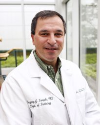 G. J. Tsongalis合作论文数Audrey & Theodor Geisel Sch Med Dartmouth, Lebanon, NH USA9Viral M. Patel合作论文数8Divya Sharma合作论文数8Cindy Wassef合作论文数Department of Dermatology, Rutgers Robert Wood Johnson Medical School8Debra S. Heller合作论文数8Karl Hoegler合作论文数7Douglas H Atmatzidis合作论文数7Gizem Tumer合作论文数7Paul D. Langer合作论文数7Javier Rojas合作论文数6John Elizabeth S合作论文数6R. Malliah合作论文数5Alyssa Swearingen合作论文数5WS TANZ合作论文数5Philip J. Cohen合作论文数5S. G. Nychay合作论文数5Tiffany Jow合作论文数5Spandana Maddukuri合作论文数5Hon-Reen Kuo合作论文数5DD PARRISH合作论文数5
 Thu Minh Truong合作论文数5Steven Skula合作论文数5R Kapila合作论文数5Sarkissian Navér合作论文数5M.L. Mesa合作论文数5Jennifer L. Parish合作论文数5




